Low-Dose 3D Bonding Mapping Through "Soft" Core-Loss EELS Tomography and Unsupervised Deep Learning
Pith reviewed 2026-06-27 11:22 UTC · model grok-4.3
The pith
DIPm-TV from soft Fe-M EELS edges produces 1 nm 3D oxidation-state volumes of nanocubes from nine projections.
A machine-rendered reading of the paper's core claim, the machinery that carries it, and where it could break.
Core claim
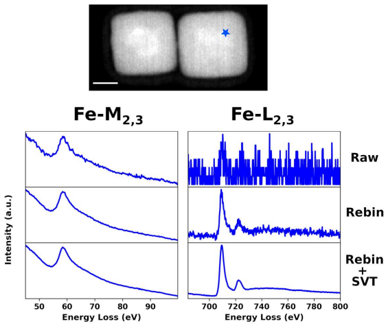
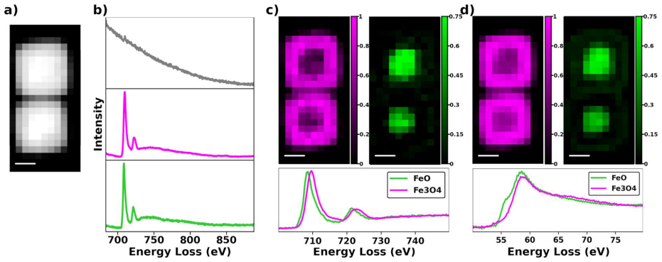
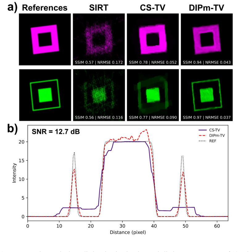
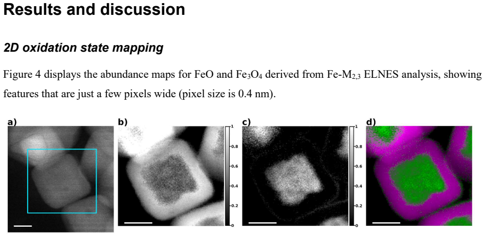
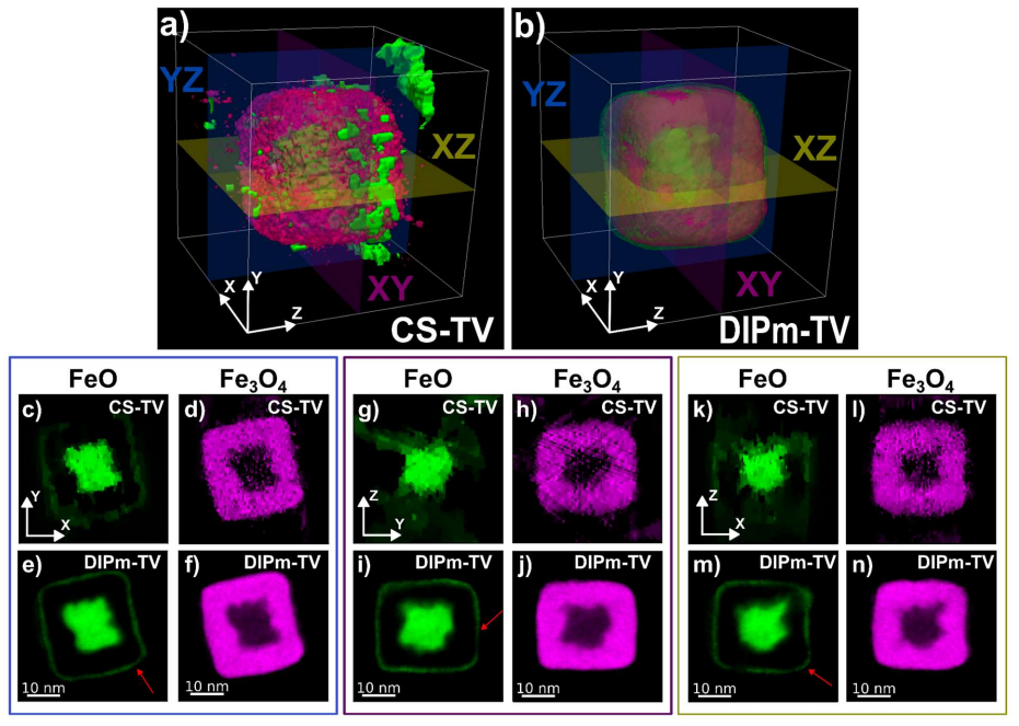
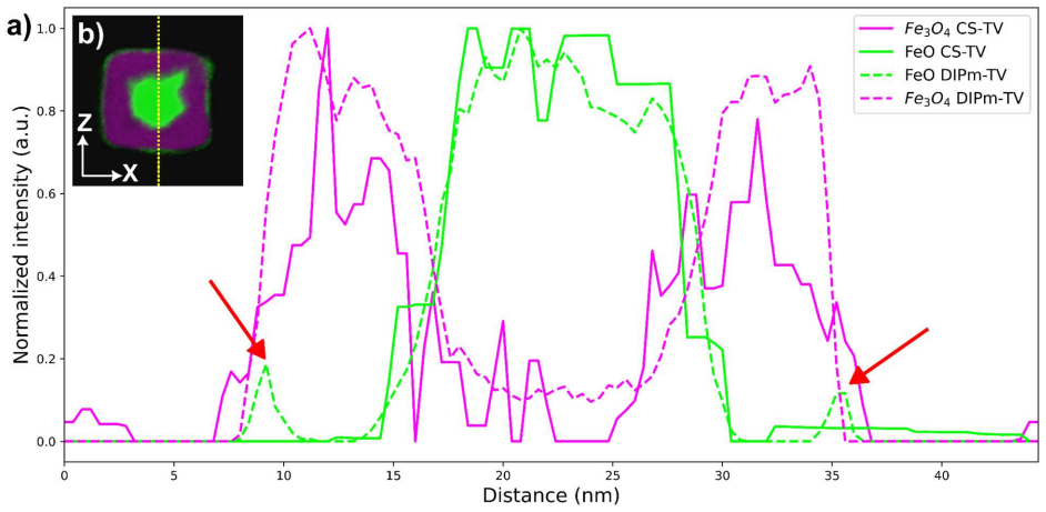
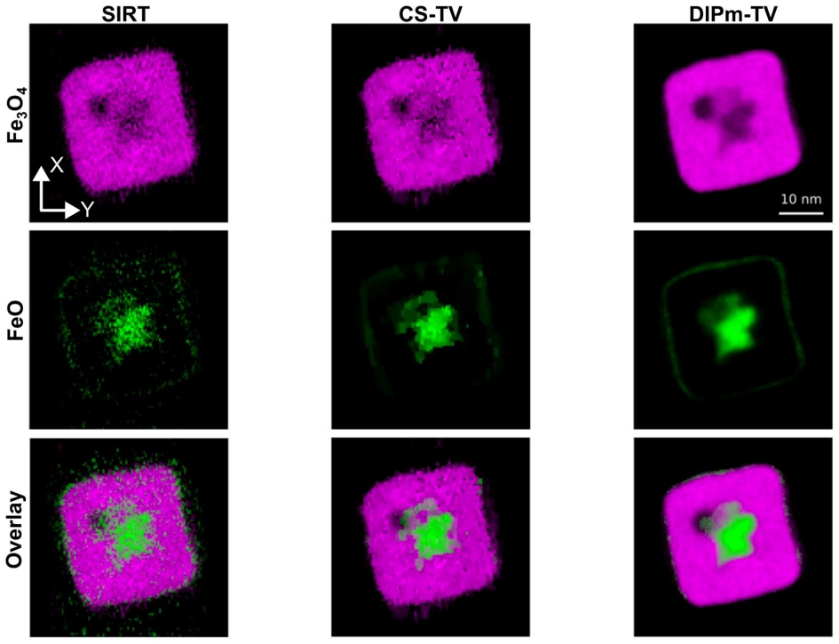
Using Fe-M2,3 core-loss EELS maps that are fifty times more dose-efficient than conventional L edges, the DIPm-TV algorithm jointly reconstructs multiple spectroscopic channels under total-variation regularization and recovers ~1 nm isotropic oxidation-state volumes from as few as nine projections; these volumes preserve cubic morphology, recover the thin outer FeO shell, and reveal a small internal void that conventional reconstruction methods cannot access.
What carries the argument
DIPm-TV (multi-channel deep image prior with total variation regularization), an unsupervised method that reconstructs multiple EELS channels together by exploiting their spatial correlations under sparse-view, low-dose conditions.
If this is right
- High-quality spectroscopic tomograms are obtained from only nine projections spanning -70° to +70° without HAADF-STEM signal or symmetry constraints.
- Fe-M maps exhibit improved signal-to-noise and spatial resolution relative to conventional Fe-L maps.
- The reconstructed oxidation-state volumes recover the outer FeO shell and a small internal void while preserving cubic shape.
- The same pipeline supplies a pathway for low-dose two- and three-dimensional analytical mapping of other beam-sensitive materials using shallow core-loss edges.
Where Pith is reading between the lines
- The method could be tested on other core-loss edges or on different nanoparticle compositions to check how broadly the correlation assumption holds.
- Because the reconstruction is unsupervised, it may be combined with existing tilt-series acquisition schemes that already limit total dose.
- If the internal void is real, similar low-dose volumes could be used to study porosity evolution during synthesis or annealing of oxide nanoparticles.
- Extending the approach to four or more simultaneous EELS channels might allow simultaneous mapping of multiple oxidation states or bonding types in a single low-dose run.
Load-bearing premise
Spatial correlations across the different EELS channels are strong enough for DIPm-TV to produce faithful three-dimensional volumes from only nine projections without HAADF data or symmetry constraints.
What would settle it
Re-running the simulated nine-projection experiment after deliberately breaking spatial correlations between channels and observing that DIPm-TV no longer recovers the known morphology, shell, or void.
Figures

read the original abstract
Resolving the 3D chemical configuration of beam-sensitive nanomaterials at high spatial resolution remains a persistent frontier in scanning transmission electron microscopy (STEM). The main limitation lies in the trade-off between high electron dose required for analytical signals and the large number of projections needed for tomographic reconstruction. Here, we achieve dose-efficient 3D bonding mapping of FeO/Fe$_3$O$_4$ core-shell nanocubes with high resolution via electron energy loss spectroscopy (EELS). Our approach relies on two developments. First, a standardless "soft" core-loss EELS methodology exploiting Fe-M$_{2,3}$ edges provides ${\sim}50\times$ higher dose efficiency than conventional Fe-L$_{2,3}$ edges, using the latter only as a source of FeO and Fe$_3$O$_4$ standards. Second, we introduce multi-channel deep image prior with total variation regularization (DIPm-TV), an unsupervised method for spectroscopic tomography that jointly reconstructs multiple channels by exploiting spatial correlations under sparse-view and low-dose conditions. Using simulated datasets, high-quality reconstructions are obtained from as few as nine projections over $-70^\circ$ to $+70^\circ$, without HAADF-STEM signal or symmetry constraints. Applied to FeO/Fe$_3$O$_4$ nanocubes, Fe-M$_{2,3}$ EELS maps show improved SNR and spatial resolution, revealing a thin outer FeO shell surrounding the magnetite shell. DIPm-TV yields ${\sim}1$ nm isotropic resolution oxidation-state volumes preserving cubic morphology, recovering the outer FeO shell, and revealing a small internal void, features not accessible with conventional reconstruction methods. This work establishes a pathway for low-dose 2D and 3D analytical mapping of beam-sensitive materials using shallow core-loss edges, enabling orders-of-magnitude dose reduction while maintaining spectral fidelity and reliable 3D information.
Editorial analysis
A structured set of objections, weighed in public.
Referee Report
Summary. The paper claims that combining 'soft' core-loss EELS (Fe-M2,3 edges) with an unsupervised multi-channel deep image prior plus total variation (DIPm-TV) reconstruction enables ~50x dose-efficient 3D oxidation-state mapping of FeO/Fe3O4 nanocubes. From only nine projections over ±70° (no HAADF signal or symmetry), the method is said to deliver ~1 nm isotropic resolution volumes that preserve cubic morphology, recover a thin outer FeO shell, and reveal a small internal void—features inaccessible to conventional tomography. Validation is presented on simulated data and one experimental nanocube case.
Significance. If the central experimental claims hold under rigorous validation, the work would be significant for beam-sensitive nanomaterials: it demonstrates a pathway to 3D chemical bonding maps at high resolution with orders-of-magnitude lower dose, using shallow edges and joint multi-channel reconstruction. The unsupervised DIPm-TV approach and standardless soft-EELS strategy address key dose-projection trade-offs in analytical STEM tomography.
major comments (2)
- [Abstract and Results] Abstract and Results (experimental nanocube case): the claim that DIPm-TV recovers a thin outer FeO shell and small internal void at ~1 nm resolution from nine projections rests on the unverified assumption that multi-channel spatial correlations dominate over regularization artifacts. No quantitative fidelity metric (FSC, feature-size error, or comparison to higher-projection or HAADF-constrained reconstructions) is reported on the real data, leaving open the possibility that the reported morphology and void are induced by the DIPm-TV prior or TV term rather than data-driven.
- [Method] Method (DIPm-TV description): the joint reconstruction exploits correlations across EELS channels under sparse-view conditions, yet the manuscript provides no ablation on the relative contribution of the deep image prior versus TV regularization, nor tests against alternative regularizers or simulated data with known ground-truth voids and shells at the reported dose and angular sampling. This is load-bearing for the experimental feature-recovery claim.
minor comments (1)
- [Abstract] The abstract states 'without HAADF-STEM signal or symmetry constraints' for both simulated and experimental cases; clarify whether any alignment or tilt-axis refinement was performed on the experimental tilt series.
Simulated Author's Rebuttal
We thank the referee for the constructive feedback. We address the two major comments below, agreeing that additional validation would strengthen the manuscript while noting practical constraints on experimental data acquisition.
read point-by-point responses
-
Referee: [Abstract and Results] Abstract and Results (experimental nanocube case): the claim that DIPm-TV recovers a thin outer FeO shell and small internal void at ~1 nm resolution from nine projections rests on the unverified assumption that multi-channel spatial correlations dominate over regularization artifacts. No quantitative fidelity metric (FSC, feature-size error, or comparison to higher-projection or HAADF-constrained reconstructions) is reported on the real data, leaving open the possibility that the reported morphology and void are induced by the DIPm-TV prior or TV term rather than data-driven.
Authors: We agree that quantitative fidelity metrics such as FSC cannot be computed for the experimental case due to the absence of ground truth. Validation in the manuscript rests on the inability of conventional SIRT to recover the shell or void, consistency with 2D EELS maps, and agreement with expected core-shell chemistry from prior literature. The unsupervised DIPm-TV formulation is intended to let data-driven correlations dominate. We will revise the Results and Discussion sections to include a dedicated paragraph on potential regularization effects and any feasible quantitative checks (e.g., inter-channel consistency). However, higher-projection or HAADF-constrained experimental comparisons are not possible without exceeding the low-dose limit required for this beam-sensitive material. revision: partial
-
Referee: [Method] Method (DIPm-TV description): the joint reconstruction exploits correlations across EELS channels under sparse-view conditions, yet the manuscript provides no ablation on the relative contribution of the deep image prior versus TV regularization, nor tests against alternative regularizers or simulated data with known ground-truth voids and shells at the reported dose and angular sampling. This is load-bearing for the experimental feature-recovery claim.
Authors: We concur that explicit ablations would better substantiate the method. Although the manuscript already demonstrates high-quality results on simulated data from nine projections, we will expand the supplementary information with: (i) ablations that isolate the DIP versus TV contributions, (ii) comparisons against alternative regularizers, and (iii) new simulations that embed known thin outer shells and internal voids at the experimental dose and angular sampling. These additions will directly address the load-bearing nature of the feature-recovery claim. revision: yes
- We cannot acquire additional experimental projections or HAADF data on the same nanocube without substantially increasing the total electron dose, which would damage the beam-sensitive FeO/Fe3O4 material and contradict the low-dose premise of the work.
Circularity Check
No circularity: derivation chain is self-contained and externally validated.
full rationale
The paper introduces DIPm-TV as an unsupervised multi-channel reconstruction method, validates it on independent simulated datasets under controlled sparse-view conditions, and then applies it to experimental EELS data. No load-bearing step reduces by definition or self-citation to the target sample's features; the central claims rest on the method's exploitation of spatial correlations across channels, which is tested separately from the experimental nanocube results. The use of Fe-L edges only as standards for Fe-M edges is a calibration step, not a fitted prediction of the reported 3D volumes.
Axiom & Free-Parameter Ledger
free parameters (1)
- DIPm-TV regularization parameters
axioms (1)
- domain assumption Fe-M2,3 core-loss edges provide bonding information equivalent to Fe-L2,3 edges when calibrated with standards from the latter
Reference graph
Works this paper leans on
-
[1]
Use of ICA in Fe-M2,3 edges and SVD results An initial strategy for the discerning of the FeO and Fe3O4 ELNES features in the Fe-M2,3 edges was employing a similar analysis routine to the one shown in the main text for the Fe-L2,3 edges, consisting of a denoising via SVD and the implementation of BSS using 3 components for the background and each Fe speci...
1999
-
[2]
Determination of the Fe-M2,3 EELS edges for FeO and Fe3O4 We consider that all possible spectra within the F-CL SPIM can be expressed as a combination of a power law background and the normalized, background-extracted Fe-L2,3 references gathered from the dataset (here called LFeO and LFe3O4). Similarly, all possible spectra within the F-LL SPIM can be exp...
-
[3]
Dose estimation The estimation for the dose used two experimental parameters: the number of counts of a pixel in the SPIM where there was only vacuum (or, in the case that was not feasible, on top of the substrate), which we will call 𝐶் and the estimated size of the nanocube being probed. The probe size was estimated as a combination of the defocus of th...
-
[4]
Initial volume estimations In order to have an initial estimation of the FeO/Fe3O4 volume ratio, a very simple approximation was employed, by taking measurements of the width and height of the shell and the core in the X-Y plane, and approximating their Z measurements as the mean of these two values, assuming a cubic shape. 43
-
[5]
For the experimental data, 2000 iterations were conducted to minimize noise and mitigate overfitting
DIPm-TV training and network architecture: The simulated data reconstructions were performed using 1000 iterations, selecting the reconstruction with the lowest loss. For the experimental data, 2000 iterations were conducted to minimize noise and mitigate overfitting. The network input noise is sampled from a uniform distribution. All reconstructions, mod...
2000
-
[6]
DIPm-TV simulation: Figure S4 compares SIRT, CS-TV and DIPm-TV reconstructions of the simulated core-shell phantom across different noise levels. At the lowest noise level (Figure S4e-j), the predominant source of artefacts is the combination of the large tilt increment and the mild missing wedge (aligned vertically in these figures). While CS-TV provides...
discussion (0)
Sign in with ORCID, Apple, or X to comment. Anyone can read and Pith papers without signing in.