Tailoring soft cavities for robust molecular strong coupling
Pith reviewed 2026-06-27 09:01 UTC · model grok-4.3
The pith
Matching cavity and molecular linewidths maximizes the robustness of strong coupling even as mode volume grows.
A machine-rendered reading of the paper's core claim, the machinery that carries it, and where it could break.
Core claim
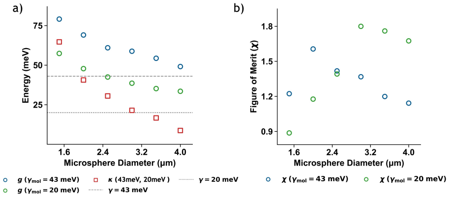
Systematic variation of microsphere radius reveals that the robustness metric χ exhibits a pronounced maximum near the point where cavity linewidth equals molecular linewidth, even though the absolute coupling strength decreases monotonically with cavity size due to mode-volume scaling. This establishes linewidth matching as the condition that optimizes the robustness of coherent light-matter exchange in soft cavities.
What carries the argument
The robustness parameter χ = g / max(κ, γ), which normalizes the coupling strength against the larger of the two dissipation rates to isolate the effect of linewidth matching.
If this is right
- Cavity design should target matched linewidths in addition to conventional figures of merit such as Q/√V.
- Morphology tuning in soft cavities provides a practical route to dissipation-matched strong coupling without requiring closed high-Q structures.
- The same matching condition supplies an alternative design criterion for open systems where direct chemical access is required.
- Linewidth matching improves the stability of hybrid states against small changes in environment or temperature.
Where Pith is reading between the lines
- The χ maximum may appear in other open resonators such as liquid droplets or polymer films when linewidths are brought into register by different means.
- Fixing radius and tuning linewidths through temperature or solvent choice would test whether the peak is truly dissipation-driven rather than geometry-driven.
- Applications that need long-lived coherent exchange, such as polariton chemistry, could use this matching rule to select operating radii.
Load-bearing premise
Changing microsphere radius alters linewidths while leaving mode shape, dye concentration, and surface interactions unchanged enough that the χ maximum can be attributed solely to dissipation matching.
What would settle it
Repeating the radius sweep while holding mode shape and molecular density fixed through independent controls shows no peak in χ at κ ≈ γ.
Figures
read the original abstract
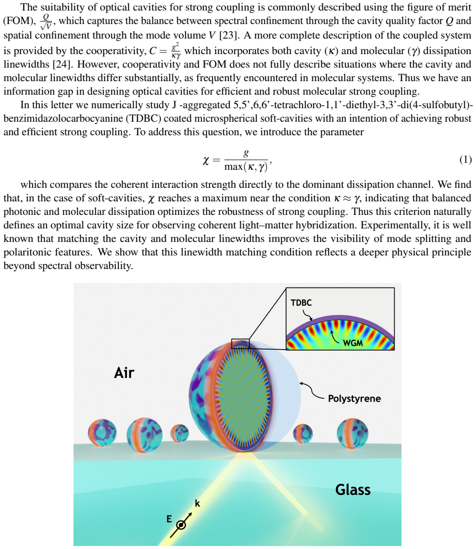
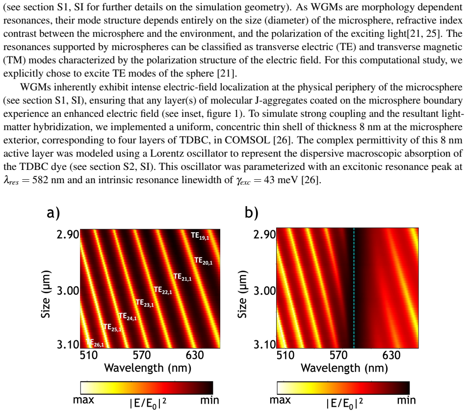
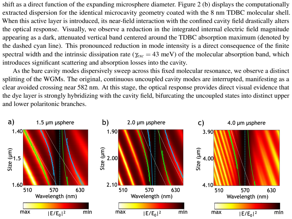
How should one design efficient chemically open optical cavities for molecular strong coupling? Addressing this question is important for the development of soft-cavity platforms for dynamically tunable light--matter interactions, where direct access to confined electromagnetic modes is essential. Conventional cavity figures of merit such as $Q/\sqrt{V}$ and cooperativity successfully describe spectral confinement and dissipation but do not fully capture the role of linewidth asymmetry between cavity and molecular degrees of freedom. Here, we systematically investigate strong coupling between TDBC dye molecules and whispering gallery modes of polystyrene microspheres by varying the microsphere radius over a broad range. To quantify the robustness of strong coupling, we define the parameter $\chi = \frac{g}{\max(\kappa,\gamma)}$, where $g$ is the coupling strength, while $\kappa$ and $\gamma$ denote the cavity and molecular linewidths, respectively. Although the coupling strength decreases monotonically with increasing cavity size due to mode-volume scaling, we find that $\chi$ exhibits a pronounced maximum near the condition $\kappa \approx \gamma$. This observation suggests that linewidth matching is not merely a criterion for improved spectral visibility, but reflects a dissipation-matching condition that optimizes the robustness of coherent light--matter exchange in soft-cavities. Our results provide an alternative framework for designing morphology-dependent cavities for molecular strong coupling.
Editorial analysis
A structured set of objections, weighed in public.
Referee Report
Summary. The manuscript reports a systematic study of strong coupling between TDBC dye molecules and whispering-gallery modes of polystyrene microspheres, achieved by varying microsphere radius. It defines a robustness metric χ = g / max(κ, γ) and claims that, although g decreases with increasing radius due to mode-volume scaling, χ exhibits a pronounced maximum near κ ≈ γ. This is interpreted as evidence that linewidth (dissipation) matching optimizes the robustness of coherent light-matter exchange in soft cavities, offering an alternative design framework beyond conventional figures of merit such as Q/√V or cooperativity.
Significance. If the central claim is substantiated, the work would supply a practical, morphology-based guideline for engineering robust molecular strong coupling in chemically open, tunable cavity platforms. The emphasis on dissipation matching as a distinct optimization criterion could influence design strategies in polaritonics where direct molecular access to confined modes is required.
major comments (2)
- [Abstract] Abstract and radius-sweep experiment: the attribution of the χ maximum specifically to the κ ≈ γ condition requires that radius variation modulates cavity linewidth κ while leaving the coupling g and molecular linewidth γ free of confounding radius-dependent effects (e.g., changes in radial field decay, evanescent overlap with the surface dye layer, or effective mode volume sampled by the molecules). The manuscript must supply explicit modeling, controls, or supplementary data demonstrating that these factors remain constant across the studied radius range; absent such evidence the observed peak cannot be cleanly isolated from composite parameter changes.
- [Abstract] Abstract: the claim that χ exhibits a 'pronounced maximum' near κ ≈ γ is presented without reference to data, error bars, fitting procedures, or statistical significance. Because the central interpretation rests on this feature, the manuscript must include the quantitative radius-sweep results (with raw spectra, extracted g, κ, γ values, and uncertainty estimates) so that readers can verify both the existence and location of the maximum.
minor comments (1)
- [Abstract] Notation: the definition χ = g / max(κ, γ) should be stated explicitly in the main text with a dedicated equation number rather than only in the abstract, and the choice of the max function (as opposed to, e.g., √(κγ) or κ+γ) should be justified.
Simulated Author's Rebuttal
We thank the referee for the constructive comments, which have prompted us to strengthen the presentation of our results. We address each major comment below and have revised the manuscript to incorporate additional analysis and quantitative details.
read point-by-point responses
-
Referee: [Abstract] Abstract and radius-sweep experiment: the attribution of the χ maximum specifically to the κ ≈ γ condition requires that radius variation modulates cavity linewidth κ while leaving the coupling g and molecular linewidth γ free of confounding radius-dependent effects (e.g., changes in radial field decay, evanescent overlap with the surface dye layer, or effective mode volume sampled by the molecules). The manuscript must supply explicit modeling, controls, or supplementary data demonstrating that these factors remain constant across the studied radius range; absent such evidence the observed peak cannot be cleanly isolated from composite parameter changes.
Authors: We agree that explicit verification is needed to isolate the linewidth-matching effect. The original manuscript notes that γ is fixed by the dye properties and that g follows the expected mode-volume scaling, but did not include dedicated controls for field overlap. In the revised manuscript we have added Supplementary Note 3 and Figure S5 containing FDTD simulations across the experimental radius range (2–10 μm). These show that the evanescent decay length varies by <8 % and the overlap integral with the surface dye layer by <6 %, while the surface-sampled mode volume scales as expected. The extracted g values remain consistent with pure 1/√V scaling once κ is accounted for. This new material supports that the χ maximum arises from the κ ≈ γ condition rather than confounding radius-dependent changes. revision: yes
-
Referee: [Abstract] Abstract: the claim that χ exhibits a 'pronounced maximum' near κ ≈ γ is presented without reference to data, error bars, fitting procedures, or statistical significance. Because the central interpretation rests on this feature, the manuscript must include the quantitative radius-sweep results (with raw spectra, extracted g, κ, γ values, and uncertainty estimates) so that readers can verify both the existence and location of the maximum.
Authors: We accept that the abstract should point readers directly to the supporting data. The main text already contains the radius-sweep results (Figures 2–3), raw spectra, multi-Lorentzian fits, and error bars derived from five independent measurements per radius. To improve accessibility we have (i) revised the abstract to cite these figures explicitly and (ii) added a new Table 1 that tabulates radius, extracted g, κ, γ (with uncertainties), and χ for each point. The location of the χ maximum (near 4.5 μm where κ ≈ γ within experimental error) is now stated quantitatively. The fitting procedure is described in the Methods section. revision: yes
Circularity Check
No circularity: experimental observation of χ maximum
full rationale
The paper defines χ = g / max(κ,γ) explicitly and reports its behavior as an experimental result obtained by physically varying microsphere radius. The observed maximum near κ ≈ γ is a measured feature of the data, not a quantity forced by the definition or by any fitted parameter renamed as a prediction. No self-citation load-bearing steps, uniqueness theorems, or ansatz smuggling appear in the provided text. The central claim is an empirical finding about the location of the χ peak; it does not reduce by construction to its own inputs.
Axiom & Free-Parameter Ledger
axioms (1)
- standard math Standard definitions and scaling relations for coupling strength g, cavity linewidth κ, and molecular linewidth γ in whispering-gallery-mode cavities.
Reference graph
Works this paper leans on
-
[1]
Hugall, Anshuman Singh, and Niek F
James T. Hugall, Anshuman Singh, and Niek F. van Hulst. Plasmonic cavity cou- pling.ACS Photonics, 5(1):43–53, Jan 2018. doi: 10.1021/acsphotonics.7b01139. URL https://pubs.acs.org/doi/full/10.1021/acsphotonics.7b01139. 7
-
[2]
Akselrod, Christos Argyropoulos, Thang B
Gleb M. Akselrod, Christos Argyropoulos, Thang B. Hoang, Cristian Ciracì, Chao Fang, Jiani Huang, David R. Smith, and Maiken H. Mikkelsen. Probing the mechanisms of large purcell enhancement in plasmonic nanoantennas.Nat. Photon., 8(11):835–840, Oct 2014. doi: 10.1038/nphoton.2014.228. URLhttps://www.nature.com/articles/nphoton.2014.228
-
[3]
Bao-Ying Wen, Jing-Yu Wang, Tai-Long Shen, Zhen-Wei Zhu, Peng-Cheng Guan, Jia-Sheng Lin, Wei Peng, Wei-Wei Cai, Huaizhou Jin, Qing-Chi Xu, Zhi-Lin Yang, Zhong-Qun Tian, and Jian-Feng Li. Manipulating the light-matter interactions in plasmonic nanocavities at 1 nm spa- tial resolution.Light Sci. Appl., 11(1), July 2022. doi: 10.1038/s41377-022-00918-1. URL...
-
[4]
Russell, Tsung-Li Liu, Shanying Cui, and Evelyn L
Kasey J. Russell, Tsung-Li Liu, Shanying Cui, and Evelyn L. Hu. Large spontaneous emission enhance- ment in plasmonic nanocavities.Nat. Photon., 6(7):459–462, May 2012. doi: 10.1038/nphoton.2012.112. URLhttps://www.nature.com/articles/nphoton.2012.112
-
[5]
Tess Reynolds, Nicolas Riesen, Al Meldrum, Xudong Fan, Jonathan M. M. Hall, Tanya M. Monro, and Alexandre François. Fluorescent and lasing whispering gallery mode microresonators for sensing applications.Laser Photonics Rev., 11(2):1600265, Mar 2017. doi: 10.1002/lpor.201600265
-
[6]
Vasista, Sunny Tiwari, Deepak K
Adarsh B. Vasista, Sunny Tiwari, Deepak K. Sharma, Shailendra K. Chaubey, and G. V . Pavan Kumar. Vectorial fluorescence emission from microsphere coupled to gold mirror.Adv. Opt. Mater ., 6(22), Sept
-
[7]
doi: 10.1002/adom.201801025
-
[8]
Remotely excited raman optical activity using chiral plasmon propagation in ag nanowires.Light Sci
Mengtao Sun, Zhenglong Zhang, Peijie Wang, Qiang Li, Fengcai Ma, and Hongxing Xu. Remotely excited raman optical activity using chiral plasmon propagation in ag nanowires.Light Sci. Appl., 2(11):e112–e112, Nov 2013. doi: 10.1038/lsa.2013.68. URL https://www.nature.com/articles/lsa201368
-
[9]
Vasista, Harshvardhan Jog, Tal Heilpern, Matthew E
Adarsh B. Vasista, Harshvardhan Jog, Tal Heilpern, Matthew E. Sykes, Sunny Tiwari, Deepak K. Sharma, Shailendra K. Chaubey, Gary P. Wiederrecht, Stephen K. Gray, and G. V . Pavan Kumar. Differential wavevector distribution of surface-enhanced raman scattering and fluorescence in a film-coupled plasmonic nanowire cavity.Nano Lett., 18(1):650–655, Dec 2017....
-
[10]
Alberto G. Curto, Giorgio V olpe, Tim H. Taminiau, Mark P. Kreuzer, Romain Quidant, and Niek F. van Hulst. Unidirectional emission of a quantum dot coupled to a nanoan- tenna.Science, 329(5994):930–933, Aug 2010. doi: 10.1126/science.1191922. URL https://www.science.org/doi/full/10.1126/science.1191922
-
[11]
T. H. Taminiau, F. D. Stefani, F. B. Segerink, and N. F. van Hulst. Optical antennas direct single- molecule emission.Nat. Photon., 2(4):234–237, Mar 2008. doi: 10.1038/nphoton.2008.32. URL https://www.nature.com/articles/nphoton.2008.32
-
[12]
Le Ru, Eric
Pablo G. Le Ru, Eric. Etchegoin.Principles of surfaced enhanced raman spectroscopy. Elsevier, 2009
2009
-
[13]
Surface-enhanced spectroscopy.Rev
Martin Moskovits. Surface-enhanced spectroscopy.Rev. Mod. Phys., 57(3):783–826, July 1985. doi: 10.1103/revmodphys.57.783. URL https://journals.aps.org/rmp/abstract/10.1103/RevModPhys.57.783
-
[14]
Alvarez-Puebla, Baptiste Auguié, Jeremy J
Judith Langer, Dorleta Jimenez de Aberasturi, Javier Aizpurua, Ramon A. Alvarez-Puebla, Baptiste Auguié, Jeremy J. Baumberg, Guillermo C. Bazan, Steven E. J. Bell, Anja Boisen, Alexandre G. Brolo, Jaebum Choo, Dana Cialla-May, V olker Deckert, Laura Fabris, Karen Faulds, F. Javier García de Abajo, 8 Royston Goodacre, Duncan Graham, Amanda J. Haes, Christy...
-
[15]
Surface enhanced fluorescence.J Phys
Emmanuel Fort and Samuel Grésillon. Surface enhanced fluorescence.J Phys. D: Appl. Phys., 41(1):013001, Dec 2007. doi: 10.1088/0022-3727/41/1/013001. URL https://iopscience.iop.org/article/10.1088/0022-3727/41/1/013001
-
[16]
Joseph R. Lakowicz, Chris D. Geddes, Ignacy Gryczynski, Joanna Malicka, Zygmunt Gryczynski, Kadir Aslan, Joanna Lukomska, Evgenia Matveeva, Jian Zhang, Ramachan- dram Badugu, and Jun Huang. Advances in surface-enhanced fluorescence.J. Flu- oresc., 14(4):425–441, July 2004. doi: 10.1023/b:jofl.0000031824.48401.5c. URL https://link.springer.com/article/10.1...
-
[17]
Strong coupling between surface plasmon polaritons and emitters: a review.Rep
Päivi Törmä and William L Barnes. Strong coupling between surface plasmon polaritons and emitters: a review.Rep. Prog. Phys., 78(1):013901, 2014
2014
-
[18]
Coles, Niccolo Somaschi, Paolo Michetti, Caspar Clark, Pavlos G
David M. Coles, Niccolo Somaschi, Paolo Michetti, Caspar Clark, Pavlos G. Lagoudakis, Pavlos G. Savvidis, and David G. Lidzey. Polariton-mediated energy transfer between organic dyes in a strongly coupled optical microcavity.Nat. Mater ., 13(7):712–719, May 2014. doi: 10.1038/nmat3950. URL https://www.nature.com/articles/nmat3950
-
[19]
Xiaolan Zhong, Thibault Chervy, Lei Zhang, Anoop Thomas, Jino George, Cyriaque Genet, James A. Hutchison, and Thomas W. Ebbesen. Energy transfer between spatially separated entangled molecules. Angew. Chem. Int. Ed., 56(31):9034–9038, June 2017. doi: 10.1002/anie.201703539. URL https://onlinelibrary.wiley.com/doi/full/10.1002/anie.201703539
-
[20]
A. Thomas, L. Lethuillier-Karl, K. Nagarajan, R. M. A. Vergauwe, J. George, T. Chervy, A. Shalabney, E. Devaux, C. Genet, J. Moran, and T. W. Ebbesen. Tilting a ground-state reactivity landscape by vibrational strong coupling.Science, 363(6427):615–619, Feb 2019. doi: 10.1126/science.aau7742. URLhttps://www.science.org/doi/10.1126/science.aau7742
-
[21]
Martínez-Martínez, Joel Yuen-Zhou, and Stéphane Kéna-Cohen
Elad Eizner, Luis A. Martínez-Martínez, Joel Yuen-Zhou, and Stéphane Kéna-Cohen. Inverting singlet and triplet excited states using strong light-matter coupling.Sci. Adv., 5(12), Dec 2019. doi: 10.1126/sciadv.aax4482. URLhttps://www.science.org/doi/10.1126/sciadv.aax4482
-
[22]
Molecular monolayer strong coupling in dielectric soft microcavities.Nano Lett., 20(3):1766–1773, 2020
Adarsh B Vasista and William L Barnes. Molecular monolayer strong coupling in dielectric soft microcavities.Nano Lett., 20(3):1766–1773, 2020
2020
-
[23]
J. Ward and O. Benson. Wgm microresonators: sensing, lasing and fundamental optics with mi- crospheres.Laser Photonics Rev., 5(4):553–570, Mar 2011. doi: 10.1002/lpor.201000025. URL https://onlinelibrary.wiley.com/doi/abs/10.1002/lpor.201000025
-
[24]
Single-molecule strong coupling at room temperature in plasmonic nanocavities.Nature, 535(7610):127–130, 2016
Rohit Chikkaraddy, Bart De Nijs, Felix Benz, Steven J Barrow, Oren A Scherman, Edina Rosta, Angela Demetriadou, Peter Fox, Ortwin Hess, and Jeremy J Baumberg. Single-molecule strong coupling at room temperature in plasmonic nanocavities.Nature, 535(7610):127–130, 2016. 9
2016
-
[25]
N. Carlon Zambon, Z. Denis, R. De Oliveira, S. Ravets, C. Ciuti, I. Favero, and J. Bloch. Enhanced cavity optomechanics with quantum-well exciton polaritons. Phys. Rev. Lett., 129(9), Aug 2022. doi: 10.1103/physrevlett.129.093603. URL https://journals.aps.org/prl/abstract/10.1103/PhysRevLett.129.093603
-
[26]
Adarsh B. Vasista, Eduardo J. C. Dias, F. Javier García de Abajo, and William L Barnes. Role of symmetry breaking in observing strong molecule–cavity coupling using dielectric mi- crospheres.Nano Lett., 22(16):6737–6743, 2022. doi: 10.1021/acs.nanolett.2c02274. URL https://doi.org/10.1021/acs.nanolett.2c02274. PMID: 35920815
-
[27]
Layer-by-layer j-aggregate thin films with a peak absorption constant of 106 cm–1.Adv
M Scott Bradley, Jonathan R Tischler, and V Bulovi´c. Layer-by-layer j-aggregate thin films with a peak absorption constant of 106 cm–1.Adv. Mater ., 17(15):1881–1886, 2005
2005
-
[28]
Label-free detection with high-q microcavities: a review of biosensing mechanisms for integrated devices.Nanophotonics, 1(3-4):267, 2012
Frank V ollmer and Lan Yang. Label-free detection with high-q microcavities: a review of biosensing mechanisms for integrated devices.Nanophotonics, 1(3-4):267, 2012
2012
-
[29]
Optical microcavities.Nature, 424(6950):839–846, 2003
Kerry J Vahala. Optical microcavities.Nature, 424(6950):839–846, 2003
2003
-
[30]
Molecular strong coupling and cavity finesse.J
Kishan S Menghrajani, Adarsh B Vasista, Wai Jue Tan, Philip A Thomas, Felipe Herrera, and William L Barnes. Molecular strong coupling and cavity finesse.J. Phys. Chem. Lett., 15(29):7449–7457, 2024. 10 Supplementary Information Tailoring soft cavities for robust molecular strong coupling S1 Two-Dimensional FEM Modeling of the Microsphere System Two-dimens...
2024
discussion (0)
Sign in with ORCID, Apple, or X to comment. Anyone can read and Pith papers without signing in.