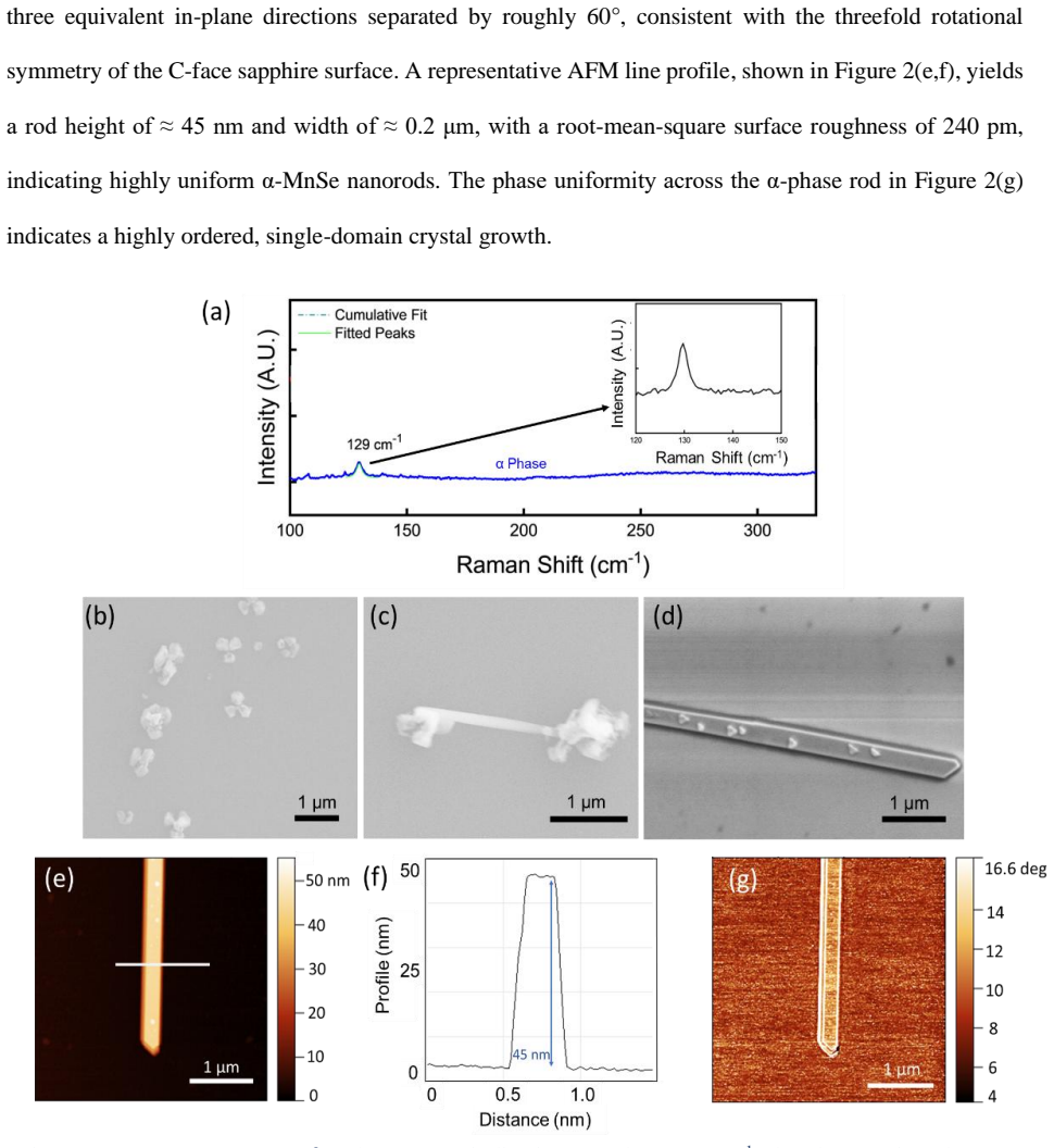
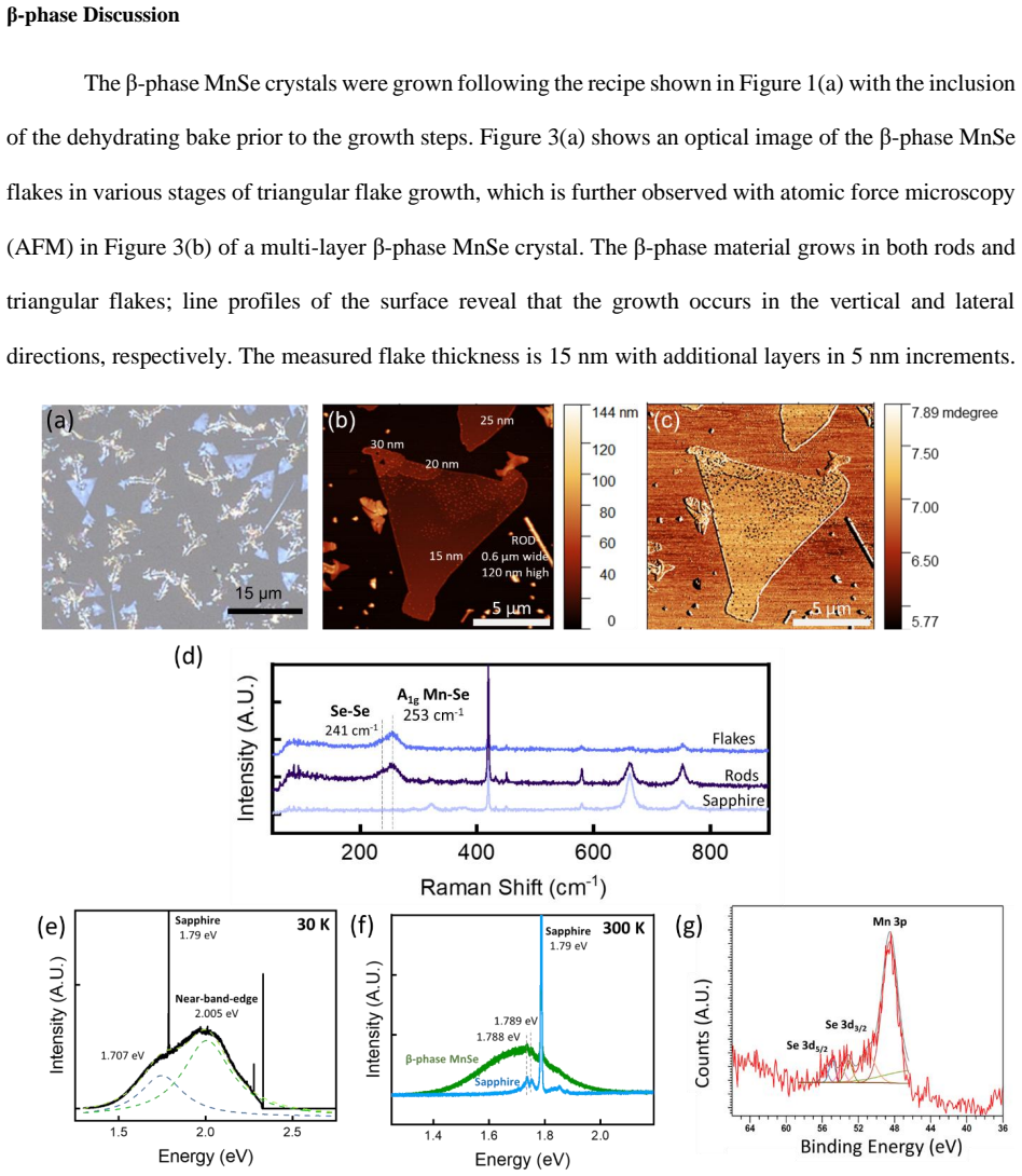
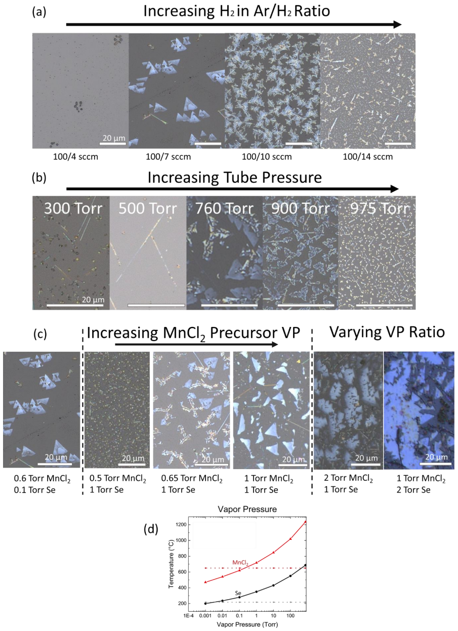
Controllable Growth and Characterization of {α}- and {β}-phase MnSe by Chemical Vapor Deposition
Pith reviewed 2026-06-26 20:03 UTC · model grok-4.3
The pith
CVD process grows phase-pure α-MnSe nanorods and β-MnSe flakes with a Néel temperature of 53 K.
A machine-rendered reading of the paper's core claim, the machinery that carries it, and where it could break.
Core claim
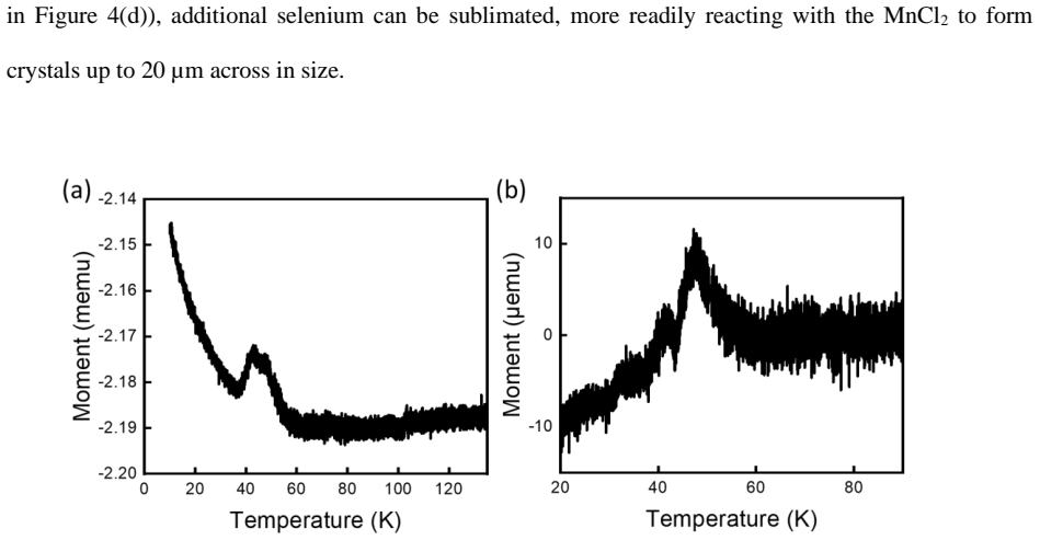
A three-zone CVD process with elemental Se and MnCl2 in Ar/H2 atmosphere produces phase-pure α-MnSe nanorods and β-MnSe triangular flakes up to 20 μm laterally and 15-30 nm thick. Precursor vapor pressure dominates lateral size control. The β-phase exhibits a photoluminescence bandgap of approximately 2.0 eV. Vibrating-sample magnetometry on the β-phase films reveals a Néel temperature of 53 K, supplying evidence of antiferromagnetism in the multilayer regime.
What carries the argument
The three-zone chemical vapor deposition process with elemental Se and MnCl2 precursors, in which precursor vapor pressure rather than H2 partial pressure controls lateral flake size and selects between α- and β-phases.
If this is right
- Phase-pure α-MnSe nanorods and β-MnSe flakes can be grown selectively by adjusting precursor vapor pressure.
- β-MnSe multilayer films display antiferromagnetic ordering with a Néel temperature of 53 K.
- The β-phase films possess an optical bandgap of approximately 2.0 eV.
- MnSe films grown this way form a tunable platform for 2D spintronic and optoelectronic devices.
- Lateral size of β-phase flakes reaches up to 20 μm while thickness stays in the 15-30 nm range.
Where Pith is reading between the lines
- Monolayer or few-layer β-MnSe might exhibit the ferromagnetism with Curie temperature near 250 K that theory predicts, once the multilayer antiferromagnetic state is suppressed by reduced thickness.
- The demonstrated air stability and phase control could allow direct integration of MnSe flakes into van der Waals heterostructures without additional encapsulation.
- Varying the substrate or adding a second precursor zone could extend the method to other transition-metal chalcogenides that also show competing magnetic phases.
- Device prototypes could test whether the 53 K Néel temperature sets a practical operating limit for spintronic elements based on these multilayer films.
Load-bearing premise
The vibrating-sample magnetometry signal originates solely from the MnSe film without contributions from the sapphire substrate or impurities, and Raman spectroscopy provides unambiguous phase identification for the multilayer films.
What would settle it
Repeating the vibrating-sample magnetometry measurement on an identical bare sapphire substrate or on the same substrate after the MnSe film has been removed and observing no transition at 53 K would show whether the magnetic signal belongs to the film.
Figures
read the original abstract
Manganese selenide (MnSe) is a promising air-stable two-dimensional magnetic semiconductor for which theory predicts robust ferromagnetism in monolayers with Curie temperatures approaching 250 K. However, the crystallographic phases and magnetic properties of thin-film MnSe grown by scalable methods remain poorly understood. Here, we demonstrate the controllable growth of ${\alpha}$- and ${\beta}$-phase MnSe on C-face sapphire using a three-zone chemical vapor deposition process with elemental Se and ${MnCl_{2}}$ precursors in an $Ar/{H_{2}}$ atmosphere. Using Raman spectroscopy, atomic force microscopy, scanning electron microscopy, and X-ray photoelectron spectroscopy, we show that our process yields phase-pure ${\alpha}$-MnSe nanorods and ${\beta}$-MnSe triangular flakes with lateral sizes up to 20 ${\mu}m$ and thicknesses of 15-30 nm. Low-temperature photoluminescence of the ${\beta}$-phase films reveals a bandgap of approximately 2.0 eV. Systematic variation of growth parameters shows that precursor vapor pressure, rather than ${H_{2}}$ partial pressure, is the dominant factor controlling lateral flake size. Vibrating-sample magnetometry measurements reveal a $N{\'e}el$ temperature of 53 K in the ${\beta}$-phase films, providing clear evidence of antiferromagnetism in the multilayer regime and establishing MnSe as a tunable platform for 2D spintronic and optoelectronic devices.
Editorial analysis
A structured set of objections, weighed in public.
Referee Report
Summary. The manuscript presents a three-zone CVD process using MnCl2 and Se precursors to grow phase-pure α-MnSe nanorods and β-MnSe triangular flakes (lateral sizes up to 20 μm, thicknesses 15-30 nm) on C-face sapphire. Raman, AFM, SEM, and XPS are used to establish morphology and phase identity; low-temperature PL gives a ~2.0 eV bandgap for β-MnSe. Growth-parameter studies identify precursor vapor pressure as the dominant control on flake size. VSM data on the β-phase films are reported to show a Néel temperature of 53 K, interpreted as evidence of antiferromagnetism in the multilayer regime.
Significance. If the VSM signal can be unambiguously attributed to the MnSe film, the work supplies a scalable, phase-selective growth route for an air-stable 2D magnetic semiconductor and demonstrates that β-MnSe multilayers exhibit antiferromagnetic order below 53 K. The explicit mapping of growth conditions to lateral size is a practical contribution for device-oriented synthesis.
major comments (2)
- [Abstract; magnetometry section] Abstract and magnetometry results: the Néel-temperature claim of 53 K rests on VSM data whose origin is not secured against substrate or impurity contributions. No bare-sapphire control runs, thickness-series scaling of the moment, or quantitative impurity limits (e.g., Fe or Mn-oxide detection thresholds from XPS) are described, yet the films are only 15-30 nm thick. This directly undermines the central antiferromagnetism conclusion.
- [Raman spectroscopy; growth and characterization sections] Raman and phase-purity claims for the β-MnSe magnetometry samples: multilayer films (15-30 nm) can exhibit strain-induced peak shifts or mixed-domain spectra that overlap with α-phase signatures. The manuscript does not report complementary XRD or cross-sectional TEM on the same flakes used for VSM, leaving the phase assignment load-bearing but incompletely validated.
minor comments (2)
- [Growth-parameter section] The abstract states that 'precursor vapor pressure, rather than H2 partial pressure, is the dominant factor,' but the main text would benefit from a table or figure explicitly showing the parameter sweep and extracted flake-size statistics.
- [AFM/SEM sections] Error bars, number of measured flakes, and substrate-temperature uniformity data are not mentioned in the characterization sections; these details would improve reproducibility.
Simulated Author's Rebuttal
We thank the referee for their careful and constructive review of our manuscript. We address each major comment below with clarifications and indicate planned revisions.
read point-by-point responses
-
Referee: [Abstract; magnetometry section] Abstract and magnetometry results: the Néel-temperature claim of 53 K rests on VSM data whose origin is not secured against substrate or impurity contributions. No bare-sapphire control runs, thickness-series scaling of the moment, or quantitative impurity limits (e.g., Fe or Mn-oxide detection thresholds from XPS) are described, yet the films are only 15-30 nm thick. This directly undermines the central antiferromagnetism conclusion.
Authors: We agree that additional controls would strengthen the attribution of the magnetic signal to the MnSe film. The observed transition temperature of 53 K matches literature values for antiferromagnetic ordering in β-MnSe, and XPS spectra show no detectable Fe or other magnetic impurities. However, the current manuscript does not include bare-substrate controls or explicit thickness-dependent scaling. We will revise the magnetometry section to discuss possible contributions more explicitly and include any available control data. revision: partial
-
Referee: [Raman spectroscopy; growth and characterization sections] Raman and phase-purity claims for the β-MnSe magnetometry samples: multilayer films (15-30 nm) can exhibit strain-induced peak shifts or mixed-domain spectra that overlap with α-phase signatures. The manuscript does not report complementary XRD or cross-sectional TEM on the same flakes used for VSM, leaving the phase assignment load-bearing but incompletely validated.
Authors: Phase assignment for the β-MnSe samples relies on Raman spectra showing characteristic β-phase peaks without α-phase overlap, supported by morphology from AFM and SEM. We acknowledge that XRD or TEM on the exact VSM samples would provide stronger validation against strain or mixed phases. In the revised manuscript we will add XRD patterns from representative β-MnSe films grown under identical conditions. revision: yes
Circularity Check
No circularity: experimental report with direct measurements only
full rationale
The manuscript is a materials-growth and characterization study. It reports CVD synthesis parameters, Raman/AFM/SEM/XPS/PL spectra, and VSM magnetization curves. No equations, fitted models, or derivations appear; the Néel temperature is stated as a direct VSM observation. No self-citation load-bearing steps, ansatz smuggling, or renaming of known results occur. The central claims rest on raw experimental data rather than any reduction to prior inputs by construction.
Axiom & Free-Parameter Ledger
axioms (1)
- domain assumption Raman spectroscopy, XPS, and other techniques can reliably identify α and β phases of MnSe
Reference graph
Works this paper leans on
-
[1]
Berkelbach & D.R
T.C. Berkelbach & D.R. Reichman. Optical and Exitonic Properties of Atomically Thin Transition - Metal Dichalcogenides. Annu Rev Condens. Phys 9, 379 (2018)
2018
-
[2]
Hung, T. L. et al. Pressure Induced Superconductivity in MnSe. Nat. Commun. 12, 5436 (2021)
2021
-
[3]
Mouchliadis, L. et al. Probing valley population imbalance in transition metal dichalcogenides via temperature-dependent second harmonic generation imaging. Npj 2D Mater. Appl. 5, 1–9 (2021)
2021
-
[4]
Zhang, X.-X., You, Y., Zhao, S. Y. F. & Heinz, T. F. Experimental Evidence for Dark Excitons in Monolayer WSe 2. Phys. Rev. Lett. 115, 257403 (2015)
2015
-
[5]
Chun, H. J. et al. Morphology-Tuned Growth of α -MnSe One-Dimensional Nanostructures. J. Phys. Chem. C 111, 519–525 (2007)
2007
-
[6]
& Kim, S
Sahoo, S., Pazhamalai, P., Krishnamoorthy, K. & Kim, S. -J. Hydrothermally prepared α -MnSe nanoparticles as a new pseudocapacitive electrode material for supercapacitor. Electrochimica Acta 268, 403–410 (2018)
2018
-
[7]
& Wang, L
Zhu, M., Xu, H., Tan, Z. & Wang, L. Synthesis of uniform two -dimensional non-layered α-MnSe by molecular sieves modified chemical vapor deposition. Results Phys. 47, 106321 (2023). 13
2023
-
[8]
Zou, J. et al. Controlled growth of ultrathin ferromagnetic β -MnSe semiconductor. SmartMat 3, 482– 490 (2022)
2022
-
[9]
Li, N. et al. Controlled synthesis and Raman study of a 2D antiferromagnetic P-type semiconductor: α- MnSe. Nanoscale 13, 6953–6964 (2021)
2021
-
[10]
Grzybowski, M. J. et al. Wurtzite vs. rock -salt MnSe epitaxy: electronic and altermagnetic properties. Nanoscale 16, 6259–6267 (2024)
2024
-
[11]
Aapro, M. et al. Synthesis and Properties of Monolayer MnSe with Unusual Atomic Structure and Antiferromagnetic Ordering. ACS Nano 15, 13794–13802 (2021)
2021
-
[12]
O’Hara, D. J. et al. Room Temperature Intrinsic Ferromagnetism in Epitaxial Manganese Selenide Films in the Monolayer Limit. Nano Lett. 18, 3125–3131 (2018)
2018
-
[13]
Sattar, S., Islam, M. F. & Canali, C. M. Monolayer MnX and Janus X MnY (X, Y= S, Se, Te): A New Family of 2D Antiferromagnetic Semiconductors. Phys. Rev. B 106, 085410 (2022)
2022
-
[14]
Popović, Z. V. & Milutinović, A. Far-infrared reflectivity and Raman scattering study of α-MnSe. Phys. Rev. B 73, 155203 (2006)
2006
-
[15]
& Smereka, P
Baskaran, A. & Smereka, P. Mechanisms of Stranski-Krastanov growth. J. Appl. Phys. 111, 044321 (2012)
2012
-
[16]
Soonmin, H. et al. Raman Investigations of Metal Chalcogenide Thin Films (A Short Review). Orient. J. Chem. 35, 01–07 (2019)
2019
-
[17]
Reshchikov, M. A. Measurement and analysis of photoluminescence in GaN. J. Appl. Phys. 129, 121101 (2021)
2021
-
[18]
Nguyen, N. L. et al. Understanding native defect induced photoluminescence in Zn2SnO4. Phys. Rev. B 107, L060102 (2023)
2023
-
[19]
-Q., Chen, J
Zhang, B. -Q., Chen, J. -S., Niu, H. -L., Mao, C. -J. & Song, J. -M. Synthesis of ultrathin WSe2 nanosheets and their high -performance catalysis for conversion of amines to imines. Nanoscale 10, 20266–20271 (2018). 14
2018
-
[20]
Castle, J. E. Practical surface analysis by Auger and X-ray photoelectron spectroscopy. D. Briggs and M. P. Seah (Editors). John Wiley and Sons Ltd, Chichester, 1983, 533 pp. Surf. Interface Anal. 6, 302–302 (1984)
1983
-
[21]
Sadler, E. C. et al. Role of H2 in the Substrate -Directed Synthesis of Size -tunable MoSe2 Nanoribbons for Exciton Engineering. ACS Appl. Nano Mater. 5, 11423–11428 (2022)
2022
-
[22]
& Shin, N
Hwang, Y. & Shin, N. Hydrogen-assisted step-edge nucleation of MoSe 2 monolayers on sapphire substrates. Nanoscale 11, 7701–7709 (2019)
2019
-
[23]
D., Brewer, J
Morris, G. D., Brewer, J. H., Dunsiger, S. R. & Montour, M. Antiferromagnetism in solid oxygen. Hyperfine Interact. 104, 381–385 (1997)
1997
-
[24]
Huang, B. et al. Layer-dependent ferromagnetism in a van der Waals crystal down to the monolayer limit. Nature 546, 270–273 (2017)
2017
-
[25]
& Sun, Q
Kan, M., Adhikari, S. & Sun, Q. Ferromagnetism in MnX2 (X = S, Se) monolayers. Phys. Chem. Chem. Phys. 16, 4990–4994 (2014)
2014
discussion (0)
Sign in with ORCID, Apple, or X to comment. Anyone can read and Pith papers without signing in.