μMatch: Foundation Models for Semi-supervised Learning and Domain Adaptation in EM
Pith reviewed 2026-06-26 14:45 UTC · model grok-4.3
The pith
Foundation models pre-trained outside EM can be adapted via student-teacher semi-supervised learning to improve segmentation of mitochondria, nuclei and neurites.
A machine-rendered reading of the paper's core claim, the machinery that carries it, and where it could break.
Core claim
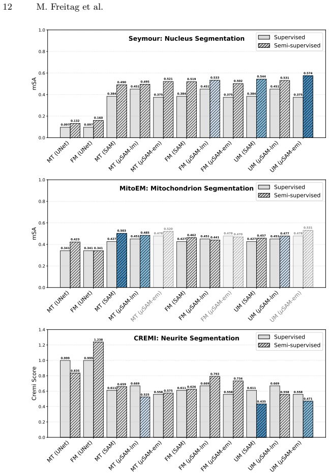
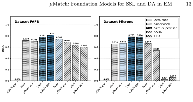
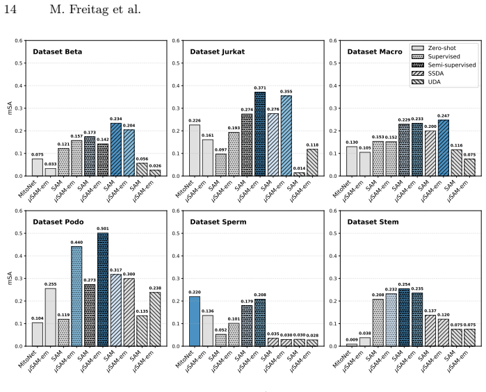
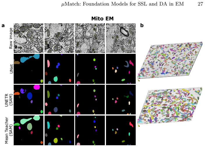
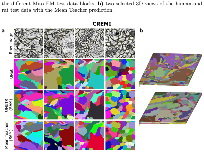
μMatch implements state-of-the-art student-teacher semi-supervised methods and evaluates multiple foundation models on challenging EM segmentation tasks including mitochondrion, nucleus and neurite segmentation. The results demonstrate consistent improvements over strong baselines and indicate that these models can be transferred to diverse EM tasks despite limited annotations and differences in image characteristics.
What carries the argument
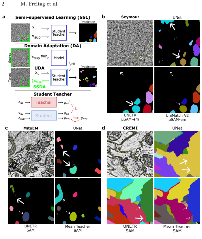
The μMatch framework that adapts foundation models with student-teacher semi-supervised learning for EM domain adaptation and segmentation.
Load-bearing premise
Foundation models pre-trained on non-EM images can transfer effectively to EM via student-teacher learning despite differences in image characteristics and limited annotations.
What would settle it
A controlled experiment on a held-out EM dataset in which the adapted foundation-model pipelines produce no accuracy gain or produce lower accuracy than standard supervised training on the same limited labels.
Figures

read the original abstract
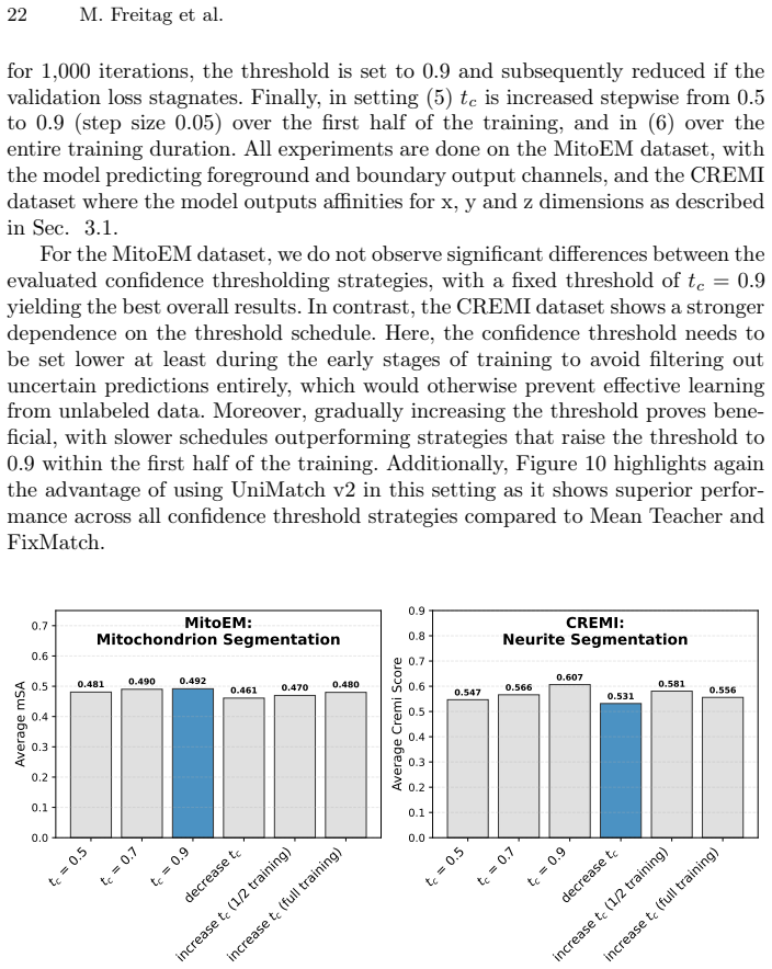
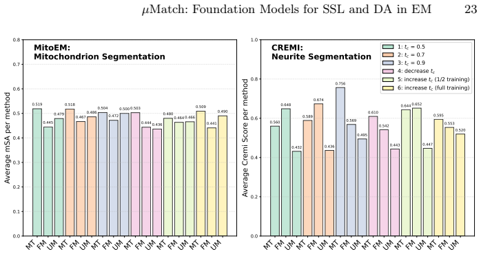
Vision foundation models have substantially advanced computer vision, enabling state-of-the-art performance in zero- and few-shot settings. They have been successfully applied to biomedical imaging tasks ranging from organ segmentation in computed tomography to cell segmentation in light microscopy. Electron microscopy (EM) is a central modality for analyzing cellular ultrastructure due to its nanometer-scale resolution. However, the application of foundation models in EM has so far been limited to specific organelles, such as mitochondria, largely due to the diversity of segmentation tasks and the scarcity of comprehensively annotated data. As a result, EM segmentation still predominantly relies on supervised learning, requiring extensive manual annotation and limiting ultrastructural analysis. To address this gap, we propose $\mu$Match, a framework for semi-supervised learning and domain adaptation that leverages foundation models. We implement state-of-the-art student-teacher-based methods and evaluate multiple foundation models (SAM, SAM2, $\mu$SAM, DINOv2/v3) on challenging EM tasks, including mitochondrion, nucleus, and neurite segmentation. Our results demonstrate consistent improvements over strong baselines and highlight a path toward substantially reducing the annotation effort in EM.
Editorial analysis
A structured set of objections, weighed in public.
Referee Report
Summary. The paper proposes μMatch, a student-teacher semi-supervised learning and domain adaptation framework that adapts vision foundation models (SAM, SAM2, μSAM, DINOv2/v3) to EM segmentation tasks including mitochondrion, nucleus, and neurite segmentation. It claims consistent improvements over strong baselines and a path toward substantially reducing annotation effort in EM.
Significance. If the results hold, the work could meaningfully advance EM analysis by showing that general-purpose foundation models can be transferred to a domain with extreme annotation scarcity and distinctive imaging characteristics, potentially enabling larger-scale ultrastructural studies without proportional increases in manual labeling.
major comments (3)
- [Abstract] Abstract: the claim of 'consistent improvements over strong baselines' supplies no quantitative metrics, dataset details, baseline descriptions, or ablation studies, preventing verification that the data support the central empirical claim.
- [Methods] Methods (student-teacher framework): the approach assumes foundation models pre-trained outside EM can supply sufficiently accurate initial features or pseudo-labels despite domain shift in noise, contrast, and scale; no analysis of initial teacher accuracy, pseudo-label quality, or confirmation bias is presented.
- [Experiments] Experiments: no ablation isolates the contribution of foundation-model initialization versus the SSL machinery alone, nor tests whether gains persist when the teacher is initialized from scratch or from an EM-only model, directly testing the transfer assumption.
minor comments (1)
- [Abstract] The abstract would be strengthened by including at least one key quantitative result (e.g., Dice or IoU improvement) to ground the 'consistent improvements' statement.
Simulated Author's Rebuttal
We thank the referee for the constructive comments, which highlight areas where the manuscript can be strengthened. We address each major comment point by point below, indicating the revisions we will make.
read point-by-point responses
-
Referee: [Abstract] Abstract: the claim of 'consistent improvements over strong baselines' supplies no quantitative metrics, dataset details, baseline descriptions, or ablation studies, preventing verification that the data support the central empirical claim.
Authors: We agree that the abstract would benefit from including key quantitative results to support the central claim. In the revised manuscript, we will update the abstract to report specific metrics (e.g., Dice score improvements on mitochondrion, nucleus, and neurite segmentation tasks), name the primary datasets, and briefly reference the baselines and foundation models evaluated. Full details and ablations remain in the Experiments section. revision: yes
-
Referee: [Methods] Methods (student-teacher framework): the approach assumes foundation models pre-trained outside EM can supply sufficiently accurate initial features or pseudo-labels despite domain shift in noise, contrast, and scale; no analysis of initial teacher accuracy, pseudo-label quality, or confirmation bias is presented.
Authors: The methods section describes the student-teacher adaptation but does not provide a dedicated analysis of initial teacher performance or pseudo-label evolution. We will add a new subsection with quantitative evaluation of initial foundation-model accuracy on EM data, pseudo-label quality metrics across training iterations, and discussion of measures taken to mitigate confirmation bias. revision: yes
-
Referee: [Experiments] Experiments: no ablation isolates the contribution of foundation-model initialization versus the SSL machinery alone, nor tests whether gains persist when the teacher is initialized from scratch or from an EM-only model, directly testing the transfer assumption.
Authors: We acknowledge that the existing experiments compare foundation models against supervised and SSL baselines but do not include the requested controls. In the revision we will add ablations initializing the teacher from random weights and from an EM-only pretrained model, allowing direct isolation of the foundation-model transfer contribution versus the SSL framework alone. revision: yes
Circularity Check
No derivation chain present; empirical evaluation only
full rationale
The paper is framed entirely as an empirical study: it proposes a framework, implements existing student-teacher SSL methods, evaluates multiple foundation models on EM segmentation tasks, and reports performance improvements over baselines. No equations, derivations, fitted parameters presented as predictions, or load-bearing self-citations appear in the provided text. The central claims rest on experimental comparisons rather than any mathematical reduction to inputs. This matches the default expectation for non-circular empirical work.
Axiom & Free-Parameter Ledger
axioms (1)
- domain assumption Vision foundation models pre-trained on non-EM data provide transferable features that student-teacher methods can adapt to EM segmentation tasks
Reference graph
Works this paper leans on
-
[1]
Circuit reconstruction from electron microscopy images (cremi) challenge.https: //cremi.org/, accessed: 2026-03-04
2026
-
[2]
Nature 640(8058), 435–447 (2025)
Functional connectomics spanning multiple areas of mouse visual cortex. Nature 640(8058), 435–447 (2025)
2025
-
[3]
Archit, A., Freckmann, L., Nair, S., Khalid, N., Hilt, P., Rajashekar, V., Freitag, M., Teuber, C., Spitzner, M., Tapia Contreras, C., Buckley, G., von Haaren, S., Gupta, S., Grade, M., Wirth, M., Schneider, G., Dengel, A., Ahmed, S., Pape, C.: Segment Anything for Microscopy. Nature Methods22(3), 579–591 (Mar 2025). https://doi.org/10.1038/s41592- 024- 0...
-
[4]
IEEE Transactions on Medical Imaging (2025)
Archit, A., Freckmann, L., Pape, C.: Medicosam: Robust improvement of sam for medical imaging. IEEE Transactions on Medical Imaging (2025)
2025
-
[5]
Archit, A., Pape, C.: Probabilistic Domain Adaptation for Biomedical Image Seg- mentation (Mar 2023).https://doi.org/10.48550/arXiv.2303.11790,http: //arxiv.org/abs/2303.11790, arXiv:2303.11790 [cs]
-
[6]
Nature methods14(2), 101–102 (2017)
Beier, T., Pape, C., Rahaman, N., Prange, T., Berg, S., Bock, D.D., Cardona, A., Knott, G.W., Plaza, S.M., Scheffer, L.K., et al.: Multicut brings automated neurite segmentation closer to human performance. Nature methods14(2), 101–102 (2017)
2017
-
[7]
IEEE Transactions on Medical Imaging39(4), 1256–1267 (Apr 2020)
Bermudez-Chacon, R., Altingovde, O., Becker, C., Salzmann, M., Fua, P.: Visual Correspondences for Unsupervised Domain Adaptation on Electron Microscopy Images. IEEE Transactions on Medical Imaging39(4), 1256–1267 (Apr 2020). https://doi.org/10.1109/TMI.2019.2946462,https://ieeexplore.ieee.org/ document/8863400/
-
[8]
https://doi.org/10.48550/arXiv.2106.04732,http://arxiv.org/abs/2106
Berthelot, D., Roelofs, R., Sohn, K., Carlini, N., Kurakin, A.: AdaMatch: A Uni- fied Approach to Semi-Supervised Learning and Domain Adaptation (Mar 2022). https://doi.org/10.48550/arXiv.2106.04732,http://arxiv.org/abs/2106. 04732, arXiv:2106.04732 [cs]
-
[9]
Cell Systems14(1), 58–71 (2023)
Conrad, R., Narayan, K.: Instance segmentation of mitochondria in electron mi- croscopy images with a generalist deep learning model trained on a diverse dataset. Cell Systems14(1), 58–71 (2023)
2023
-
[10]
Nature634(8032), 124–138 (2024)
Dorkenwald, S., Matsliah, A., Sterling, A.R., Schlegel, P., Yu, S.C., McKellar, C.E., Lin, A., Costa, M., Eichler, K., Yin, Y., et al.: Neuronal wiring diagram of an adult brain. Nature634(8032), 124–138 (2024)
2024
-
[11]
Neuroinformatics20(2), 437–450 (2022)
Franco-Barranco, D., Muñoz-Barrutia, A., Arganda-Carreras, I.: Stable deep neu- ral network architectures for mitochondria segmentation on electron microscopy volumes. Neuroinformatics20(2), 437–450 (2022)
2022
-
[12]
IEEE transactions on pattern analysis and machine intelligence41(7), 1669–1680 (2018)
Funke, J., Tschopp, F., Grisaitis, W., Sheridan, A., Singh, C., Saalfeld, S., Turaga, S.C.: Large scale image segmentation with structured loss based deep learning for connectome reconstruction. IEEE transactions on pattern analysis and machine intelligence41(7), 1669–1680 (2018)
2018
-
[13]
Journal of Cell Biology222(2), e202208005 (2022) 16 M
Gallusser, B., Maltese, G., Di Caprio, G., Vadakkan, T.J., Sanyal, A., Somerville, E.,Sahasrabudhe,M.,O’connor,J.,Weigert,M.,Kirchhausen,T.:Deepneuralnet- work automated segmentation of cellular structures in volume electron microscopy. Journal of Cell Biology222(2), e202208005 (2022) 16 M. Freitag et al
2022
-
[14]
arXiv preprint arXiv:2502.00408 (2025)
Griebel, T., Archit, A., Pape, C.: Segment anything for histopathology. arXiv preprint arXiv:2502.00408 (2025)
arXiv 2025
-
[15]
In: Proceedings of the IEEE/CVF winter conference on applications of computer vi- sion
Hatamizadeh, A., Tang, Y., Nath, V., Yang, D., Myronenko, A., Landman, B., Roth, H.R., Xu, D.: Unetr: Transformers for 3d medical image segmentation. In: Proceedings of the IEEE/CVF winter conference on applications of computer vi- sion. pp. 574–584 (2022)
2022
-
[16]
Nature599(7883), 141–146 (2021)
Heinrich, L., Bennett, D., Ackerman, D., Park, W., Bogovic, J., Eckstein, N., Petruncio, A., Clements, J., Pang, S., Xu, C.S., et al.: Whole-cell organelle seg- mentation in volume electron microscopy. Nature599(7883), 141–146 (2021)
2021
-
[17]
Nature methods21(2), 213–216 (2024)
Hirling, D., Tasnadi, E., Caicedo, J., Caroprese, M.V., Sjögren, R., Aubreville, M., Koos, K., Horvath, P.: Segmentation metric misinterpretations in bioimage analysis. Nature methods21(2), 213–216 (2024)
2024
-
[18]
Medical image analysis94, 103143 (2024)
Hörst, F., Rempe, M., Heine, L., Seibold, C., Keyl, J., Baldini, G., Ugurel, S., Siveke, J., Grünwald, B., Egger, J., et al.: Cellvit: Vision transformers for precise cell segmentation and classification. Medical image analysis94, 103143 (2024)
2024
-
[19]
IEEE Transactions on Information Theory , author =
Huang, W., Chen, C., Xiong, Z., Zhang, Y., Chen, X., Sun, X., Wu, F.: Semi- Supervised Neuron Segmentation via Reinforced Consistency Learning. IEEE Transactions on Medical Imaging41(11), 3016–3028 (Nov 2022).https://doi. org/10.1109/TMI.2022.3176050,https://ieeexplore.ieee.org/document/ 9777694/
-
[20]
Journal of classification2(1), 193– 218 (1985)
Hubert, L., Arabie, P.: Comparing partitions. Journal of classification2(1), 193– 218 (1985)
1985
-
[21]
bioRxiv p
Januszewski, M., Jain, V.: Segmentation-enhanced cyclegan. bioRxiv p. 548081 (2019)
2019
-
[22]
Nature methods15(8), 605–610 (2018)
Januszewski, M., Kornfeld, J., Li, P.H., Pope, A., Blakely, T., Lindsey, L., Maitin- Shepard, J., Tyka, M., Denk, W., Jain, V.: High-precision automated reconstruc- tion of neurons with flood-filling networks. Nature methods15(8), 605–610 (2018)
2018
-
[23]
Advances in neural information processing sys- tems31(2018)
Kohl, S., Romera-Paredes, B., Meyer, C., De Fauw, J., Ledsam, J.R., Maier-Hein, K., Eslami, S., Jimenez Rezende, D., Ronneberger, O.: A probabilistic u-net for segmentation of ambiguous images. Advances in neural information processing sys- tems31(2018)
2018
-
[24]
IEEE transactions on medical imaging 40(12), 3801–3811 (2021)
Lee, K., Lu, R., Luther, K., Seung, H.S.: Learning and segmenting dense voxel embeddings for 3d neuron reconstruction. IEEE transactions on medical imaging 40(12), 3801–3811 (2021)
2021
-
[25]
Li, M., Chen, C., Liu, X., Huang, W., Zhang, Y., Xiong, Z.: Advanced deep net- worksfor3dmitochondriainstancesegmentation.In:2022IEEE19thInternational Symposium on Biomedical Imaging (ISBI). pp. 1–5. IEEE (2022)
2022
-
[26]
bioRxiv pp
Liu, P., Shen, B., Liu, L., Wang, Q., Zhang, S., Bhardwaj, A., Arganda-Carreras, I., Narayan, K., Wei, D.: Mitoem 2.0: A benchmark for challenging 3d mitochondria instance segmentation from em images. bioRxiv pp. 2025–11 (2025)
2025
-
[27]
Nature communications15(1), 654 (2024)
Ma, J., He, Y., Li, F., Han, L., You, C., Wang, B.: Segment anything in medical images. Nature communications15(1), 654 (2024)
2024
-
[28]
Frontiers in Bioinformatics3, 1308708 (2023)
Machireddy, A., Thibault, G., Loftis, K.G., Stoltz, K., Bueno, C.E., Smith, H.R., Riesterer, J.L., Gray, J.W., Song, X.: Segmentation of cellular ultrastructures on sparsely labeled 3d electron microscopy images using deep learning. Frontiers in Bioinformatics3, 1308708 (2023)
2023
-
[29]
Matskevych, A., Wolny, A., Pape, C., Kreshuk, A.: From Shallow to Deep: Exploit- ing Feature-Based Classifiers for Domain Adaptation in Semantic Segmentation. Frontiers in Computer Science4, 805166 (Mar 2022).https://doi.org/10.3389/ fcomp.2022.805166,https://www.frontiersin.org/articles/10.3389/fcomp. 2022.805166/full µMatch: Foundation Models for SSL an...
-
[30]
In: Learning Theory and Kernel Machines: 16th Annual Conference on Learning Theory and 7th Kernel Workshop, COLT/Kernel 2003, Washington, DC, USA, August 24-27,
Meilă, M.: Comparing clusterings by the variation of information. In: Learning Theory and Kernel Machines: 16th Annual Conference on Learning Theory and 7th Kernel Workshop, COLT/Kernel 2003, Washington, DC, USA, August 24-27,
2003
-
[31]
Proceedings. pp. 173–187. Springer (2003)
2003
-
[32]
bioRxiv pp
Mu, S., Yu, S.c., Turner, N.L., McKellar, C.E., Dorkenwald, S., Collman, F., Kool- man, S., Moore, M., Morejohn, S., Silverman, B., et al.: 3d reconstruction of cell nuclei in a full drosophila brain. bioRxiv pp. 2021–11 (2021)
2021
-
[33]
org/doi/10.1091/mbc.E24-11-0519
Muth, S., Moschref, F., Freckmann, L., Mutschall, S., Hojas-Garcia-Plaza, I., Bahr, J.N., Petrovic, A., Do, T.T., Schwarze, V., Archit, A., Weyand, K., Michanski, S., Maus, L., Imig, C., Hintze, A., Brose, N., Wichmann, C., Fernandez-Busnadiego, R., Moser, T., Rizzoli, S.O., Cooper, B.H., Pape, C.: SynapseNet: Deep learning for automaticsynapsereconstruct...
-
[34]
BioRxiv pp
Pachitariu,M.,Rariden,M.,Stringer,C.:Cellpose-sam:superhumangeneralization for cellular segmentation. BioRxiv pp. 2025–04 (2025)
2025
-
[35]
Nature Communications15(1), 3982 (2024)
Parlakgül, G., Pang, S., Artico, L.L., Min, N., Cagampan, E., Villa, R., Goncalves, R.L., Lee, G.Y., Xu, C.S., Hotamışlıgil, G.S., et al.: Spatial mapping of hepatic er and mitochondria architecture reveals zonated remodeling in fasting and obesity. Nature Communications15(1), 3982 (2024)
2024
-
[36]
bioRxiv pp
Peale, D.R., Hess, H., Lee, P., Cardona, A., Bock, D.D., Schneider-Mizell, C., Fetter, R.D., Lee, W.P., Robinson, C.G., Iyer, N., et al.: itome volumetric serial sectioning apparatus for tem. bioRxiv pp. 2024–07 (2024)
2024
-
[37]
Qiu, D., Xiong, S., Yi, J., Peng, J.: Weakly-Supervised Cross-Domain Segmenta- tion of Electron Microscopy with Sparse Point Annotation (Mar 2024).https: //doi.org/10.48550/arXiv.2404.00667,http://arxiv.org/abs/2404.00667, arXiv:2404.00667 [cs]
-
[38]
In: 2022 IEEE International Conference on Bioinformatics and Biomedicine (BIBM)
Qiu, D., Yi, J., Peng, J.: WDA-Net: Weakly-Supervised Domain Adaptive Seg- mentation of Electron Microscopy. In: 2022 IEEE International Conference on Bioinformatics and Biomedicine (BIBM). pp. 1132–1137. IEEE, Las Vegas, NV, USA (Dec 2022).https://doi.org/10.1109/BIBM55620.2022.9995167,https: //ieeexplore.ieee.org/document/9995167/
-
[39]
Roels, J., Hennies, J., Saeys, Y., Philips, W., Kreshuk, A.: Domain Adap- tive Segmentation in Volume Electron Microscopy Imaging (Dec 2018).https: //doi.org/10.48550/arXiv.1810.09734,http://arxiv.org/abs/1810.09734, arXiv:1810.09734 [cs]
work page internal anchor Pith review Pith/arXiv arXiv doi:10.48550/arxiv.1810.09734 2018
-
[40]
Rumberger, J.L., Franzen, J., Hirsch, P., Albrecht, J.P., Kainmueller, D.: AC- TIS: Improving data efficiency by leveraging semi-supervised Augmentation Con- sistency Training for Instance Segmentation. In: 2023 IEEE/CVF International Conference on Computer Vision Workshops (ICCVW). pp. 3792–3801. IEEE, Paris, France (Oct 2023).https://doi.org/10.1109/ICC...
-
[41]
07685,http://arxiv.org/abs/2001.07685, arXiv:2001.07685 [cs]
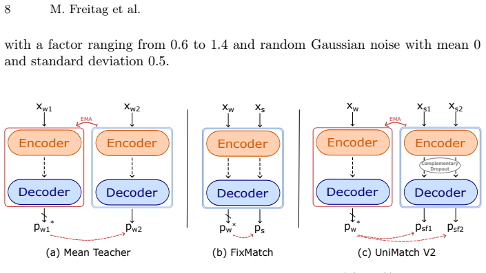
Sohn, K., Berthelot, D., Li, C.L., Zhang, Z., Carlini, N., Cubuk, E.D., Kurakin, A., Zhang, H., Raffel, C.: FixMatch: Simplifying Semi-Supervised Learning with Con- sistency and Confidence (Nov 2020).https://doi.org/10.48550/arXiv.2001. 07685,http://arxiv.org/abs/2001.07685, arXiv:2001.07685 [cs]
-
[42]
Traffic22(7), 240–253 (2021) 18 M
Spiers, H., Songhurst, H., Nightingale, L., De Folter, J., Community, Z.V., Hutch- ings, R., Peddie, C.J., Weston, A., Strange, A., Hindmarsh, S., et al.: Deep learning for automatic segmentation of the nuclear envelope in electron microscopy data, trained with volunteer segmentations. Traffic22(7), 240–253 (2021) 18 M. Freitag et al
2021
-
[43]
Tarvainen, A., Valpola, H.: Mean teachers are better role models: Weight-averaged consistency targets improve semi-supervised deep learning results (Apr 2018). https://doi.org/10.48550/arXiv.1703.01780,http://arxiv.org/abs/1703. 01780, arXiv:1703.01780 [cs]
work page internal anchor Pith review Pith/arXiv arXiv doi:10.48550/arxiv.1703.01780 2018
-
[44]
Cell184(18), 4819–4837 (2021)
Vergara, H.M., Pape, C., Meechan, K.I., Zinchenko, V., Genoud, C., Wanner, A.A., Mutemi, K.N., Titze, B., Templin, R.M., Bertucci, P.Y., et al.: Whole-body inte- gration of gene expression and single-cell morphology. Cell184(18), 4819–4837 (2021)
2021
-
[45]
Radiology: Artificial Intelligence 5(5), e230024 (2023)
Wasserthal, J., Breit, H.C., Meyer, M.T., Pradella, M., Hinck, D., Sauter, A.W., Heye, T., Boll, D.T., Cyriac, J., Yang, S., et al.: Totalsegmentator: robust segmen- tation of 104 anatomic structures in ct images. Radiology: Artificial Intelligence 5(5), e230024 (2023)
2023
-
[46]
In: International Conference on Med- ical Image Computing and Computer-Assisted Intervention
Wei, D., Lin, Z., Franco-Barranco, D., Wendt, N., Liu, X., Yin, W., Huang, X., Gupta, A., Jang, W.D., Wang, X., et al.: Mitoem dataset: large-scale 3d mitochon- dria instance segmentation from em images. In: International Conference on Med- ical Image Computing and Computer-Assisted Intervention. pp. 66–76. Springer (2020)
2020
-
[47]
Science379(6636), eadd9330 (2023)
Winding, M., Pedigo, B.D., Barnes, C.L., Patsolic, H.G., Park, Y., Kazimiers, T., Fushiki, A., Andrade, I.V., Khandelwal, A., Valdes-Aleman, J., et al.: The connectome of an insect brain. Science379(6636), eadd9330 (2023)
2023
-
[48]
Frontiers in neuroanatomy12, 92 (2018)
Xiao, C., Chen, X., Li, W., Li, L., Wang, L., Xie, Q., Han, H.: Automatic mito- chondria segmentation for em data using a 3d supervised convolutional network. Frontiers in neuroanatomy12, 92 (2018)
2018
-
[49]
Yang, L., Qi, L., Feng, L., Zhang, W., Shi, Y.: Revisiting Weak-to-Strong Consistency in Semi-Supervised Semantic Segmentation (Mar 2023).https:// doi.org/10.48550/arXiv.2208.09910,http://arxiv.org/abs/2208.09910, arXiv:2208.09910 [cs]
-
[50]
Rethinking VLMs and LLMs for Image Classification.arXiv e-prints, art
Yang, L., Zhao, Z., Zhao, H.: UniMatch V2: Pushing the Limit of Semi-Supervised Semantic Segmentation (Jan 2025).https://doi.org/10.48550/arXiv.2410. 10777,http://arxiv.org/abs/2410.10777, arXiv:2410.10777 [cs]
-
[51]
In: Proceedings of the IEEE interna- tional conference on computer vision
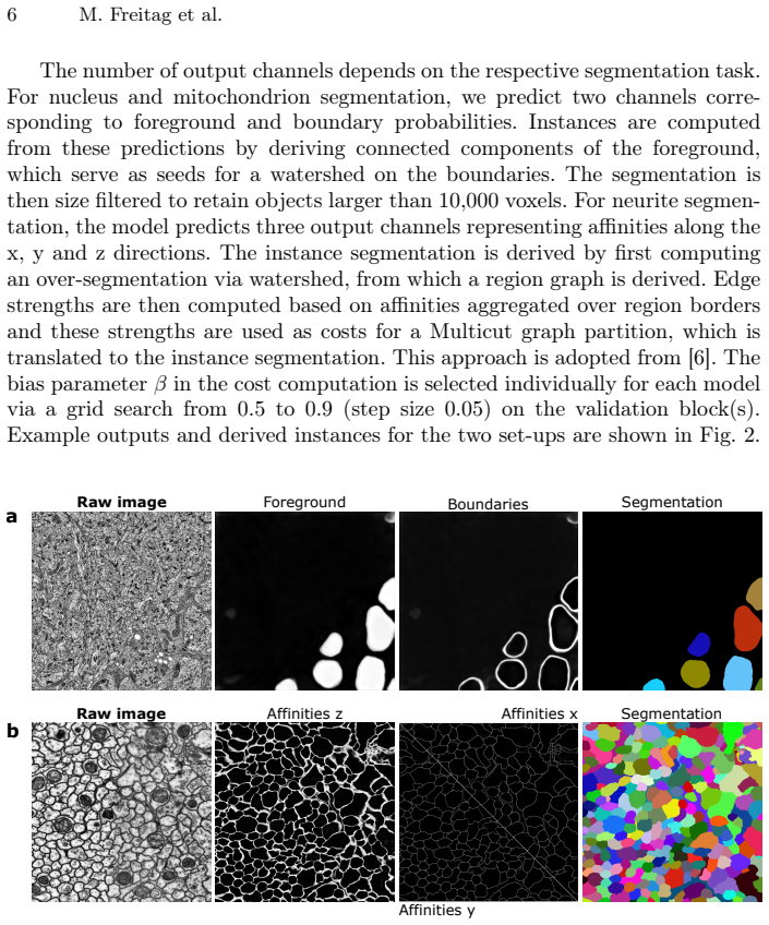
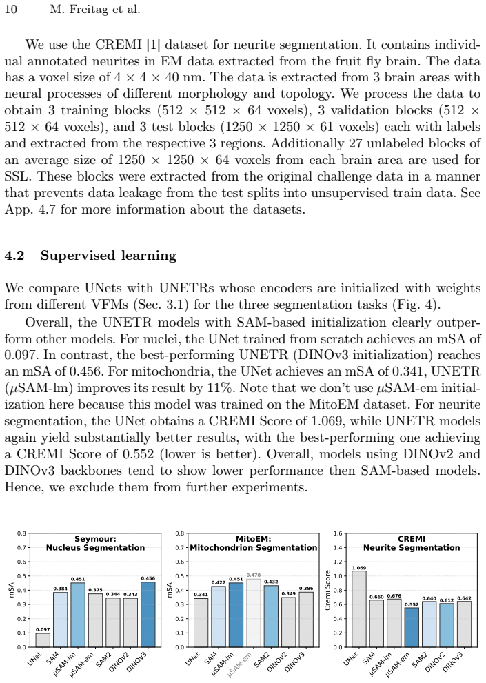
Zhu, J.Y., Park, T., Isola, P., Efros, A.A.: Unpaired image-to-image translation using cycle-consistent adversarial networks. In: Proceedings of the IEEE interna- tional conference on computer vision. pp. 2223–2232 (2017) µMatch: Foundation Models for SSL and DA in EM 19 Appendix 4.6 Metrics mSA is calculated as the mean of segmentation accuracies over mu...
2017
discussion (0)
Sign in with ORCID, Apple, or X to comment. Anyone can read and Pith papers without signing in.