Scaling up fine-grained intracranial vessel annotations in computed tomography angiography
Pith reviewed 2026-06-26 12:22 UTC · model grok-4.3
The pith
Including a generic artery class for minor vessels improves fine-grained segmentation of 20 specific brain arteries in CTA scans.
A machine-rendered reading of the paper's core claim, the machinery that carries it, and where it could break.
Core claim
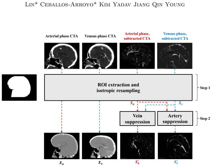
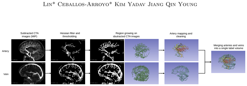
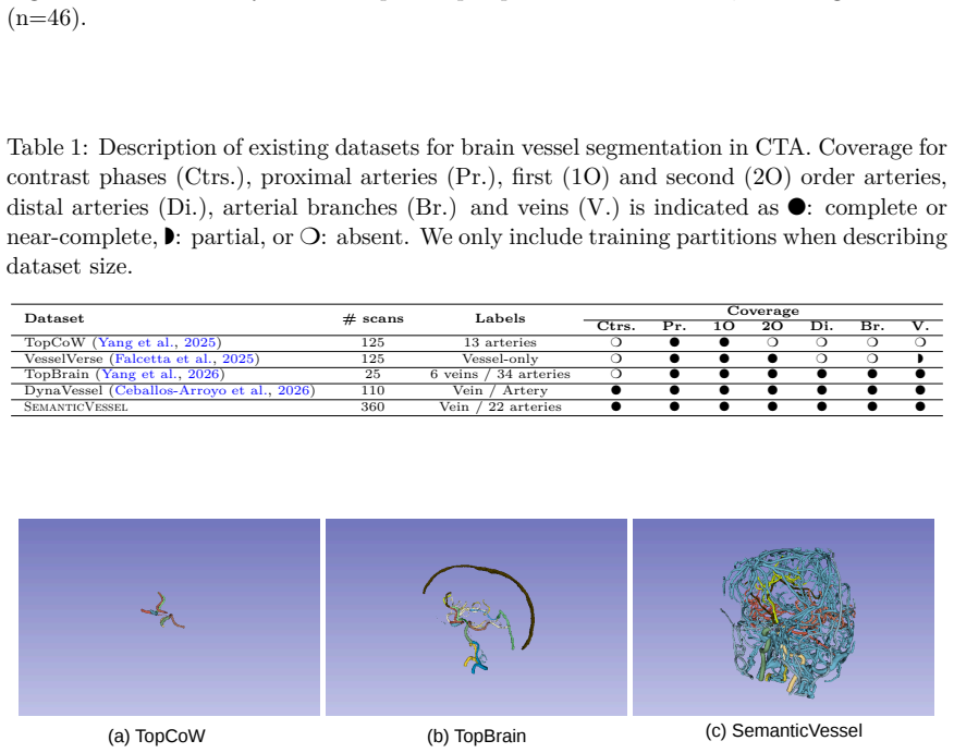
The central claim is that models trained with the additional generic artery class produce better fine-grained segmentations across the board. The dataset is built by using intensity-guided region growing on 4D-CTA to segment major vascular territories, followed by expert refinement into 20 unique arterial classes plus the merged generic class; labels are transferred across phases of the same acquisition series to increase dataset size at no extra cost.
What carries the argument
The SemanticVessel dataset construction, which merges minor arteries into a generic arterial class and reuses single-phase labels across the multiple phases of each 4D-CTA series.
If this is right
- Training sets that retain minor arteries as a single extra class can be used to improve accuracy on the labeled major arteries.
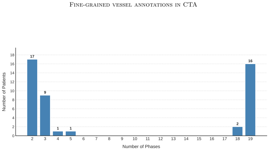
- Multi-phase 4D-CTA acquisitions become a low-cost source of additional training examples once one phase is annotated.
- The same label-reuse strategy can enlarge other vascular segmentation datasets derived from dynamic contrast scans.
- Downstream clinical tools for vessel analysis gain from models that better handle the full range of artery sizes present in real scans.
Where Pith is reading between the lines
- The same merging of minor structures into a generic class could be tested on vein segmentation or on non-brain vascular territories.
- If label reuse across phases works reliably, the method could be applied to other 4D imaging modalities such as dynamic contrast-enhanced MRI.
- Performance gains from the generic class might be largest on the smallest or most variable arterial territories, which could be checked by per-class metrics.
- The dataset size increase from phase reuse makes it feasible to train larger models or to perform more extensive data augmentation without new manual work.
Load-bearing premise
Labels created for one phase of a 4D-CTA series can be directly reused on other phases of the same series without introducing meaningful spatial or intensity mismatches caused by vessel motion or changing contrast.
What would settle it
An experiment that measures segmentation performance on the 20 fine-grained classes when the generic artery class is omitted versus included and finds no consistent improvement, or direct measurement of label overlap across phases showing large spatial mismatches.
Figures
read the original abstract
In this work, we present SemanticVessel, a dataset for fine-grained brain vessel segmentation in computed tomography angiography scans. Based on the detailed contrast provided by dynamic 4D-CTA scans, we generate segmentation traces for arteries and veins. We then use intensity-guided region growing to obtain segmentations of the majority of vascular territories in the human brain, which are refined and annotated with 20 unique arterial classes by an expert radiologist. Unlike existing datasets, where minor arteries are discarded as background content, we merge these minor arteries into a generic arterial class. Due to the multiple-phase acquisition of dynamic 4D-CTA, labels for a single phase can be re-used for other phases in the same series, greatly increasing the size of our dataset with no additional annotation cost. The results show that models trained with the additional generic artery class produce better fine-grained segmentations across the board. We will make our code, annotation GUI, and model weights available to the scientific community. Code, weights, and data will be made available on https://github.com/alceballosa/robust-vessel-segmentation
Editorial analysis
A structured set of objections, weighed in public.
Referee Report
Summary. The manuscript presents SemanticVessel, a dataset for fine-grained intracranial vessel segmentation in 4D-CTA scans. Annotations are generated using intensity-guided region growing on dynamic scans, refined by an expert into 20 arterial classes, with minor arteries merged into a generic arterial class. Labels from one phase are reused across other phases in the series to scale the dataset without additional annotation effort. The key result is that including the generic artery class leads to improved fine-grained segmentation performance.
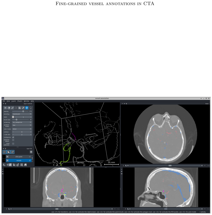
Significance. If the empirical results hold, this work provides a scalable approach to creating large annotated datasets for vessel segmentation and demonstrates the value of including a generic class for minor arteries, which are often discarded. The commitment to releasing code, annotation GUI, model weights, and data supports reproducibility and community use in medical image analysis.
major comments (2)
- [Abstract] Abstract: The claim that 'models trained with the additional generic artery class produce better fine-grained segmentations across the board' is asserted without any quantitative metrics, baseline comparisons, statistical tests, or details on evaluation methodology, preventing assessment of the central empirical claim.
- [Methods (label reuse description)] Methods (label reuse description): The reuse of labels from a single phase across other phases in 4D-CTA is presented as introducing 'no additional annotation cost' and enabling dataset scaling, but no quantitative validation (such as overlap metrics between phases or assessment of vessel motion/contrast variation effects) is provided. This is load-bearing, as misalignment could introduce label noise that confounds attribution of performance gains to the generic class rather than data quality issues.
minor comments (1)
- [Abstract] Abstract: The final two sentences both announce data/code release; this repetition can be consolidated for clarity.
Simulated Author's Rebuttal
We thank the referee for their detailed review and constructive comments. We address each major comment point by point below.
read point-by-point responses
-
Referee: [Abstract] Abstract: The claim that 'models trained with the additional generic artery class produce better fine-grained segmentations across the board' is asserted without any quantitative metrics, baseline comparisons, statistical tests, or details on evaluation methodology, preventing assessment of the central empirical claim.
Authors: We agree that the abstract would benefit from including key quantitative details to support the central claim. The full manuscript reports quantitative improvements (including Dice scores and other metrics) from including the generic artery class, along with baseline comparisons and evaluation methodology in the Experiments section. We will revise the abstract to concisely include representative metrics, mention the evaluation protocol, and note the observed gains. revision: yes
-
Referee: [Methods (label reuse description)] Methods (label reuse description): The reuse of labels from a single phase across other phases in 4D-CTA is presented as introducing 'no additional annotation cost' and enabling dataset scaling, but no quantitative validation (such as overlap metrics between phases or assessment of vessel motion/contrast variation effects) is provided. This is load-bearing, as misalignment could introduce label noise that confounds attribution of performance gains to the generic class rather than data quality issues.
Authors: We acknowledge that explicit quantitative validation of label reuse would strengthen the methodology and help rule out confounding noise. Although the intensity-guided region growing and dynamic 4D-CTA acquisition are designed to handle contrast variations, and intra-series vessel positions are relatively stable, the submitted version does not include overlap metrics. We will add a validation analysis with overlap metrics (e.g., Dice scores between phases on a subset) to the Methods section. revision: yes
Circularity Check
No circularity: empirical dataset construction and model evaluation only
full rationale
The paper presents a new dataset (SemanticVessel) built from 4D-CTA scans via intensity-guided region growing and expert annotation, followed by standard supervised training of segmentation models. No derivations, equations, fitted parameters renamed as predictions, or self-citation chains appear in the abstract or described content. The central claim (better fine-grained segmentations when including a generic artery class) rests on empirical comparison of trained models, not on any self-referential reduction or imported uniqueness theorem. This is a standard dataset+benchmark paper with no load-bearing circular steps.
Axiom & Free-Parameter Ledger
axioms (2)
- domain assumption Intensity-guided region growing produces accurate segmentations of the majority of intracranial vascular territories in 4D-CTA
- domain assumption Expert radiologist review yields reliable ground-truth labels for the 20 arterial classes
Reference graph
Works this paper leans on
-
[1]
Pablo Villablanca, Reza Jahan, Gary Duckwiler, Monica Tillis, Chelsea Kidwell, Jeffrey Saver, and James Sayre
Suzie Bash, J. Pablo Villablanca, Reza Jahan, Gary Duckwiler, Monica Tillis, Chelsea Kidwell, Jeffrey Saver, and James Sayre. Intracranial vascular stenosis and occlusive disease: evaluation with CT angiography, MR angiography, and digital subtraction angiography. AJNR Am J Neuroradiol, 26 0 (5): 0 1012--1021, May 2005. ISSN 0195-6108
2005
-
[2]
Alberto M. Ceballos-Arroyo et al. Robust automatic brain vessel segmentation in 3D CTA scans using dynamic 4D-CTA data. arXiv preprint arXiv:2602.00391, 2026. URL https://arxiv.org/abs/2602.00391
arXiv 2026
-
[3]
Hippe, Niranjan Balu, Quan Yuan, Kristi Pimentel, Thomas S
Li Chen, Mahmud Mossa-Basha, Jie Sun, Daniel S. Hippe, Niranjan Balu, Quan Yuan, Kristi Pimentel, Thomas S. Hatsukami, Jenq-Neng Hwang, and Chun Yuan. Quantification of morphometry and intensity features of intracranial arteries from 3D TOF MRA using the intracranial artery feature extraction ( iCafe ): A reproducibility study. Magn Reson Imaging, 57: 0 2...
-
[4]
Ting Chen, Wei You, Liyuan Zhang, Wanxing Ye, Junqiang Feng, Jing Lu, Jian Lv, Yudi Tang, Dachao Wei, Siming Gui, et al. Automated anatomical labeling of the intracranial arteries via deep learning in computed tomography angiography. Frontiers in Physiology, 14: 0 1310357, 2024. doi:10.3389/fphys.2023.1310357
-
[5]
Xiaodan Chen, Yun Liu, Huazhang Tong, Yonghai Dong, Dongyang Ma, Lei Xu, and Cheng Yang. Meta-analysis of computed tomography angiography versus magnetic resonance angiography for intracranial aneurysm. Medicine (Baltimore), 97 0 (20): 0 e10771, May 2018. ISSN 0025-7974. doi:10.1097/MD.0000000000010771. URL https://pmc.ncbi.nlm.nih.gov/articles/PMC5976319/
-
[6]
Daniele Falcetta, Vincenzo Marciano, Kaiyuan Yang, Jon Cleary, Loïc Legris, Massimiliano Domenico Rizzaro, Ioannis Pitsiorlas, Hava Chaptoukaev, Benjamin Lemasson, Bjoern Menze, and Maria A. Zuluaga. VesselVerse: A Dataset and Collaborative Framework for Vessel Annotation . In proceedings of Medical Image Computing and Computer Assisted Intervention -- MI...
2025
-
[7]
Canas, Lorenzo Suppa, Matteo Pentassuglia, Jon Cleary, Marc Modat, Sébastien Ourselin, and Maria A
Daniele Falcetta, Liane S. Canas, Lorenzo Suppa, Matteo Pentassuglia, Jon Cleary, Marc Modat, Sébastien Ourselin, and Maria A. Zuluaga. An automated framework for large-scale graph-based cerebrovascular analysis, 2026. URL https://arxiv.org/abs/2512.03869
Pith/arXiv arXiv 2026
-
[8]
Fan Fu, Jianyong Wei, Miao Zhang, Fan Yu, Yueting Xiao, Dongdong Rong, Yi Shan, Yan Li, Cheng Zhao, Fangzhou Liao, Zhenghan Yang, Yuehua Li, Yingmin Chen, Ximing Wang, and Jie Lu. Rapid vessel segmentation and reconstruction of head and neck angiograms using 3D convolutional neural network. Nat Commun, 11 0 (1): 0 4829, September 2020. ISSN 2041-1723. doi...
-
[9]
Hamadache, Clara Lisazo, Cansu Yalcin, Uma M
Rachika E. Hamadache, Clara Lisazo, Cansu Yalcin, Uma M. Lal-Trehan Estrada, Valeriia Abramova, Adri \`a Casamitjana, Arnau Oliver, and Xavier Llad \'o . Topology-aware multiclass segmentation of the Circle of Willis from MRA and CTA images. Computers in Biology and Medicine, 204: 0 111516, 2026. doi:10.1016/j.compbiomed.2026.111516
-
[10]
Adam Hilbert, Vince I. Madai, Ela M. Akay, Orhun U. Aydin, Jonas Behland, Jan Sobesky, Ivana Galinovic, Ahmed A. Khalil, Abdel A. Taha, Jens Wuerfel, Petr Dusek, Thoralf Niendorf, Jochen B. Fiebach, Dietmar Frey, and Michelle Livne. BRAVE - NET : Fully Automated Arterial Brain Vessel Segmentation in Patients With Cerebrovascular Disease . Front. Artif. In...
-
[11]
Fabian Isensee, Paul F. Jaeger, Simon A. A. Kohl, Jens Petersen, and Klaus H. Maier-Hein. nnU - Net : a self-configuring method for deep learning-based biomedical image segmentation. Nat Methods, 18 0 (2): 0 203--211, February 2021. ISSN 1548-7105. doi:10.1038/s41592-020-01008-z. URL https://www.nature.com/articles/s41592-020-01008-z
-
[12]
Ron Kikinis, Steve D. Pieper, and Kirby G. Vosburgh. 3D Slicer: A Platform for Subject-Specific Image Analysis, Visualization, and Clinical Support, pages 277--289. Springer New York, New York, NY, 2014. ISBN 978-1-4614-7657-3. URL https://doi.org/10.1007/978-1-4614-7657-3_19
-
[13]
Yannick Kirchhoff, Maximilian R. Rokuss, Saikat Roy, Balint Kovacs, Constantin Ulrich, Tassilo Wald, Maximilian Zenk, Philipp Vollmuth, Jens Kleesiek, Fabian Isensee, and Klaus Maier-Hein. Skeleton Recall Loss for Connectivity Conserving and Resource Efficient Segmentation of Thin Tubular Structures . In Aleš Leonardis, Elisa Ricci, Stefan Roth, Olga Russ...
-
[14]
Segment anything in medical images
Jun Ma, Yuting He, Feifei Li, Lin Han, Chenyu You, and Bo Wang. Segment anything in medical images. Nat Commun, 15 0 (1): 0 654, January 2024. ISSN 2041-1723. doi:10.1038/s41467-024-44824-z. URL https://www.nature.com/articles/s41467-024-44824-z
-
[15]
van de Leemput, Mathias Prokop, Ewoud J
Midas Meijs, Ajay Patel, Sil C. van de Leemput, Mathias Prokop, Ewoud J. van Dijk, Frank-Erik de Leeuw, Frederick J. A. Meijer, Bram van Ginneken, and Rashindra Manniesing. Robust Segmentation of the Full Cerebral Vasculature in 4D CT of Suspected Stroke Patients . Sci Rep, 7 0 (1): 0 15622, November 2017. ISSN 2045-2322. doi:10.1038/s41598-017-15617-w. U...
-
[16]
Yuqin Min, Jing Li, Shouqiang Jia, Yuehua Li, and Shengdong Nie. Automated Cerebrovascular Segmentation and Visualization of Intracranial Time -of- Flight Magnetic Resonance Angiography Based on Deep Learning . J Imaging Inform Med, 38 0 (2): 0 703--716, August 2024. ISSN 2948-2925. doi:10.1007/s10278-024-01215-6. URL https://pmc.ncbi.nlm.nih.gov/articles...
-
[17]
Patel, Aakash Patel, Sricharan S
Tatsat R. Patel, Aakash Patel, Sricharan S. Veeturi, Munjal Shah, Muhammad Waqas, Andre Monteiro, Ammad A. Baig, Nandor Pinter, Elad I. Levy, Adnan H. Siddiqui, and Vincent M. Tutino. Evaluating a 3D deep learning pipeline for cerebral vessel and intracranial aneurysm segmentation from computed tomography angiography-digital subtraction angiography image ...
-
[18]
C. Douglas Phillips and Lori A. Bubash. CT angiography and MR angiography in the evaluation of extracranial carotid vascular disease. Radiol Clin North Am, 40 0 (4): 0 783--798, July 2002. ISSN 0033-8389. doi:10.1016/s0033-8389(02)00017-9
-
[19]
Bifurcation matching for consistent cerebral vessel labeling in CTA of stroke patients
Leonhard Rist, Oliver Taubmann, Florian Thamm, Hendrik Ditt, Michael S \"u hling, and Andreas Maier. Bifurcation matching for consistent cerebral vessel labeling in CTA of stroke patients. International Journal of Computer Assisted Radiology and Surgery, 18 0 (3): 0 509--516, 2023. doi:10.1007/s11548-022-02750-9
-
[20]
Paetzold, Fengbei Liu, and Mert R
Rachit Saluja, Asli Cihangir, Ruining Deng, Johannes C. Paetzold, Fengbei Liu, and Mert R. Sabuncu. BackSplit : The Importance of Sub -dividing the Background in Biomedical Lesion Segmentation , November 2025. URL http://arxiv.org/abs/2511.19394. arXiv:2511.19394 [cs]
Pith/arXiv arXiv 2025
-
[21]
Paetzold, Anjany Sekuboyina, Ivan Ezhov, Alexander Unger, Andrey Zhylka, Josien P
Suprosanna Shit, Johannes C. Paetzold, Anjany Sekuboyina, Ivan Ezhov, Alexander Unger, Andrey Zhylka, Josien P. W. Pluim, Ulrich Bauer, and Bjoern H. Menze. clDice - a Novel Topology - Preserving Loss Function for Tubular Structure Segmentation . In 2021 IEEE / CVF Conference on Computer Vision and Pattern Recognition ( CVPR ) , pages 16555--16564, Nashvi...
arXiv 2021
-
[22]
Adaptive constrained constructive optimisation for complex vascularisation processes
Gonzalo Daniel Maso Talou, Soroush Safaei, Peter John Hunter, and Pablo Javier Blanco. Adaptive constrained constructive optimisation for complex vascularisation processes. Scientific Reports, 11 0 (1): 0 6180, March 2021. ISSN 2045-2322. URL https://www.nature.com/articles/s41598-021-85434-9. Publisher: Nature Publishing Group
2021
-
[23]
Florian Thamm et al. An algorithm for the labeling and interactive visualization of the cerebrovascular system of ischemic strokes. Biomedical Physics & Engineering Express, 8 0 (6): 0 065016, 2022. doi:10.1088/2057-1976/ac9107
-
[24]
Nicholas J. Tustison, Philip A. Cook, Andrew J. Holbrook, Hans J. Johnson, John Muschelli, Gabriel A. Devenyi, Jeffrey T. Duda, Sandhitsu R. Das, Nicholas C. Cullen, Daniel L. Gillen, Michael A. Yassa, James R. Stone, James C. Gee, and Brian B. Avants. The ANTsX ecosystem for quantitative biological and medical imaging. Scientific Reports, 11 0 (1): 0 906...
-
[25]
Henk van Voorst, Jiahang Su, Praneeta R. Konduri, Charles B. L. M. Majoie, Yvo B. W. E. M. Roos, Bart J. Emmer, Henk A. Marquering, Bob D. de Vos, Matthan W. A. Caan, Ivana Išgum, and MR CLEAN Registry collaborators . Deep generative models for vessel segmentation in CT angiography of the brain. Comput Biol Med, 202: 0 111432, February 2026. ISSN 1879-053...
-
[26]
Radiology: Artificial Intelligence (Jul 2023), https://pubs.rsna.org/doi/full/10.1148/ryai.230024
Jakob Wasserthal, Hanns-Christian Breit, Manfred T. Meyer, Maurice Pradella, Daniel Hinck, Alexander W. Sauter, Tobias Heye, Daniel T. Boll, Joshy Cyriac, Shan Yang, Michael Bach, and Martin Segeroth. TotalSegmentator : Robust Segmentation of 104 Anatomic Structures in CT Images . Radiology: Artificial Intelligence, 5 0 (5): 0 e230024, September 2023. doi...
-
[27]
Benchmarking the CoW with the TopCoW challenge: Topology-aware anatomical segmentation of the circle of Willis for CTA and MRA , 2025
Kaiyuan Yang, Fabio Musio, Yihui Ma, Norman Juchler, et al. Benchmarking the CoW with the TopCoW challenge: Topology-aware anatomical segmentation of the circle of Willis for CTA and MRA , 2025
2025
-
[28]
Kaiyuan Yang, Pengcheng Shi, Houjing Huang, Fabio Musio, Hakim Baazaoui, Orhun Utku Aydin, Adam Hilbert, Rachika E. Hamadache, Cansu Yalcin, Minghui Zhang, Daniele Falcetta, Ezequiel de la Rosa, Suprosanna Shit, Chinmay Prabhakar, Bastian Wittmann, Maximilian R. Rokuss, Yannick Kirchhoff, Rami Al-Maskari, Luciano Höher, Norman Juchler, Adrià Casamitjana, ...
-
[29]
Minghui Zhang et al. Topology-aware exploration of Circle of Willis for CTA and MRA : Segmentation, detection, and classification. arXiv preprint arXiv:2410.15614, 2024. URL https://arxiv.org/abs/2410.15614
arXiv 2024
-
[30]
Langtao Zhou, Huiting Wu, Guanghua Luo, and Hong Zhou. Deep learning-based 3D cerebrovascular segmentation workflow on bright and black blood sequences magnetic resonance angiography. Insights Imaging, 15 0 (1): 0 81, March 2024. ISSN 1869-4101. doi:10.1186/s13244-024-01657-0
discussion (0)
Sign in with ORCID, Apple, or X to comment. Anyone can read and Pith papers without signing in.