Rendering Novel Views of MRI Using 3D Gaussian Splatting
Pith reviewed 2026-06-26 01:04 UTC · model grok-4.3
The pith
Adapting 3D Gaussian Splatting to sparse spinal MRIs produces resampled views that improve accuracy of stenosis severity gradings.
A machine-rendered reading of the paper's core claim, the machinery that carries it, and where it could break.
Core claim
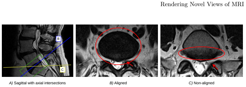
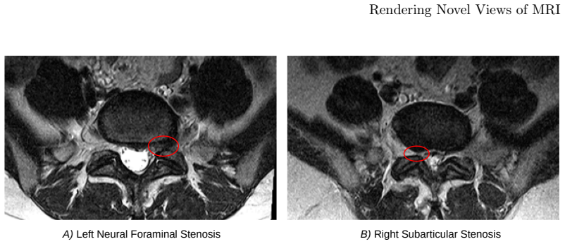
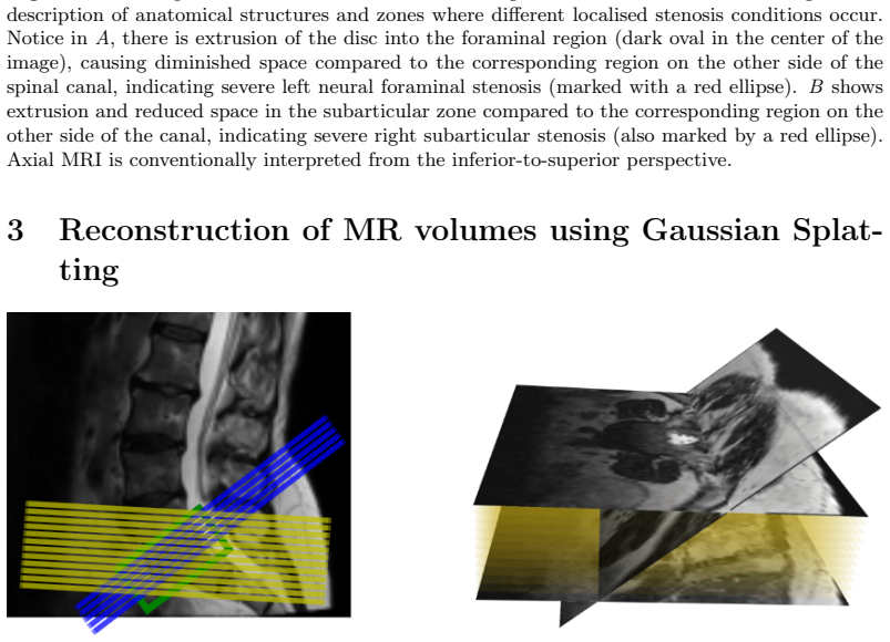
By starting from sparse anisotropic MRIs and using 3D Gaussian Splatting to create a volumetric model, the method allows sampling and rendering of novel view planes that are optimally aligned with the target spinal anatomy. When these resampled scans are used to predict stenosis grades, they yield higher accuracy across conditions than raw scans missing complete in-plane anatomy or scans resampled via inverse-distance weighted voxel interpolation.
What carries the argument
3D Gaussian Splatting adapted for volumetric reconstruction from sparse MRIs, representing the scene as a collection of 3D Gaussians that are rendered into novel aligned 2D views.
If this is right
- Gaussian Splatting resampling produces higher stenosis grading accuracy than raw anisotropic scans.
- It also outperforms voxel interpolation resampling across all tested stenosis conditions.
- The novel views supply complete in-plane anatomy where the original slices do not.
- Existing sparse MRI data can be repurposed for improved clinical grading without new acquisitions.
Where Pith is reading between the lines
- The approach could be tested on other anisotropic modalities such as CT to check for similar grading gains.
- If the Gaussians preserve fine intensity detail, the method might incidentally support limited super-resolution in the rendered planes.
- Deployment would need checks that the splatting step does not systematically shift intensity distributions used in grading.
Load-bearing premise
The Gaussian Splatting reconstruction must produce views whose intensities and geometry match the true anatomy closely enough that grading gains come from better plane alignment rather than from reconstruction artifacts.
What would settle it
Acquire high-resolution isotropic reference scans in the aligned planes and compare stenosis gradings from the Gaussian Splatting renders against those references; if accuracy does not exceed that of the raw scans or voxel interpolation, the claim is false.
Figures

read the original abstract
The objective of this paper is to improve radiological gradings measured on MRIs of spines, by resampling scans so that the new view planes are better aligned with the target anatomy than the original sparse images. To this end, we adapt 3D Gaussian Splatting to form a volumetric reconstruction starting from sparse anisotropic MRIs, and imaging planes aligned with the anatomy relevant for clinical evaluation are then sampled and rendered. The novel view plane is optimal for diagnostic radiological grading of the target anatomy, whereas the original MRI is not. The resampled scans are then used to predict ordinal severity grades of localised stenosis conditions in spinal MRIs. We compare our method against Voxel Interpolation resampling, which takes the average of inverse-distance weighted nearest neighbour intensities for each target coordinate. Experiments show that across all stenosis conditions, resampled scans using Gaussian Splatting produce more accurate stenosis gradings compared to the raw scans which do not include the complete anatomy in-plane, as well as images resampled using Voxel Interpolation.
Editorial analysis
A structured set of objections, weighed in public.
Referee Report
Summary. The paper adapts 3D Gaussian Splatting to reconstruct a volumetric representation from sparse anisotropic spinal MRIs, then renders novel imaging planes aligned with the target anatomy for stenosis grading. It compares the resulting gradings against those from the original raw scans and from voxel-interpolation resampling, claiming superior accuracy for the Gaussian Splatting approach across all tested stenosis conditions.
Significance. If the empirical claim holds with proper controls and quantitative validation, the work could provide a practical route to obtaining anatomy-aligned views from existing anisotropic acquisitions without additional scanning, which would be relevant for spinal MRI diagnostics where plane misalignment is common.
major comments (2)
- [Abstract] Abstract: the central claim that 'resampled scans using Gaussian Splatting produce more accurate stenosis gradings' is asserted without any reported accuracy numbers, dataset size, number of stenosis conditions, statistical tests, or implementation details; this absence makes the empirical result impossible to evaluate or reproduce.
- [Abstract] Abstract: no description is given of how stenosis grades were obtained (e.g., reader protocol, number of readers, ground-truth definition), so it is impossible to determine whether the reported improvement is attributable to better plane alignment or to other factors.
Simulated Author's Rebuttal
We thank the referee for these comments on the abstract. We agree that the abstract requires additional quantitative and methodological details to allow proper evaluation of the claims, and we will revise it in the next version of the manuscript.
read point-by-point responses
-
Referee: [Abstract] Abstract: the central claim that 'resampled scans using Gaussian Splatting produce more accurate stenosis gradings' is asserted without any reported accuracy numbers, dataset size, number of stenosis conditions, statistical tests, or implementation details; this absence makes the empirical result impossible to evaluate or reproduce.
Authors: We agree that the abstract should report key quantitative results to support the claim. In the revised manuscript we will expand the abstract to include the observed accuracy improvements across stenosis conditions, the dataset size, the number of conditions evaluated, and references to the statistical tests and implementation details already present in the methods and results sections. revision: yes
-
Referee: [Abstract] Abstract: no description is given of how stenosis grades were obtained (e.g., reader protocol, number of readers, ground-truth definition), so it is impossible to determine whether the reported improvement is attributable to better plane alignment or to other factors.
Authors: We acknowledge the absence of this information from the abstract. We will revise the abstract to include a concise summary of the grading process (reader protocol, number of readers, and ground-truth definition) as described in the methods section, so readers can assess whether the improvement is attributable to plane alignment. revision: yes
Circularity Check
No significant circularity
full rationale
The paper describes an empirical adaptation of 3D Gaussian Splatting for MRI resampling followed by comparative experiments on stenosis grading accuracy. No derivation chain, self-referential equations, fitted parameters presented as predictions, or load-bearing self-citations are present. The central claim rests on observable grading improvements versus raw scans and voxel interpolation, which is externally falsifiable and does not reduce to its own inputs by construction.
Axiom & Free-Parameter Ledger
Reference graph
Works this paper leans on
-
[1]
Y. Bao, T. Ding, J. Huo, Y. Liu, Y. Li, W. Li, Y. Gao, and J. Luo. 3D Gaussian Splatting: Survey, Technologies, Challenges, and Opportunities.IEEE Transactions on Circuits and Systems for Video Technology, 35(7):6832–6852, July 2025
2025
-
[2]
Blondiaux and C
E. Blondiaux and C. Garel. Fetal cerebral imaging – ultrasound vs. MRI: An update. Acta Radiologica, 54(9):1046–1054, Nov. 2013
2013
-
[3]
Bonilla, S
S. Bonilla, S. Zhang, D. Psychogyios, D. Stoyanov, F. Vasconcelos, and S. Bano. Gaussian Pancakes: Geometrically-Regularized 3D Gaussian Splatting for Realistic Endoscopic Reconstruction. In M. G. Linguraru, Q. Dou, A. Feragen, S. Gian- narou, B. Glocker, K. Lekadir, and J. A. Schnabel, editors,Medical Image Computing and Computer Assisted Intervention – ...
2024
-
[4]
Y. Cai, Y. Liang, J. Wang, A. Wang, Y. Zhang, X. Yang, Z. Zhou, and A. Yuille. Radiative gaussian splatting for efficient X-ray novel view synthesis. InECCV, pages 283–299. 2024
2024
-
[5]
L. S. de Vries, M. J. N. L. Benders, and F. Groenendaal. Imaging the premature brain: Ultrasound or MRI?Neuroradiology, 55(2):13–22, Sept. 2013
2013
-
[6]
M. C. Eid, A. I. Namburete, and J. F. Henriques. UltraGauss: Ultrafast gaussian reconstruction of 3D ultrasound volumes. InICLR, 2026
2026
-
[7]
B. Fei, J. Xu, R. Zhang, Q. Zhou, W. Yang, and Y. He. 3D Gaussian Splatting as a New Era: A Survey.IEEE Transactions on Visualization and Computer Graphics, 31(8):4429–4449, Aug. 2025
2025
-
[8]
Nano banana 2 generative AI image model
Google DeepMind. Nano banana 2 generative AI image model. Image generated using the Nano Banana 2 model in Gemini. Accessed: 27 May 2026]
2026
-
[9]
C. Guo, G. Pleiss, Y. Sun, and K. Q. Weinberger. On calibration of modern neural networks. InICML, pages 1321–1330. PMLR, 2017
2017
-
[10]
J. Guo, J. Wang, D. Kang, W. Dong, W. Wang, and Y.-h. Liu. Free-surgs: Sfm-free 3D gaussian splatting for surgical scene reconstruction. InMICCAI, pages 350–360, 2024
2024
-
[11]
K.-T. Hong, Y. Cho, C. H. Kang, K.-S. Ahn, H. Lee, J. Kim, S. J. Hong, B. H. Kim, and E. Shim. Lumbar spine computed tomography to magnetic resonance imaging synthesis using generative adversarial network: Visual turing test.Diagnostics, 12(2):530, 2022
2022
-
[12]
J. P. Hornak.The basics of MRI. Rochester Institute of Technology, 1996
1996
-
[13]
Huang, L
Y. Huang, L. Bai, B. Cui, K. Yuan, G. Wang, M. I. Hoque, N. Padoy, N. Navab, and H. Ren. SurgTPGS: Semantic 3D surgical scene understanding with text promptable gaussian splatting. InMICCAI, pages 584–594, 2025
2025
-
[14]
Iddrisu, S
K. Iddrisu, S. Malec, and A. Crimi. 3d reconstructions of brain from mri scans using neural radiance fields. In L. Rutkowski, R. Scherer, M. Korytkowski, W. Pedrycz, R. Tadeusiewicz, and J. M. Zurada, editors,Artificial Intelligence and Soft Computing, pages 207–218, Cham, 2023. Springer Nature Switzerland
2023
-
[15]
Jamaludin, T
A. Jamaludin, T. Kadir, and A. Zisserman. SpineNet: automated classification and evidence visualization in spinal MRIs.Medical image analysis, 41:63–73, 2017
2017
-
[16]
Y. Jia, A. Gholipour, Z. He, and S. K. Warfield. A new sparse representation framework for reconstruction of an isotropic high spatial resolution mr volume from orthogonal anisotropic resolution scans.IEEE transactions on medical imaging, 36(5):1182–1193, 2017
2017
-
[17]
Kastryulin, J
S. Kastryulin, J. Zakirov, N. Pezzotti, and D. V. Dylov. Image quality assessment for magnetic resonance imaging.IEEE Access, 11:14154–14168, 2023. 16 Rendering Novel Views of MRI
2023
-
[18]
J. N. Katz, Z. E. Zimmerman, H. Mass, and M. C. Makhni. Diagnosis and management of lumbar spinal stenosis.JAMA, 327(17):1688–1699, 2022
2022
-
[19]
Kerbl, G
B. Kerbl, G. Kopanas, T. Leimkühler, G. Drettakis, et al. 3d gaussian splatting for real-time radiance field rendering.ACM Trans. Graph., 42(4):139–1, 2023
2023
-
[20]
K. H. Kim, E.-C. Lee, Y. D. Yoon, D.-W. Shin, H.-W. Koo, and B.-J. Lee. Translation of computed tomography images to T2-weighted magnetic resonance images of lum- bar spine using generative adversarial networks on sagittal images.Scientific Reports, 15(1):18385, 2025
2025
-
[21]
Landauer and K
F. Landauer and K. Trieb. Diagnostic limitations and aspects of the lumbosacral transitional vertebrae (LSTV).Applied Sciences, 12(21):10830, 2022
2022
-
[22]
S. Lee, J. W. Lee, J. S. Yeom, K.-J. Kim, H.-J. Kim, S. K. Chung, and H. S. Kang. A practical mri grading system for lumbar foraminal stenosis.American Journal of Roentgenology, 194(4):1095–1098, 2010
2010
-
[23]
Lin, R.-M
S.-I. Lin, R.-M. Lin, and L.-W. Huang. Disability in patients with degenerative lumbar spinal stenosis.Archives of physical medicine and rehabilitation, 87(9):1250–1256, 2006
2006
-
[24]
S. Liu, M. Yang, T. Xing, and R. Yang. A survey of 3D reconstruction: the evolution from multi-view geometry to NeRF and 3DGS.Sensors, 25(18):5748, 2025
2025
-
[25]
J.-T. Lu, S. Pedemonte, B. Bizzo, S. Doyle, K. P. Andriole, M. H. Michalski, R. G. Gonzalez, and S. R. Pomerantz. Deep spine: automated lumbar vertebral segmentation, disc-level designation, and spinal stenosis grading using deep learning. InMLHC, pages 403–419. PMLR, 2018
2018
-
[26]
K. Marzol, I. Kolton, W. Smolak-DyĹL’ewska, J. Kaleta, M. Mazur, P. Spurek, et al. MedGS: Gaussian splatting for multi-modal 3D medical imaging.arXiv preprint arXiv:2509.16806, 2025
Pith/arXiv arXiv 2025
-
[27]
J. L. Melancia, A. F. Francisco, and J. L. Antunes. Spinal stenosis.Handbook of clinical neurology, 119:541–549, 2014
2014
-
[28]
Spinal stenosis symptoms, diagnosis & treatment, 2019
Miami Neuroscience Center. Spinal stenosis symptoms, diagnosis & treatment, 2019. Published: 18 October 2019; last modified: 8 July 2020
2019
-
[29]
Mildenhall, P
B. Mildenhall, P. P. Srinivasan, M. Tancik, J. T. Barron, R. Ramamoorthi, and R. Ng. Nerf: Representing scenes as neural radiance fields for view synthesis. InECCV, 2020
2020
-
[30]
H. J. Park, S. Kim, S. Lee, N. Park, E. Chung, M. Rho, H. Kwon, and S. Kook. A practical MRI grading system for cervical foraminal stenosis based on oblique sagittal images.The British journal of radiology, 86(1025), 2013
2013
-
[31]
M. S. Park, S.-H. Moon, H.-M. Lee, T.-H. Kim, J. K. Oh, S. Y. Lee, J. B. Oh, and K. D. Riew. Diagnostic value of oblique magnetic resonance images for evaluating cervical foraminal stenosis.The Spine Journal, 15(4):607–611, 2015
2015
-
[32]
R. Y. Park, R. Windsor, A. Jamaludin, and A. Zisserman. Multi-view and multimodal radiological grading using spinal MRIs. InML-CDS Workshop, MICCAI 2025, 2025
2025
-
[33]
T. J. Richards, A. E. Flanders, E. Colak, L. M. Prevedello, R. L. Ball, F. Kitamura, J. Mongan, M. Vazirabad, H.-M. Lin, A. Kendell, et al. The RSNA lumbar degenerative imaging spine classification (LumbarDISC) dataset.Radiology: Artificial Intelligence, page e250480, 2026
2026
-
[34]
Sąsiadek and J
M. Sąsiadek and J. Jacków-Nowicka. Degenerative disease of the spine: How to re- late clinical symptoms to radiological findings.Advances in Clinical and Experimental Medicine, 33(1):91–98, 2024
2024
-
[35]
Sobański, R
D. Sobański, R. Staszkiewicz, M. Stachura, M. Gadzieliński, and B. O. Grabarek. Pre- sentation, diagnosis, and management of lower back pain associated with spinal steno- sis: a narrative review.Medical Science Monitor: International Medical Journal of Experimental and Clinical Research, 29:e939237–1, 2023. 17 R. Park, M. Eid et al
2023
-
[36]
Takahashi, A
K. Takahashi, A. K. Yadav, K. Hashimoto, T. Tsubakino, T. Aizawa, and Y. Tanaka. Foraminal stenosis at L5–S1 as an overlooked pathology of bilateral radiculopathy: A case series.Journal of Orthopaedic Case Reports, 12(6):13, 2022
2022
-
[37]
N. A. Tawfik, A. T. Ahmed, T. E. El-Shafei, and M. R. Habba. Diagnostic value of spinal ultrasound compared to MRI for diagnosis of spinal anomalies in pediatrics. Egyptian Journal of Radiology and Nuclear Medicine, 51(1):18, Jan. 2020
2020
-
[38]
Van Reeth, I
E. Van Reeth, I. W. Tham, C. H. Tan, and C. L. Poh. Super-resolution in magnetic resonance imaging: a review.Concepts in Magnetic Resonance Part A, 40(6):306–325, 2012
2012
-
[39]
J. C. van Rijn, N. Klemetsö, J. B. Reitsma, C. B. Majoie, F. J. Hulsmans, W. C. Peul, J. Stam, P. M. Bossuyt, and G. J. den Heeten. Observer variation in MRI evaluation of patients suspected of lumbar disk herniation.American Journal of Roentgenology, 184(1):299–303, 2005
2005
-
[40]
Verheijen, T
E. Verheijen, T. Kapogiannis, D. Munteh, J. Chabros, M. Staring, T. Smith, and C. Vleggeert-Lankamp. Artificial intelligence for segmentation and classification in lumbar spinal stenosis: an overview of current methods.European Spine Journal, 34(3):1146–1155, 2025
2025
-
[41]
Windsor, A
R. Windsor, A. Jamaludin, T. Kadir, and A. Zisserman. Context-aware transformers for spinal cancer detection and radiological grading. InMICCAI, pages 271–281, 2022
2022
-
[42]
Windsor, A
R. Windsor, A. Jamaludin, T. Kadir, and A. Zisserman. Automated detection, labelling and radiological grading of clinical spinal MRIs.Scientific Reports, 14(1):14993, 2024
2024
-
[43]
R. Zha, T. J. Lin, Y. Cai, J. Cao, Y. Zhang, and H. Li. R^2-gaussian: Rectifying radiative gaussian splatting for tomographic reconstruction. InNeurIPS, volume 37, pages 44907–44934, 2024
2024
-
[44]
Zhang, P
R. Zhang, P. Isola, A. A. Efros, E. Shechtman, and O. Wang. The unreasonable effectiveness of deep features as a perceptual metric. InCVPR, pages 586–595, 2018. 18
2018
discussion (0)
Sign in with ORCID, Apple, or X to comment. Anyone can read and Pith papers without signing in.