Three-dimensional visualization of threading dislocation in GaN by polarized-light microscopy
Pith reviewed 2026-06-29 04:15 UTC · model grok-4.3
The pith
Threading dislocations in GaN tilt 3.3 degrees from the c-axis perpendicular to their Burgers vectors.
A machine-rendered reading of the paper's core claim, the machinery that carries it, and where it could break.
Core claim
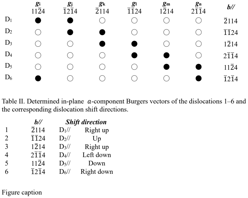
Threading dislocations in GaN wafers imaged by polarized-light microscopy display focal-depth-dependent in-plane shifts that reveal their three-dimensional inclination. Dislocations sharing the same in-plane Burgers vector component show matching trace geometries and inclination directions. These dislocations tilt approximately 3.3 degrees from the c-axis in directions perpendicular to their Burgers vectors, consistent with climb-mediated motion that accommodates strain relaxation during crystal growth.
What carries the argument
Focal-depth-dependent in-plane shifts under polarized-light microscopy, which directly map the three-dimensional inclination angles of threading dislocations across large wafer areas.
If this is right
- Large-area nondestructive three-dimensional mapping of threading dislocations becomes feasible in GaN wafers.
- Dislocations with identical in-plane Burgers vector components exhibit consistent inclination behavior.
- Strain relaxation in ammonothermal GaN growth proceeds through climb motion of threading dislocations.
Where Pith is reading between the lines
- The optical shift method could apply to defect characterization in other hexagonal crystals where similar microscopy contrast exists.
- Adjusting growth parameters to minimize the observed climb tilts might reduce overall dislocation density.
- Electrical or optical device metrics could be correlated with these specific inclination angles in future studies.
Load-bearing premise
The focal-depth-dependent in-plane shifts seen in the microscope images accurately reflect the true three-dimensional tilt angles and directions of the dislocations without optical artifacts or projection distortions.
What would settle it
Transmission electron microscopy or synchrotron tomography on matched wafer regions showing tilt angles or directions that differ from 3.3 degrees and the perpendicular-to-Burgers-vector rule.
Figures
read the original abstract
We demonstrate high-throughput three-dimensional imaging of threading dislocations in ammonothermal GaN wafers using polarized-light microscopy with collimated LED illumination. Threading dislocations exhibited focal-depth-dependent in-plane shifts, enabling visualization of their three-dimensional inclination behavior over large areas. Synchrotron radiation X-ray topography indicated that dislocations with identical in-plane Burgers vector components tended to exhibit similar trace geometries and inclination directions. The threading dislocations were found to be tilted by approximately 3.3{\deg} from the c-axis in directions perpendicular to their Burgers vectors, indicating climb-mediated motion associated with strain relaxation during crystal growth. These results demonstrate a simple nondestructive approach for large-area three-dimensional characterization of dislocations in GaN wafers.
Editorial analysis
A structured set of objections, weighed in public.
Referee Report
Summary. The paper demonstrates high-throughput three-dimensional imaging of threading dislocations in ammonothermal GaN wafers via polarized-light microscopy with collimated LED illumination. Dislocations show focal-depth-dependent in-plane image shifts that are used to visualize their 3D inclinations over large areas. Synchrotron X-ray topography reveals that dislocations sharing the same in-plane Burgers-vector component exhibit similar trace geometries and inclination directions. The authors report that the dislocations are tilted by approximately 3.3° from the c-axis in directions perpendicular to their Burgers vectors, which they interpret as evidence of climb-mediated motion linked to strain relaxation during growth. The work positions the optical method as a simple nondestructive alternative for large-area dislocation characterization.
Significance. If the conversion from observed focal-depth shifts to true geometric inclination angles is shown to be free of optical artifacts, the approach would offer a practical, scalable route to map dislocation distributions and orientations in GaN wafers without destructive sectioning. Such capability is relevant for understanding and mitigating defects that limit performance in GaN-based power and optoelectronic devices. The reported correlation between Burgers-vector direction and inclination sense provides a testable link to growth-induced strain relaxation mechanisms.
major comments (3)
- [Abstract] Abstract: the reported tilt of approximately 3.3° is presented without error bars, the number of dislocations sampled, or any description of the fitting or averaging procedure used to extract the angle from the focal-depth-dependent shifts.
- [Abstract] Abstract: the mapping from observed in-plane image displacements versus focal depth to a true inclination angle of 3.3° is not validated by any independent three-dimensional metrology (serial sectioning, confocal imaging, or X-ray section topography) performed on the same dislocation segments; this leaves the central quantitative claim dependent on an untested assumption that the shifts are purely geometric.
- [Abstract] Abstract: no optical model, ray-tracing simulation, or analysis of possible depth-dependent refraction, birefringence weighting, or depth-of-field effects is supplied to demonstrate that the measured lateral shifts equal the geometric inclination rather than imaging artifacts.
minor comments (1)
- The abstract would be strengthened by stating the typical dislocation density and wafer thickness to place the reported tilt in experimental context.
Simulated Author's Rebuttal
We thank the referee for the constructive comments. We address each major comment point by point below and have revised the manuscript to improve quantitative reporting and address methodological concerns where feasible.
read point-by-point responses
-
Referee: [Abstract] Abstract: the reported tilt of approximately 3.3° is presented without error bars, the number of dislocations sampled, or any description of the fitting or averaging procedure used to extract the angle from the focal-depth-dependent shifts.
Authors: We agree that the abstract should report these details for transparency. The 3.3° value was obtained via linear fits to focal-depth versus in-plane shift data followed by averaging across multiple dislocations. In the revised manuscript we have updated the abstract to include the standard deviation, the number of dislocations sampled, and a brief description of the fitting and averaging procedure. revision: yes
-
Referee: [Abstract] Abstract: the mapping from observed in-plane image displacements versus focal depth to a true inclination angle of 3.3° is not validated by any independent three-dimensional metrology (serial sectioning, confocal imaging, or X-ray section topography) performed on the same dislocation segments; this leaves the central quantitative claim dependent on an untested assumption that the shifts are purely geometric.
Authors: We acknowledge that independent three-dimensional metrology performed on the identical dislocation segments would provide stronger validation of the quantitative angle. The synchrotron X-ray topography data confirm that inclination directions correlate with Burgers-vector components, supporting the geometric interpretation, but do not supply an independent angle measurement on the same segments. We have added a discussion of this limitation and the underlying geometric assumption to the revised manuscript. revision: partial
-
Referee: [Abstract] Abstract: no optical model, ray-tracing simulation, or analysis of possible depth-dependent refraction, birefringence weighting, or depth-of-field effects is supplied to demonstrate that the measured lateral shifts equal the geometric inclination rather than imaging artifacts.
Authors: We have added a simple geometric model in the methods section of the revised manuscript that relates observed in-plane shifts to inclination angle under the paraxial approximation. A brief analysis of depth-dependent refraction and depth-of-field effects is included, showing these contributions are negligible for the small angles involved. Full ray-tracing simulations are outside the present scope but the observed correlation between inclination sense and Burgers vector (from X-ray topography) makes systematic imaging artifacts unlikely. revision: yes
- Independent three-dimensional metrology (serial sectioning, confocal imaging, or X-ray section topography) performed on the exact same dislocation segments to quantitatively validate the 3.3° tilt angle.
Circularity Check
No circularity: purely experimental observations with no derivations or self-referential steps
full rationale
The paper consists entirely of experimental reports from polarized-light microscopy (focal-depth shifts) and synchrotron X-ray topography (Burgers vectors and trace geometries). The reported 3.3° tilt is stated as a direct finding from observed in-plane shifts versus depth, with no equations, fitted parameters, models, or self-citations that reduce any result to its own inputs by construction. No self-definitional loops, fitted-input predictions, or ansatzes appear. The work is self-contained against external benchmarks (direct imaging correlation) and receives the default non-circularity finding.
Axiom & Free-Parameter Ledger
Reference graph
Works this paper leans on
-
[1]
1 R. A. Oliver, Mater. Sci. Technol. 32, 737 (2016). 2 X. Fan, J. Shi, Y . Chen, G. Miao, H. Jiang, and H. Song, Micromachines 15, 1188 (2024). 3 H. Amano, Y . Baines, E. Beam, M. Borga, T. Bouchet, P. R. Chalker, et al., J. Phys. D: Appl. Phys. 51, 163001 (2018). 4 M. Meneghini, R. A. Khadar, C. De Santi, L. Nela, I. Abid, M. Buffolo, et al., J. Appl. Ph...
2016
-
[2]
Status Solidi B 257, 1900553 (2020)
Amano, Phys. Status Solidi B 257, 1900553 (2020). 14 A. Kawata, K. Murayama, S. Sumitani, and S. Harada, Jpn. J. Appl. Phys. 60, SBBD06 (2021). 16 15 S. Harada and K. Murayama, J. Appl. Crystallogr. 55, 1029 (2022). 16 S. Harada, Y . Matsubara, and K. Murayama, Diamond Relat. Mater. 138, 110192 (2023). 17 S. Harada, Y . Matsubara, S. Hayashi, M. Kawase, K...
2020
-
[3]
Murayama, and S
Mizutani, K. Murayama, and S. Harada, Acta Mater. 290, 120923 (2025). 19 S. Harada and K. Murayama, J. Cryst. Growth 640, 127982 (2025). 20 M. Kato, H. Sato, T. Kato, K. Murata, and S. Harada, Rev. Sci. Instrum. 96, 083901 (2025). 21 L. Kirste, K. Grabianska, R. Kucharski, T. Sochacki, B. Lucznik, and M
2025
-
[4]
Bockowski, Materials 14, 5472 (2021). 22 J. Schindelin, I. Arganda-Carreras, E. Frise, V . Kaynig, M. Longair, T. Pietzsch, S
2021
-
[5]
Rueden, S
Preibisch, C. Rueden, S. Saalfeld, B. Schmid, J. Y . Tinevez, et al., Nat. Methods 9, 676 (2012). 23 L. Kirste, T. N. T. Caliste, J. L. Weyher, J. Smalc-Koziorowska, M. A. Zajac, R
2012
-
[6]
Sochacki, K
Kucharski, T. Sochacki, K. Grabianska, M. Iwinska, C. Detlefs, A. N. Danilewsky, M. Bockowski, and J. Baruchel, Materials 15, 6996 (2022). 24 M. Dudley, X. R. Huang, and W. Huang, J. Phys. D: Appl. Phys. 32, A139 (1999). 25 I. Kamata, H. Tsuchida, T. Jikimoto, and K. Izumi, J. Cryst. Growth 311, 1416 (2009). 26 F. Wu, S. Byrappa, H. Wang, Y . Chen, B. Rag...
2022
-
[7]
Chung, D
Sanchez, G. Chung, D. Hansen, S. G. Mueller, and M. J. Loboda, Mater. Res. Soc. Symp. Proc. 1433, 53 (2012). 27 J. Q. Guo, X. R. Huang, M. Dudley, V . Vetter, M. Dudley, and W. Huang, Mater. Sci. Forum 858, 15 (2016). 28 Q. Cheng, Z. Chen, S. Hu, Y . Liu, B. Raghothamachar, and M. Dudley, J. Electron. 17 Mater. 50, 4104 (2021). 29 Q. Cheng, Z. Chen, S. Hu...
2012
discussion (0)
Sign in with ORCID, Apple, or X to comment. Anyone can read and Pith papers without signing in.