Patient-Specific Articulated Digital Twins from a Single Full-Body CT Scan
Pith reviewed 2026-07-03 15:22 UTC · model grok-4.3
The pith
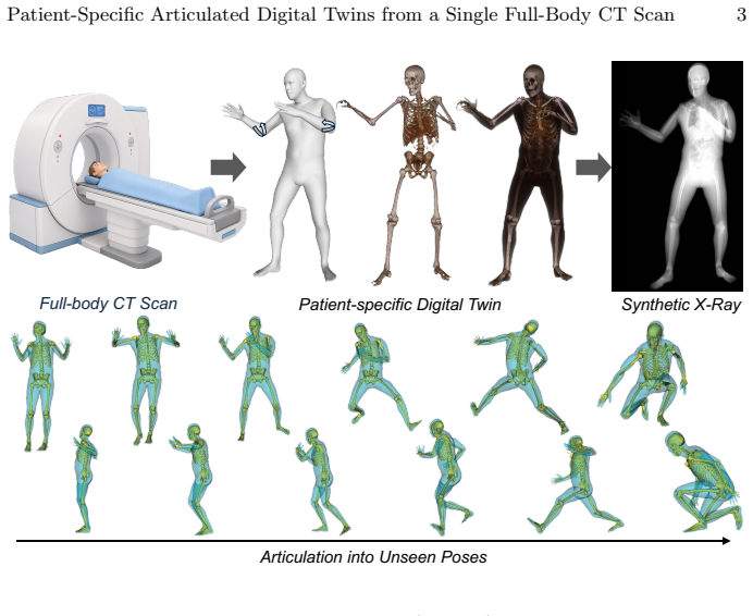
A single full-body CT scan can generate a patient-specific articulated digital twin that moves while preserving the individual's skeletal geometry.
A machine-rendered reading of the paper's core claim, the machinery that carries it, and where it could break.
Core claim
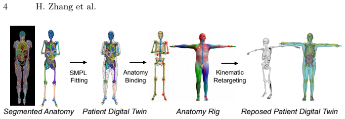
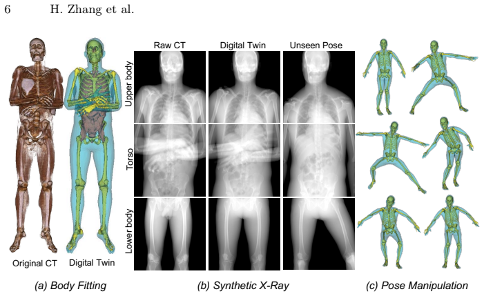
The central claim is that fitting the SMPL parametric human body model to a full-body CT scan provides a kinematic scaffold to which segmented bones and organs can be bound, enabling retargeting of pose changes while preserving patient-specific skeletal geometry. This is shown through a proof-of-concept on three subjects, with quantitative measures of fit quality and image similarity in original and new poses.
What carries the argument
The patient-aligned kinematic scaffold obtained by fitting the SMPL parametric model to the CT scan, to which segmented bones and organs are bound via an anatomy-aware rig.
If this is right
- The fitted scaffold achieves 15.8 mm chamfer distance and 95.9% skeletal enclosure on the test subjects.
- Recomposition at the original acquisition pose produces DRRs with SSIM of 0.872 and PSNR of 18.5 dB.
- Across unseen target poses the twin maintains 94.4% skeletal enclosure.
- The articulated twin supports rendering of pose-dependent DRRs for synthetic imaging studies.
Where Pith is reading between the lines
- The same single-scan construction could supply varied-pose training examples for machine-learning methods in radiographic image analysis.
- If extended, the twins might allow pre-operative rehearsal of patient positioning for procedures that depend on exact body orientation.
- Direct validation against real multi-pose CT pairs of the same individuals would test whether the retargeted geometry matches physical reality beyond the current metrics.
Load-bearing premise
The SMPL parametric model can be fitted accurately enough to the CT scan to serve as a kinematic scaffold that keeps the patient's unique skeletal geometry intact during pose retargeting.
What would settle it
Acquire a second full-body CT of one of the test subjects in a substantially different pose and measure whether the retargeted twin's bone positions and rendered DRRs match the new scan within the reported chamfer distance, enclosure, SSIM, and PSNR ranges.
Figures
read the original abstract
Patient-specific anatomical models provide individualized context for surgical planning, image-guided intervention, and algorithm development. However, most CT-derived models are static: they preserve the body configuration captured at scan time, but cannot represent how the same anatomy would appear after patient repositioning. This limitation is especially important for radiographic imaging, where appearance depends jointly on imaging geometry and patient pose. We present a proof-of-concept for constructing a patient-specific articulated digital twin from a single full-body CT scan. The method fits a parametric human body model (SMPL) to obtain a patient-aligned kinematic scaffold, binds segmented bones and organs to an anatomy-aware rig, and retargets body-pose changes while preserving skeletal geometry. On three full-body CT subjects, the fitted scaffold achieved 15.8 $\pm$ 4.0 mm chamfer distance and 95.9 $\pm$ 1.8% skeletal enclosure. Recomposition at the acquisition pose preserved major radiographic structure, with overall SSIM of 0.872 $\pm$ 0.016 and PSNR of 18.5 $\pm$ 1.4 dB across paired DRRs. Across unseen target poses, the resulting twins enabled articulation while maintaining high skeletal enclosure (94.4 $\pm$ 0.4%). As a feasibility demonstration, we render the articulated twin as pose-dependent DRRs. These results suggest the feasibility of extending static, view-controllable CT simulation toward pose-controllable anatomical twins for future synthetic imaging and positioning studies.
Editorial analysis
A structured set of objections, weighed in public.
Referee Report
Summary. The manuscript presents a proof-of-concept for constructing patient-specific articulated digital twins from a single full-body CT scan. The method fits the SMPL parametric model to obtain a patient-aligned kinematic scaffold, binds segmented bones and organs to an anatomy-aware rig, and retargets body-pose changes while preserving skeletal geometry. On three subjects, it reports chamfer distance of 15.8 ± 4.0 mm and 95.9 ± 1.8% skeletal enclosure for the fit, SSIM of 0.872 ± 0.016 and PSNR of 18.5 ± 1.4 dB for DRRs at the acquisition pose, and 94.4 ± 0.4% skeletal enclosure for unseen target poses, with rendered pose-dependent DRRs as a feasibility demonstration.
Significance. If the central claim holds under stronger validation, the work could enable pose-controllable anatomical models from static CTs, with potential value for surgical planning, image-guided interventions, and synthetic radiographic datasets. The quantitative metrics on fitting accuracy and acquisition-pose DRR similarity, combined with the use of an established parametric model (SMPL) for the scaffold, provide initial evidence of technical feasibility on a small cohort.
major comments (2)
- [Abstract] Abstract: The claim that retargeting preserves patient-specific skeletal geometry under arbitrary pose changes rests on skeletal enclosure of 94.4 ± 0.4% for unseen poses, but no ground-truth DRR or geometric fidelity metrics (e.g., organ/bone relative positions) are provided for target poses, in contrast to the acquisition-pose results (SSIM/PSNR). Skeletal enclosure alone does not confirm maintenance of radiographic appearance or anatomical relationships.
- [Methods] Methods (binding and retargeting description): No derivation details, error analysis, or validation protocol are given for how segmented bones and organs are bound to the SMPL-derived rig or how the anatomy-aware rig enforces constraints during retargeting. This leaves unclear whether the mechanism goes beyond the initial fit (chamfer distance 15.8 mm) to preserve patient-specific geometry.
minor comments (1)
- [Abstract] Abstract: The dataset of three full-body CT subjects is not described (source, acquisition parameters, or subject diversity), which would help evaluate the reported standard deviations and generalizability.
Simulated Author's Rebuttal
We thank the referee for the constructive feedback on this proof-of-concept manuscript. We address each major comment below.
read point-by-point responses
-
Referee: [Abstract] Abstract: The claim that retargeting preserves patient-specific skeletal geometry under arbitrary pose changes rests on skeletal enclosure of 94.4 ± 0.4% for unseen poses, but no ground-truth DRR or geometric fidelity metrics (e.g., organ/bone relative positions) are provided for target poses, in contrast to the acquisition-pose results (SSIM/PSNR). Skeletal enclosure alone does not confirm maintenance of radiographic appearance or anatomical relationships.
Authors: We agree that skeletal enclosure is a geometric proxy and does not substitute for direct radiographic metrics such as SSIM/PSNR or organ relative positions on target poses. Because the study uses single static CT acquisitions, no ground-truth multi-pose data exist for the same subjects, precluding those comparisons. We will revise the abstract to state the retargeting result more precisely as preservation of skeletal enclosure while explicitly noting the absence of full radiographic validation for unseen poses. revision: partial
-
Referee: [Methods] Methods (binding and retargeting description): No derivation details, error analysis, or validation protocol are given for how segmented bones and organs are bound to the SMPL-derived rig or how the anatomy-aware rig enforces constraints during retargeting. This leaves unclear whether the mechanism goes beyond the initial fit (chamfer distance 15.8 mm) to preserve patient-specific geometry.
Authors: The manuscript presents the binding and retargeting at a conceptual level. We accept that additional technical detail is warranted. In revision we will expand the Methods section with the mathematical formulation of the binding step, an error-propagation analysis for retargeting, and the explicit validation protocol used to confirm that the rig preserves the initial patient-specific fit beyond the reported Chamfer distance. revision: yes
Circularity Check
No circularity: procedural pipeline with independent metrics
full rationale
The paper presents a standard fitting-and-binding pipeline (SMPL fit to CT, bone/organ binding to rig, pose retargeting) whose outputs are evaluated by direct geometric and radiographic metrics (chamfer distance, skeletal enclosure, SSIM/PSNR on DRRs). These quantities are computed from the constructed models rather than being forced by redefinition or self-citation; the central claim is a feasibility demonstration of the pipeline itself, not a derived prediction that collapses to the input data by construction. No load-bearing self-citations, uniqueness theorems, or ansatzes appear in the provided text.
Axiom & Free-Parameter Ledger
free parameters (1)
- SMPL shape and pose parameters
axioms (2)
- domain assumption SMPL provides a suitable kinematic scaffold for human anatomy
- domain assumption Segmented bones and organs can be bound rigidly to the rig while preserving geometry
Reference graph
Works this paper leans on
-
[1]
IEEE Transactions on Pattern Analysis and Machine Intelligence (2019)
Cao, Z., Hidalgo Martinez, G., Simon, T., Wei, S., Sheikh, Y.A.: Openpose: Real- time multi-person 2d pose estimation using part affinity fields. IEEE Transactions on Pattern Analysis and Machine Intelligence (2019)
2019
-
[2]
https://doi.org/10.48550/arXiv.2405.11133
Dahal, L., Ghojoghnejad, M., Ghosh, D., Bhandari, Y., Kim, D., Ho, F.C., Tushar, F.I.,Luoa,S.,Lafata,K.J.,Abadi,E.,Samei,E.,Lo,J.Y.,Segars,W.P.:XCAT-3.0: A Comprehensive Library of Personalized Digital Twins Derived from CT Scans (Sep.). https://doi.org/10.48550/arXiv.2405.11133
-
[3]
https://doi.org/10.25827/5s8c-n515, https://doi.org/10.25827/5s8c-n515
Edgar, H.J.H., Daneshvari Berry, S., Moes, E., Adolphi, N.L., Bridges, P., Nolte, K.B.: New Mexico Decedent Image Database (2020). https://doi.org/10.25827/5s8c-n515, https://doi.org/10.25827/5s8c-n515
-
[4]
Nature machine intelligence5, 294 – 308 (2023), https://api.semanticscholar.org/CorpusID:257668838
Gao, C., Killeen, B., Hu, Y., Grupp, R., Taylor, R.H., Armand, M., Unberath, M.: Synthetic data accelerates the development of generalizable learning-based algo- rithms for x-ray image analysis. Nature machine intelligence5, 294 – 308 (2023), https://api.semanticscholar.org/CorpusID:257668838
2023
-
[5]
arXiv preprint arXiv:2312.06358 (2023)
Gopalakrishnan, V., Dey, N., Golland, P.: Intraoperative 2D/3D image registration via differentiable x-ray rendering. arXiv preprint arXiv:2312.06358 (2023)
-
[6]
In: Medical Image Computing and Com- puter Assisted Intervention 2025
Killeen, B.D., Wang, L.J., Iñígo, B., Zhang, H., Armand, M., Taylor, R.H., Os- good, G., Unberath, M.: FluoroSAM: A Language-Promptable Foundation Model for Flexible X-Ray Image Segmentation. In: Medical Image Computing and Com- puter Assisted Intervention 2025. https://doi.org/10.1007/978-3-032-04981-0_24
-
[7]
https://doi.org/10.1007/978-3-031-43996-4_13
Killeen, B.D., Zhang, H., Mangulabnan, J., Armand, M., Taylor, R.H., Osgood, G., Unberath, M.: Pelphix: Surgical Phase Recognition from X-Ray Images in Per- cutaneous Pelvic Fixation. https://doi.org/10.1007/978-3-031-43996-4_13
-
[8]
International Journal of Computer As- sisted Radiology and Surgery (2024)
Killeen, B.D., Zhang, H., Wang, L.J., Liu, Z., Kleinbeck, C., Rosen, M., Taylor, R.H., Osgood, G., Unberath, M.: Stand in surgeon’s shoes: virtual reality cross- training to enhance teamwork in surgery. International Journal of Computer As- sisted Radiology and Surgery (2024). https://doi.org/10.1007/s11548-024-03138-7
-
[9]
Loper, M., Mahmood, N., Romero, J., Pons-Moll, G., Black, M.J.: SMPL: A Skinned Multi-Person Linear Model. In: Seminal Graphics Papers: Pushing the Boundaries, Volume 2 (Aug.), https://dl.acm.org/doi/10.1145/3596711.3596800
-
[10]
AMASS: Archive of Motion Capture as Surface Shapes
Mahmood, N., Ghorbani, N., Troje, N.F., Pons-Moll, G., Black, M.J.: AMASS: Archive of Motion Capture as Surface Shapes (Apr 2019). https://doi.org/10.48550/arXiv.1904.03278
work page internal anchor Pith review Pith/arXiv arXiv doi:10.48550/arxiv.1904.03278 2019
-
[11]
https://doi.org/10.1038/s41746-025-01575-5 10 H
Mekki, Y.M., Luijten, G., Hagert, E., Belkhair, S., Varghese, C., Qadir, J., So- laiman, B., Bilal, M., Dhanda, J., Egger, J., Deng, J., Khanduja, V., Frangi, A.F., Zughaier, S.M., Stotland, M.A.: Digital twins for the era of personalized surgery 8(1), 283 (May 2025). https://doi.org/10.1038/s41746-025-01575-5 10 H. Zhang et al
-
[12]
Journal of Clinical Imaging Sci- ence13, 34
Midtgaard, M., Pedersen, M.R.V., Christensen, N.L., McKnight, K.L., Jensen, J.: Patient positioning during the radiographic procedure affects the radiological signs of acetabular retroversion - A systematic review. Journal of Clinical Imaging Sci- ence13, 34. https://doi.org/10.25259/JCIS_82_2023
-
[13]
Jour- nal of Clinical Medicine14(11) (2025)
Prządka, M., Pająk, W., Kleinrok, J., Pec, J., Michno, K., Karpiński, R., Baj, J.: Advances in 3d printing applications for personalized ortho- pedic surgery: From anatomical modeling to patient-specific implants. Jour- nal of Clinical Medicine14(11) (2025). https://doi.org/10.3390/jcm14113989, https://www.mdpi.com/2077-0383/14/11/3989
-
[14]
https://doi.org/10.48550/arXiv.2303.04923
Shetty, K., Birkhold, A., Jaganathan, S., Strobel, N., Egger, B., Kowarschik, M., Maier, A.: BOSS: Bones, Organs and Skin Shape Model (Mar 2023). https://doi.org/10.48550/arXiv.2303.04923
-
[15]
In: Medical Image Computing and Computer Assisted Intervention
Unberath, M., Zaech, J.N., Lee, S.C., Bier, B., Fotouhi, J., Armand, M., Navab, N.: DeepDRR – A Catalyst for Machine Learning in Fluoroscopy-Guided Procedures. In: Medical Image Computing and Computer Assisted Intervention. Cham (2018). https://doi.org/10.1007/978-3-030-00937-3_12
-
[16]
Ra- diology: Artificial Intelligence5(5) (2023)
Wasserthal, J., Breit, H.C., Meyer, M.T., Pradella, M., Hinck, D., Sauter, A.W., Heye, T., Boll, D.T., Cyriac, J., Yang, S., Bach, M., Segeroth, M.: Totalseg- mentator: Robust segmentation of 104 anatomic structures in ct images. Ra- diology: Artificial Intelligence5(5) (2023). https://doi.org/10.1148/ryai.230024, http://dx.doi.org/10.1148/ryai.230024
discussion (0)
Sign in with ORCID, Apple, or X to comment. Anyone can read and Pith papers without signing in.