Continuum modeling of fluidic and elastic flow during growth-driven wound closure in partial-EMT cell monolayers
Reviewed by Pith T0 review T1 audit T2 compute T3 formal T4 kernel 2026-07-08 19:43 UTCgrok-4.5pith:JGASLWXNrecord.jsonopen to challenge →
The pith
A continuum model of fluidized growth-elasticity shows that both tissue fluidity and fiber-reinforced elasticity are required to close large circular gaps in partial-EMT cell monolayers.
A machine-rendered reading of the paper's core claim, the machinery that carries it, and where it could break.
Core claim
Both tissue fluidity and the elastic properties associated with fiber reinforcement are critical for reproducing large circular gap-closure kinematics in MEC1 monolayers. Specifically, the TGF-β-treated condition has lower fluidity, a lower fluidic deformation rate, and a higher elastic deformation rate than the untreated condition, in agreement with experimental observations. The enabling device is a nonlinear Maxwell-fluid-with-growth decomposition of the observable strain rate into additive growth, elastic, and fluidic parts.
What carries the argument
The fluidized growth-elasticity framework: a nonlinear analogue of a Maxwell fluid with growth that decomposes the experimentally observable strain rate into the additive sum of growth, elastic, and fluidic strain rates. That decomposition carries the argument by converting raw tissue kinematics into separate, quantitative estimates of fluidity (inverse viscosity) and elasticity, including fiber-reinforcement effects.
If this is right
- Growth is the main driver of large circular gap closure on multi-hour timescales; elastic stretch and fluidic rearrangement only modulate the kinematics.
- TGF-β-induced partial EMT lowers tissue fluidity and raises the relative elastic deformation rate in MEC1 monolayers.
- Fiber reinforcement (elastic anisotropy) must be retained in the constitutive law to match observed closure shapes and rates.
- The same kinematic decomposition can extract fluidity and elasticity from other growth-driven tissue morphogenesis experiments that record strain-rate fields.
Where Pith is reading between the lines
- If the additive strain-rate split holds more broadly, ordinary live imaging of gap closure could serve as a non-invasive assay for how drugs or genetic changes alter tissue fluidity without separate rheometers.
- Embryonic wound healing and collective invasion after partial EMT could be re-analyzed with the same decomposition to test whether partial EMT consistently trades fluidity for elastic fiber reinforcement.
- A direct experimental check is to vary gap diameter or substrate stiffness and verify that the extracted fluidity remains a property of the treatment condition rather than of the geometry.
Load-bearing premise
The observed tissue strain rate can be cleanly split into independent additive growth, elastic, and fluidic contributions; if that additive split does not hold for partial-EMT monolayers on multi-hour millimeter scales, separate numbers for fluidity and elasticity cannot be extracted from kinematics.
What would settle it
Measure gap-closure trajectories and independent tissue rheology under TGF-β versus control; the claim fails if no combination of lower fluidity and higher elastic deformation rate inside the model can reproduce the treated kinematics, or if rheology shows the untreated monolayers are not more fluid than the treated ones.
Figures


read the original abstract
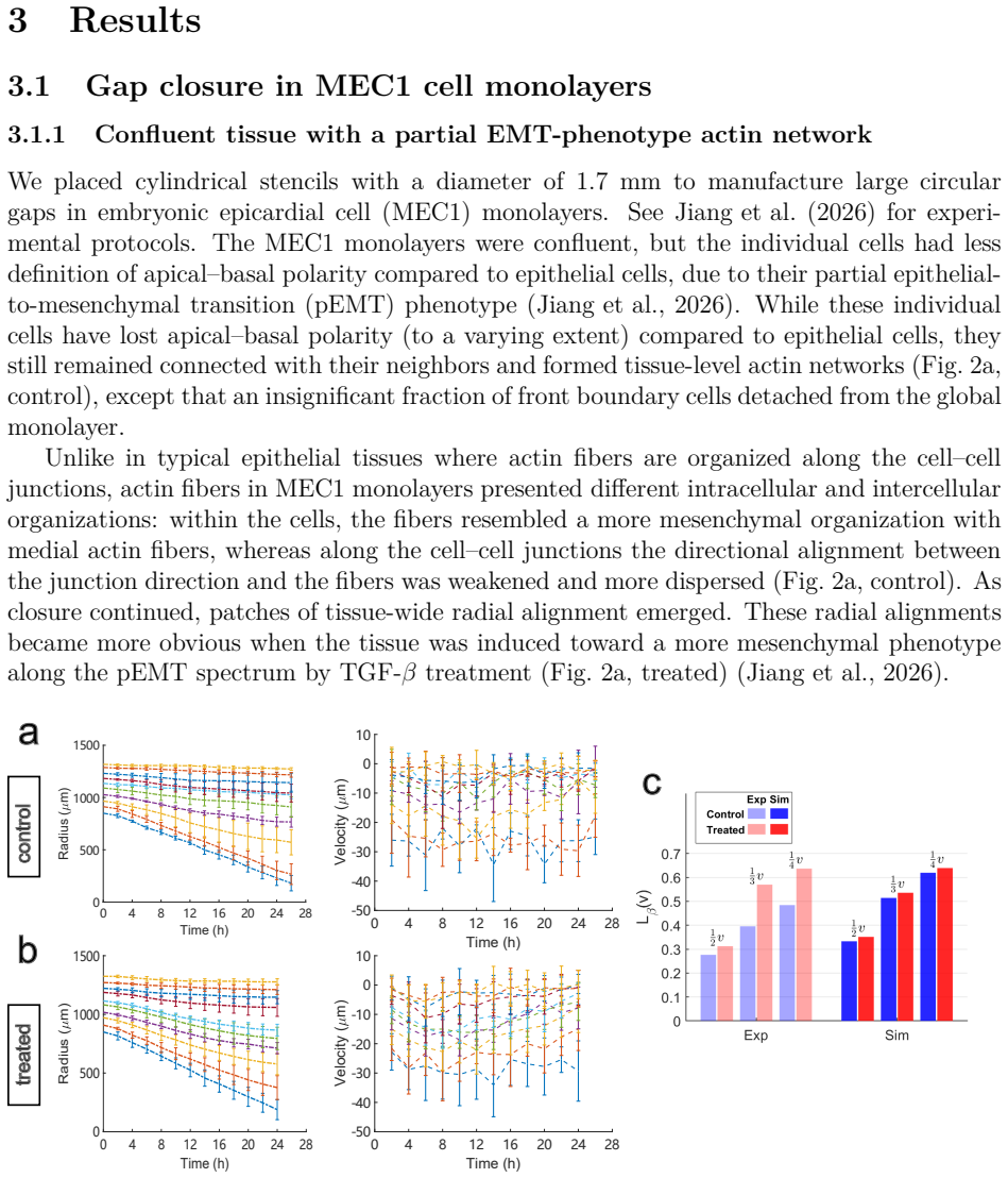
Large-scale circular gap closure occurs over a time scale on which cell growth and proliferation become important. Growth is the main driver of the closing process, while cell dynamics such as elongation and intercalation reflect elastic and fluidic contributions to tissue deformation. We develop a novel fluidized growth-elasticity framework as a nonlinear analogue of a Maxwell fluid with growth. The framework decomposes the experimentally observable strain rate into the additive sum of the growth, elastic, and fluidic strain rates, thus enabling the separate quantification of these contributions from tissue kinematics and allowing the roles of tissue elasticity and fluidity (the inverse of viscosity) to be characterized. We apply the model to large circular gaps ($\sim$1.7 mm in diameter) in confluent monolayers of mouse embryonic epicardial cells (MEC1) under two conditions, without and with TGF-$\beta$ treatment. We show that both tissue fluidity and the elastic properties associated with fiber reinforcement are critical for reproducing the closure kinematics. Specifically, we predict that the treated condition has lower fluidity, associated with a lower fluidic deformation rate and a higher elastic deformation rate than the untreated condition, in agreement with the experimental observations.
Editorial analysis
A structured set of objections, weighed in public.
Referee Report
Summary. The manuscript develops a continuum fluidized growth-elasticity model, presented as a nonlinear Maxwell-fluid-with-growth analogue, for growth-driven closure of large circular gaps (~1.7 mm) in confluent MEC1 monolayers under untreated and TGF-β-treated conditions. The framework additively decomposes the experimentally observable tissue strain-rate tensor into growth, elastic, and fluidic contributions, thereby aiming to quantify tissue fluidity (inverse viscosity) and fiber-reinforced elastic properties from kinematics alone. The central claim is that both fluidity and fiber-reinforced elasticity are required to reproduce the observed closure kinematics, and that the treated condition exhibits lower fluidity, a lower fluidic deformation rate, and a higher elastic deformation rate than the untreated condition, in agreement with experiment.
Significance. If the kinematic decomposition and parameter identification are sound, the work would supply a practical continuum tool for separating growth, elastic, and fluidic contributions during multi-hour, millimeter-scale gap closure in partial-EMT monolayers—an experimentally relevant regime where growth and proliferation matter. The comparative prediction between TGF-β-treated and untreated MEC1 monolayers is concrete and potentially useful for linking continuum parameters to partial-EMT phenotype. Credit is due for stating an explicit constitutive framework and for tying the model to a clear experimental geometry rather than remaining purely formal. The result would be of interest to tissue mechanics and active-matter communities provided the finite-strain kinematics and identification procedure are shown to be non-circular.
major comments (3)
- The load-bearing modeling step is the additive split of the observable strain-rate tensor into independent growth, elastic, and fluidic rates (nonlinear Maxwell-with-growth). In finite-strain continuum mechanics the natural structure is multiplicative (e.g., F = F_f F_e F_g or an equivalent intermediate-configuration factorization); an additive split of L holds only after specific push-forwards/pull-backs and under assumptions on intermediate configurations, objectivity, and relative rate magnitudes. For ~1.7 mm gaps closing over multi-hour timescales the deformation is large. The manuscript must state the precise intermediate configuration, the objective rates used, and the conditions under which the additive decomposition remains valid for partial-EMT monolayers. Without that justification, the extracted “fluidic” and “elastic” rates are not uniquely identified from kinematics, and the
- Relatedly, the free parameters (tissue fluidity / inverse viscosity, fiber-reinforcement elastic moduli or anisotropy parameters, and growth strain-rate parameters) appear to be identified from the same gap-closure kinematics that are then said to be “predicted” and to “agree with experiment.” The manuscript needs an explicit identification protocol: which observables fix which parameters, whether any subset is held fixed across conditions, and a sensitivity or cross-validation analysis showing that the comparative claim (treated: lower fluidity, lower fluidic rate, higher elastic rate) is robust rather than an artifact of compensating parameter trade-offs. Absent independent cell-scale intercalation, stress-relaxation, or traction data that break the circularity, the agreement with experiment does not yet establish that both fluidity and fiber reinforcement are separately required.
- The claim that both fluidity and fiber-reinforced elasticity are “critical” for reproducing closure kinematics requires a controlled ablation or reduced-model comparison (e.g., pure growth; growth + isotropic elasticity; growth + fluidity without fiber reinforcement; full model), with quantitative error metrics against the experimental radius-vs-time (or equivalent) curves for both conditions. If such comparisons exist only qualitatively, the criticality statement should be weakened or the quantitative evidence made explicit (tables of residual norms, parameter values, and confidence intervals).
minor comments (5)
- Define all symbols for the strain-rate decomposition at first use and state the reference configuration and any intermediate configurations explicitly so that readers can reconstruct the push-forward/pull-back steps.
- Report numerical values (with units and uncertainty) for the identified fluidity, elastic fiber parameters, and growth rates for both untreated and treated conditions, preferably in a single table, so that the comparative claim can be checked quantitatively.
- Clarify how “fluidic deformation rate” and “elastic deformation rate” are reduced from tensor fields to the scalar time series used in the treated-vs-untreated comparison (spatial average, edge-localized measure, etc.).
- Ensure figure captions state the experimental sample size, whether error bars are SD or SEM, and whether model curves are single-parameter-set predictions or ensemble fits.
- Position the framework relative to existing multiplicative growth–elasticity and active-viscoelastic monolayer models so that the novelty of the additive Maxwell-with-growth analogue is clear.
Simulated Author's Rebuttal
We thank the referee for a careful and constructive report. The three major comments correctly identify the load-bearing assumptions of the work: (i) the finite-strain status of the additive rate split, (ii) the identification protocol and possible circularity between fitting and “prediction,” and (iii) the need for controlled reduced-model evidence that both fluidity and fiber reinforcement are required. We agree that each point must be addressed more explicitly than in the present manuscript. In revision we will add a dedicated kinematics subsection stating intermediate configurations and objective rates, an explicit parameter-identification protocol with sensitivity analysis, and quantitative ablation comparisons (residual norms) against the experimental radius–time curves for both conditions. Where independent cell-scale or traction data are unavailable, we will state the limitation plainly and temper claims of uniqueness accordingly. We believe these revisions will place the comparative TGF-β conclusions on firmer ground without changing the core modeling framework.
read point-by-point responses
-
Referee: The load-bearing modeling step is the additive split of the observable strain-rate tensor into independent growth, elastic, and fluidic rates (nonlinear Maxwell-with-growth). In finite-strain continuum mechanics the natural structure is multiplicative (e.g., F = F_f F_e F_g); an additive split of L holds only after specific push-forwards/pull-backs and under assumptions on intermediate configurations, objectivity, and relative rate magnitudes. For ~1.7 mm gaps the deformation is large. The manuscript must state the precise intermediate configuration, the objective rates used, and the conditions under which the additive decomposition remains valid. Without that justification, the extracted “fluidic” and “elastic” rates are not uniquely identified from kinematics.
Authors: We agree that this justification is essential and is underdeveloped in the present text. Our working decomposition is performed on the Eulerian strain-rate (symmetric part of the spatial velocity gradient) obtained from the experimentally measured monolayer kinematics in the current configuration. We adopt a multiplicative factorization of the deformation gradient of the form F = F_f F_e F_g (or an equivalent intermediate-configuration ordering), and the additive split of the spatial rate of deformation then follows after the appropriate push-forwards of the intermediate growth and fluidic rates, under the standard assumptions that intermediate configurations are continuously updated by remodeling and that elastic strains remain moderate relative to the cumulative growth and fluidic rearrangements over multi-hour timescales. Elastic stress evolution will be stated with an objective rate (Oldroyd/Lie derivative consistent with the intermediate elastic configuration). In revision we will add a dedicated kinematics subsection that (i) defines the intermediate configurations explicitly, (ii) derives the conditions under which D = D_g + D_e + D_f holds in the current configuration, (iii) states the objective rates used for the elastic contribution, and (iv) discusses the regime of validity for partial-EMT monolayers closing ~1.7 mm gaps. We do not claim that the split is unique without constitutive structure; uniqueness is recovered only once the constitutive closures for growth, elasticity (including fiber reinforcement), and fluidity are imposed. That dependence will be stated clearly so that the extracted rates are understood as model-based kinematic attributions rather than model-free observables. revision: yes
-
Referee: Relatedly, the free parameters (tissue fluidity / inverse viscosity, fiber-reinforcement elastic moduli or anisotropy parameters, and growth strain-rate parameters) appear to be identified from the same gap-closure kinematics that are then said to be “predicted” and to “agree with experiment.” The manuscript needs an explicit identification protocol: which observables fix which parameters, whether any subset is held fixed across conditions, and a sensitivity or cross-validation analysis showing that the comparative claim (treated: lower fluidity, lower fluidic rate, higher elastic rate) is robust rather than an artifact of compensating parameter trade-offs. Absent independent cell-scale intercalation, stress-relaxation, or traction data that break the circularity, the agreement with experiment does not yet establish that both fluidity and fiber reinforcement are separately required.
Authors: The referee is right that the identification procedure must be made fully explicit and that circularity is a genuine concern when only gap-closure kinematics are available. In revision we will add a protocol table that maps each free parameter (or parameter group) to the observable(s) that constrain it: growth-rate parameters to measured areal expansion / proliferation-linked kinematics away from the free edge where possible; fluidity and fiber-reinforcement parameters primarily to the spatiotemporal structure of the closure (radius–time curve, near-edge strain-rate anisotropy, and residual mismatch under reduced models). We will state which parameters are held fixed across untreated and TGF-β-treated conditions and which are allowed to vary, and we will report a sensitivity / leave-one-parameter-group-out analysis of the comparative claim (treated: lower fluidity, lower fluidic rate, higher elastic rate). We acknowledge honestly that we do not have independent intercalation counts, stress-relaxation, or traction measurements that would break residual trade-offs between fluidity and elasticity. That limitation will be stated in the Discussion: the comparative ranking between conditions is supported by the kinematics under a fixed constitutive structure and by the reduced-model residuals (see next point), but absolute uniqueness of the fluidity–elasticity split cannot be claimed from kinematics alone. Language of “prediction” will be restricted to quantities not used as primary fitting targets (e.g., the relative elastic vs fluidic rate contributions and the treated/untreated ranking under shared structure). revision: yes
-
Referee: The claim that both fluidity and fiber-reinforced elasticity are “critical” for reproducing closure kinematics requires a controlled ablation or reduced-model comparison (e.g., pure growth; growth + isotropic elasticity; growth + fluidity without fiber reinforcement; full model), with quantitative error metrics against the experimental radius-vs-time (or equivalent) curves for both conditions. If such comparisons exist only qualitatively, the criticality statement should be weakened or the quantitative evidence made explicit (tables of residual norms, parameter values, and confidence intervals).
Authors: We accept this requirement. The present manuscript argues criticality largely on qualitative grounds (failure of reduced descriptions to capture the observed closure kinematics and near-edge anisotropy). In revision we will implement a controlled ablation suite for both untreated and TGF-β-treated conditions: (i) pure growth; (ii) growth + isotropic elasticity (no fluidity); (iii) growth + fluidity without fiber reinforcement; (iv) growth + fiber-reinforced elasticity without fluidity; (v) the full fluidized growth-elasticity model. For each case we will report quantitative error metrics against the experimental radius-versus-time curves (and, where informative, against the spatial strain-rate fields), together with tables of residual norms, best-fit parameter values, and uncertainty estimates (bootstrap or equivalent). The word “critical” will be retained only where the residual comparison shows a clear, statistically meaningful degradation upon removal of fluidity or of fiber reinforcement; otherwise the claim will be weakened to “necessary within the present constitutive class to match the measured kinematics at the reported residual level.” This will convert the central claim into a falsifiable, quantitative statement rather than a qualitative assertion. revision: yes
- We do not possess independent cell-scale intercalation, stress-relaxation, or traction-force data that would uniquely separate fluidity from elasticity without constitutive assumptions; residual parameter trade-offs cannot be fully eliminated from kinematics alone, and this limitation will remain after revision.
Circularity Check
Abstract frames fitted kinematic decomposition rates as a 'prediction' in agreement with the same observations; central claim still has independent model-selection content.
specific steps
-
fitted input called prediction
[Abstract, final sentence]
"Specifically, we predict that the treated condition has lower fluidity, associated with a lower fluidic deformation rate and a higher elastic deformation rate than the untreated condition, in agreement with the experimental observations."
The framework decomposes the experimentally observable strain-rate field into growth + elastic + fluidic contributions and is applied to the same gap-closure kinematics used as the target. Once fluidity and fiber-elastic parameters are chosen so that the model reproduces those trajectories for each condition, the resulting lower fluidic / higher elastic rates for the treated case are fixed by construction; labeling them a 'prediction' that agrees with experiment therefore partly restates the quality of the fit rather than an independent forecast.
full rationale
Only the abstract is available for direct quotation. It introduces a novel additive strain-rate decomposition (growth + elastic + fluidic) as a nonlinear Maxwell-with-growth analogue and states that both fluidity and fiber-reinforced elasticity are required to reproduce large-gap closure kinematics. The sole language that raises a circularity flag is the claim that the model 'predict[s]' lower treated fluidity / fluidic rate and higher elastic rate 'in agreement with the experimental observations.' In continuum tissue models of this class the fluidity and elastic parameters are typically calibrated to the observed trajectories; if that is the case here, the decomposed rates and the reported agreement are partly forced by the fit rather than being out-of-sample. That matches pattern 2 (fitted input called prediction) at moderate strength. The additive split itself is an explicit modeling assumption, not a self-definitional loop or a uniqueness theorem imported from the authors' prior work; whether the split is mechanically justified under finite strain is a correctness question, not circularity. No self-citation chain, ansatz smuggling, or renaming of a known empirical law can be exhibited from the given text. The model-selection claim (both ingredients critical) retains independent content if reduced models are shown to fail. Hence partial circularity only, score 4. Full methods would be needed to raise or lower the score further.
Axiom & Free-Parameter Ledger
free parameters (3)
- tissue fluidity (inverse viscosity)
- fiber-reinforcement elastic parameters
- growth strain-rate parameters
axioms (3)
- ad hoc to paper Observable tissue strain rate equals the additive sum of growth, elastic, and fluidic strain-rate contributions (nonlinear Maxwell fluid with growth).
- domain assumption A confluent MEC1 monolayer on the ~mm gap and multi-hour timescale may be treated as a continuum with growth, elasticity, and fluidity.
- domain assumption TGF-β treatment modulates fluidity and fiber-reinforced elastic properties within the same continuum framework rather than breaking the additive split.
Reference graph
Works this paper leans on
-
[1]
Actin cables and epidermal movement in embryonic wound healing , author=. Nature , volume=. 1992 , publisher=
work page 1992
-
[2]
Journal of biomechanics , volume=
Stress-dependent finite growth in soft elastic tissues , author=. Journal of biomechanics , volume=. 1994 , publisher=
work page 1994
-
[3]
PLoS computational biology , volume=
Stress generation, relaxation and size control in confined tumor growth , author=. PLoS computational biology , volume=. 2021 , publisher=
work page 2021
-
[4]
SIAM Journal on Applied Mathematics , volume =
Wei, Chaozhen and Wu, Min , title =. SIAM Journal on Applied Mathematics , volume =. 2023 , doi =
work page 2023
-
[5]
Journal of Mathematical Biology , volume=
Chemomechanical regulation of growing tissues from a thermodynamically-consistent framework and its application to tumor spheroid growth , author=. Journal of Mathematical Biology , volume=. 2025 , publisher=
work page 2025
-
[6]
Dynamic analysis of actin cable function during Drosophila dorsal closure , author=. Current Biology , volume=. 2002 , publisher=
work page 2002
-
[7]
Proceedings of the National Academy of Sciences , volume=
Cell crawling mediates collective cell migration to close undamaged epithelial gaps , author=. Proceedings of the National Academy of Sciences , volume=. 2012 , publisher=
work page 2012
-
[8]
Forces driving epithelial wound healing , author=. Nature physics , volume=. 2014 , publisher=
work page 2014
-
[9]
Current opinion in cell biology , volume=
Mechanics of epithelial tissues during gap closure , author=. Current opinion in cell biology , volume=. 2016 , publisher=
work page 2016
-
[10]
Towards a unified theory for morphomechanics , author=. Philosophical transactions of the royal society a: mathematical, physical and engineering sciences , volume=. 2009 , publisher=
work page 2009
-
[11]
Journal of theoretical biology , volume=
A mechanochemical model for adult dermal wound contraction and the permanence of the contracted tissue displacement profile , author=. Journal of theoretical biology , volume=. 1995 , publisher=
work page 1995
-
[12]
Differential proliferation rates generate patterns of mechanical tension that orient tissue growth , author=. The EMBO journal , volume=
-
[13]
Physical review letters , volume=
Mechanochemical active feedback generates convergence extension in epithelial tissue , author=. Physical review letters , volume=. 2023 , publisher=
work page 2023
-
[14]
PLoS computational biology , volume=
Geometry can provide long-range mechanical guidance for embryogenesis , author=. PLoS computational biology , volume=. 2017 , publisher=
work page 2017
-
[15]
Global morphogenetic flow is accurately predicted by the spatial distribution of myosin motors , author=. Elife , volume=. 2018 , publisher=
work page 2018
-
[16]
Biomechanics and modeling in mechanobiology , volume=
A morphoelastic model for dermal wound closure , author=. Biomechanics and modeling in mechanobiology , volume=. 2016 , publisher=
work page 2016
-
[17]
Recapitulation of morphogenetic cell shape changes enables wound re-epithelialisation , author=. Development , volume=. 2014 , publisher=
work page 2014
-
[18]
Nature communications , volume=
Gap geometry dictates epithelial closure efficiency , author=. Nature communications , volume=. 2015 , publisher=
work page 2015
-
[19]
Nature and anisotropy of cortical forces orienting Drosophila tissue morphogenesis , author=. Nature cell biology , volume=. 2008 , publisher=
work page 2008
-
[20]
Cell shape changes indicate a role for extrinsic tensile forces in Drosophila germ-band extension , author=. Nature cell biology , volume=. 2009 , publisher=
work page 2009
-
[21]
A positional Toll receptor code directs convergent extension in Drosophila , author=. Nature , volume=. 2014 , publisher=
work page 2014
-
[22]
Local and tissue-scale forces drive oriented junction growth during tissue extension , author=. Nature cell biology , volume=. 2015 , publisher=
work page 2015
-
[23]
Energy barriers and cell migration in densely packed tissues , author=. Soft matter , volume=. 2014 , publisher=
work page 2014
-
[24]
A density-independent rigidity transition in biological tissues , author=. Nature Physics , volume=. 2015 , publisher=
work page 2015
-
[25]
Myosin II controls junction fluctuations to guide epithelial tissue ordering , author=. Developmental cell , volume=. 2017 , publisher=
work page 2017
-
[26]
Journal of The Royal Society Interface , volume=
Re-epithelialization: advancing epithelium frontier during wound healing , author=. Journal of The Royal Society Interface , volume=. 2014 , publisher=
work page 2014
-
[27]
Philosophical Transactions of the Royal Society B: Biological Sciences , volume=
The same but different: cell intercalation as a driver of tissue deformation and fluidity , author=. Philosophical Transactions of the Royal Society B: Biological Sciences , volume=. 2018 , publisher=
work page 2018
-
[28]
Tissue fluidity promotes epithelial wound healing , author=. Nature physics , volume=. 2019 , publisher=
work page 2019
-
[29]
The influence of cell mechanics, cell-cell interactions, and proliferation on epithelial packing , author=. Current biology , volume=. 2007 , publisher=
work page 2007
-
[30]
Nature communications , volume=
Regionalized tissue fluidization is required for epithelial gap closure during insect gastrulation , author=. Nature communications , volume=. 2020 , publisher=
work page 2020
-
[31]
A fitness landscape instability governs the morphological diversity of tip-growing cells , author=. Cell Reports , volume=. 2024 , publisher=
work page 2024
-
[32]
Regulation of root hair initiation and expansin gene expression in Arabidopsis , author=. The Plant Cell , volume=. 2002 , publisher=
work page 2002
-
[33]
Molecular cloning and characterization of -expansin gene related to root hair formation in barley , author=. Plant Physiology , volume=. 2006 , publisher=
work page 2006
-
[34]
Molecular mechanisms regulating root hair tip growth: a comparison with pollen tubes , author=. Pollen tip growth , pages=. 2017 , publisher=
work page 2017
-
[35]
Proceedings of the National Academy of Sciences , volume=
Oscillations in extracellular pH and reactive oxygen species modulate tip growth of Arabidopsis root hairs , author=. Proceedings of the National Academy of Sciences , volume=. 2007 , publisher=
work page 2007
- [36]
-
[37]
Localized changes in apoplastic and cytoplasmic pH are associated with root hair development in Arabidopsis thaliana , author=. Development , volume=. 1998 , publisher=
work page 1998
-
[38]
The auxin-regulated CrRLK1L kinase ERULUS controls cell wall composition during root hair tip growth , author=. Current Biology , volume=. 2018 , publisher=
work page 2018
-
[39]
Physiologia plantarum , volume=
Roles of plasma membrane proton ATPases AHA2 and AHA7 in normal growth of roots and root hairs in Arabidopsis thaliana , author=. Physiologia plantarum , volume=. 2019 , publisher=
work page 2019
-
[40]
A role for CSLD3 during cell-wall synthesis in apical plasma membranes of tip-growing root-hair cells , author=. Nature cell biology , volume=. 2011 , publisher=
work page 2011
-
[41]
Functional analysis of the cellulose synthase-like genes CSLD1, CSLD2, and CSLD4 in tip-growing Arabidopsis cells , author=. Plant Physiology , volume=. 2008 , publisher=
work page 2008
-
[42]
Direct observation of the effects of cellulose synthesis inhibitors using live cell imaging of Cellulose Synthase (CESA) in Physcomitrella patens , author=. Scientific reports , volume=. 2018 , publisher=
work page 2018
-
[43]
Mechanics and hydraulics of pollen tube growth , author=. New Phytologist , volume=. 2021 , publisher=
work page 2021
-
[44]
The cytoskeleton and secretory vesicles in root hairs of Equisetum and Limnobium and cytoplasmic streaming in root hairs of Equisetum , author=. Annals of botany , volume=. 1987 , publisher=
work page 1987
-
[45]
Control of cell wall extensibility during pollen tube growth , author=. Molecular Plant , volume=. 2013 , publisher=
work page 2013
-
[46]
Root hair cell walls: filling in the framework , author=. Botany , volume=
-
[47]
Boundary integral and singularity methods for linearized viscous flow , author=. 1992 , publisher=
work page 1992
-
[48]
The Mathematics and Mechanics of Biological Growth , author=. 2017 , publisher=
work page 2017
-
[49]
James A. Lockhart , abstract =. An analysis of irreversible plant cell elongation , journal =. 1965 , issn =. doi:https://doi.org/10.1016/0022-5193(65)90077-9 , url =
-
[50]
PLOS Computational Biology , publisher =
Model of Fission Yeast Cell Shape Driven by Membrane-Bound Growth Factors and the Cytoskeleton , year =. PLOS Computational Biology , publisher =. doi:10.1371/journal.pcbi.1003287 , author =
-
[51]
Physical review letters , volume=
Growth of walled cells: from shells to vesicles , author=. Physical review letters , volume=. 2003 , publisher=
work page 2003
-
[52]
Rab-E and its interaction with myosin XI are essential for polarised cell growth , author=. New Phytologist , volume=. 2021 , publisher=
work page 2021
-
[53]
Journal of experimental botany , volume=
Vesicle trafficking dynamics and visualization of zones of exocytosis and endocytosis in tobacco pollen tubes , author=. Journal of experimental botany , volume=. 2008 , publisher=
work page 2008
- [54]
-
[55]
The Journal of general physiology , volume=
The topography of tip growth in a plant cell , author=. The Journal of general physiology , volume=. 1958 , publisher=
work page 1958
-
[56]
Cell surface expansion in polarly growing root hairs of Medicago truncatula , author=. Plant Physiology , volume=. 2000 , publisher=
work page 2000
-
[57]
Magnitude and direction of vesicle dynamics in growing pollen tubes using spatiotemporal image correlation spectroscopy and fluorescence recovery after photobleaching , author=. Plant physiology , volume=. 2008 , publisher=
work page 2008
-
[58]
Frontiers in plant science , volume=
Secretion and endocytosis in pollen tubes: models of tip growth in the spot light , author=. Frontiers in plant science , volume=. 2017 , publisher=
work page 2017
-
[59]
Measuring exocytosis rate using corrected fluorescence recovery after photoconversion , author=. Traffic , volume=. 2016 , publisher=
work page 2016
-
[60]
Plant Molecular Biology , pages=
Quantitative cell biology of tip growth in moss , author=. Plant Molecular Biology , pages=. 2021 , publisher=
work page 2021
-
[61]
Mapping the growth of fungal hyphae: orthogonal cell wall expansion during tip growth and the role of turgor , author=. Biophysical Journal , volume=. 2000 , publisher=
work page 2000
-
[62]
A scheme for 3-dimensional morphological reconstruction and force inference in the early C
Xu, Muzhi AND Wu, Yicong AND Shroff, Hari AND Wu, Min AND Mani, Madhav , journal =. A scheme for 3-dimensional morphological reconstruction and force inference in the early C. elegans embryo , year =
-
[63]
Inferring lateral tension distribution in wall structures of single cells , volume =
Chelladurai, Danush and Galotto, Giulia and Petitto, Jocelyn and Vidali, Luis and Wu, Min , issn =. Inferring lateral tension distribution in wall structures of single cells , volume =. European physical journal plus , number =
-
[64]
Yanagisawa, Makoto and Desyatova, Anastasia S and Belteton, Samuel A and Mallery, Eileen L and Turner, Joseph A and Szymanski, Daniel B , address =. Patterning mechanisms of cytoskeletal and cell wall systems during leaf trichome morphogenesis , volume =. Nature plants , language =
-
[65]
Zygmunt Hejnowicz and Burkhard Heinemann and Andreas Sievers , title =. Zeitschrift f\". 1977 , month = mar, publisher =. doi:10.1016/s0044-328x(77)80176-1 , url =
-
[66]
2016 , month = mar, publisher =
Nan Luo and An Yan and Zhenbiao Yang , title =. 2016 , month = mar, publisher =. doi:10.1111/tra.12380 , url =
-
[67]
The European Physical Journal Plus , volume=
Inferring relative surface elastic moduli in thin-wall models of single cells , author=. The European Physical Journal Plus , volume=. 2022 , publisher=
work page 2022
-
[68]
Systematic mapping of cell wall mechanics in the regulation of cell morphogenesis , journal =
Valeria Dav. Systematic mapping of cell wall mechanics in the regulation of cell morphogenesis , journal =. 2019 , month = jun, publisher =. doi:10.1073/pnas.1820455116 , url =
-
[69]
Jacques Dumais and Sidney L. Shaw and Charles R. Steele and Sharon R. Long and Peter M. Ray , title =. 2006 , publisher =. doi:10.1387/ijdb.052066jd , url =
-
[70]
Self-Similar Tip Growth in Filamentary Organisms , author =. Phys. Rev. Lett. , volume =. 2003 , month =. doi:10.1103/PhysRevLett.90.108101 , url =
-
[71]
Gray, D. I. and Gooday, G. W. and Prosser, J. I. Apical hyphal extension in Streptomyces coelicolor A3(2). Microbiology. 1990. doi:https://doi.org/10.1099/00221287-136-6-1077
-
[72]
Analysis of the variation in the determination of the shear modulus of the erythrocyte membrane: Effects of the constitutive law and membrane modeling , author =. Phys. Rev. E , volume =. 2012 , month =. doi:10.1103/PhysRevE.85.041917 , url =
-
[73]
Boundary integral and singularity methods for linearized viscous flow , author =. 1992 , edition =
work page 1992
- [74]
- [75]
-
[76]
On error bounds for spline interpolation , journal =. 1968 , issn =. doi:https://doi.org/10.1016/0021-9045(68)90025-7 , url =
-
[77]
Biomechanical models of hyphal growth in actinomycetes
Alain Goriely and Michael Tabor. Biomechanical models of hyphal growth in actinomycetes. Journal of Theoretical Biology. 2003. doi:https://doi.org/10.1016/S0022-5193(03)00029-8
-
[78]
Adaptive Robust Numerical Differentiation
John D'Errico. Adaptive Robust Numerical Differentiation
-
[79]
Loosening of plant cell walls by expansins , author=. Nature , volume=. 2000 , publisher=
work page 2000
-
[80]
Daniel J. Cosgrove , title =. 2005 , month = nov, publisher =. doi:10.1038/nrm1746 , url =
discussion (0)
Sign in with ORCID, Apple, or X to comment. Anyone can read and Pith papers without signing in.