REVIEW 4 major objections 4 minor 1 cited by
NeuroGRIP: Retrieval-Augmented Graph Refinement for Knowledge-Grounded EEG Seizure Diagnosis
T0 review · 4 major / 4 minor · reviewed 2026-08-02 · deepseek-v4-flash
Pith's one-line read This paper claims that retrieving clinical knowledge triplets and pruning EEG-graph edges that lack medical support consistently improves seizure detection across spatial-temporal graph networks and grounds each prediction in clinically val
desk verdict Plausible, well-motivated RAG-for-EEG idea with consistent gains, but the projection head is underspecified and there is no sparsification control, so the knowledge-grounding claim is not yet supported. read the letter →
The pith
A machine-rendered reading of the paper's core claim, the machinery that carries it, and where it could break.
The reading
What carries the argument
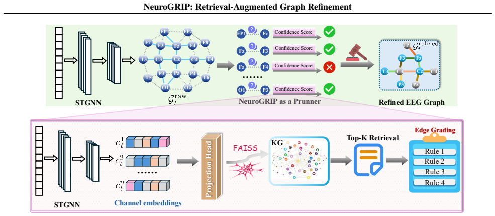
SemAlignQuery, an alignment-aware query construction module: it averages the projected embeddings of each EEG channel's k-hop neighborhood to form a query vector in knowledge-graph space. This query vector drives retrieval of medical triples (via an approximate nearest-neighbor index over a biomedical language model's embeddings), and the retrieved triples feed a multi-factor confidence score—cosine similarity, relation type/path length, source reliability, and an entity-match indicator—that decides which edges to prune. The work of this machinery is to convert an unsupervised, data-driven graph into a clinically filtered graph without retraining the backbone.
What would settle it
Replace the retrieved medical triples with random triples, or freeze the projection head at random initialization, and rerun the pipeline: if F1/AUROC gains persist unchanged, the improvement is generic sparsification rather than knowledge grounding. Alternatively, compare pruned edges against independent expert annotations of plausible functional connectivity on a subset of recordings; low agreement would falsify the clinical-plausibility claim.
Extended reading notes
Core claim
On the paper's own terms, NeuroGRIP establishes that a retrieval-augmented pruning mechanism—where clinical priors from a guideline-derived knowledge graph score each predicted EEG-graph edge—consistently improves seizure detection over purely data-driven spatial-temporal graph models. The mechanism works by projecting STGNN channel embeddings into the semantic space of medical text, retrieving the top-M knowledge triples per local neighborhood via approximate nearest-neighbor search, and grading each edge with a confidence score that combines cosine similarity, relation compatibility, and source reliability; edges below a threshold are pruned. The reported outcome is a 2-5 point improvement
Load-bearing premise
The load-bearing premise is that an unspecified projection (never trained or given a loss) can map EEG channel neighborhoods into the same semantic space as medical text, and that a missing retrieved triple is evidence that a connection is clinically implausible; if either fails, the gains would not be due to knowledge grounding.
Editorial extensions
If this is right
- If the claim holds, the refinement module can be bolted onto any STGNN backbone, providing a general, model-agnostic way to inject clinical priors into EEG graph learning.
- The larger gains on shorter clips imply that external knowledge can partially substitute for missing temporal context, which matters for early seizure detection.
- The consistent decrease in edge density suggests the benefit comes from suppressing medically unsupported connections rather than from generic graph sparsification.
- The ablation showing performance varies with the LLM used to construct the knowledge base indicates that the quality of the extracted knowledge graph is a primary driver of downstream gains.
- Because source reliability contributes to the score, the method explicitly models the trustworthiness of different knowledge sources, which is a step toward auditable clinical decisions.
Reading between the lines
- A natural extension would be to test whether the same retrieval-plus-pruning recipe transfers to other EEG analytics tasks, such as sleep staging or cognitive-load monitoring, where data-driven connectivity graphs suffer from analogous noise.
- The paper leaves open whether the pruning rule could be learned end-to-end; replacing the fixed similarity threshold with a learned calibrator might make the method robust to incomplete knowledge graphs.
- Since the knowledge graph is built from disease-level guidelines, the method's success on electrode-level connectivity depends on an unstated mapping between scalp regions and clinical concepts; making that mapping explicit and validating it against expert-labeled connectivity would strengthen the causal reading of the accuracy gains.
Signed reviews
Editorial analysis
A structured set of objections, weighed in public.
Referee Report
Summary. The paper proposes NeuroGRIP, a retrieval-augmented graph refinement method for EEG-based seizure detection. It constructs a knowledge graph from clinical guidelines using GPT-4o, uses BioBERT/FAISS to retrieve clinical triplets for STGNN-derived EEG subgraph queries, computes confidence scores from semantic similarity, source reliability, and entity matching, and prunes low-scoring graph edges. Experiments on TUSZ and CHB-MIT with five STGNN baselines at 12s and 60s clip lengths report consistent F1/Recall/AUROC gains, with ablations over framework components and LLM choices for knowledge-graph construction.
Significance. If the knowledge-driven causal claim held, NeuroGRIP would be a useful contribution: it couples external medical knowledge into graph refinement for EEG, potentially improving both accuracy and clinical interpretability. The paper has several positive features: evaluation on two public benchmarks, multiple STGNN baselines, ablations, a reproducibility-oriented code release, and a knowledge base sourced from authoritative clinical guidelines. However, the central evidence is currently incomplete. The projection head that grounds the retrieval mechanism is not specified, the edge-to-knowledge matching procedure is undefined, and the reported gains are not compared against generic sparsification controls. As a result, the main claim that medical knowledge is the driver of the observed improvement is not yet established.
major comments (4)
- [Section 3.3, Eqs. (1)-(2)] The projection head φproj is introduced as a shared map from STGNN channel embeddings to the KG embedding space, but no training objective, loss, or optimization procedure is described anywhere in the manuscript. The cosine similarity in Eq. (2) is only meaningful if this projection is actually learned to align EEG channel embeddings with BioBERT triplet embeddings; otherwise the query vector qi in Eq. (1) is arbitrary. The ablation 'w/o SemAlignQuery' in Section 4.4 does not clarify what is removed (the projection, the k-hop aggregation, or both). Please specify the training objective, the data used to fit φproj, and confirm that evaluation data are not used to fit it, so as to rule out leakage.
- [Section 3.5, Eq. (3)] The matching indicator 1_match is never operationalized. No mapping is given from EEG channel names (F3, C3, etc.) to KG entities, and the 'entity-type matching and cosine-similarity thresholding' is left undefined. The constructed KG contains clinical triplets such as (EEG, detects, interictal discharges) and (valproate, treats, generalized epilepsy), not electrode-pair connectivity statements. Moreover, qi is a subgraph-level average over the k-hop neighborhood (Eq. 1), so using the same qi in Eq. (3) for every edge in that subgraph cannot distinguish edge e_ij from other edges. Please define an edge-level matching procedure and clarify how electrode-level connectivity can be adjudicated by the constructed KG.
- [Section 4.2 and Section 4.3] There is no sparsification control. The method prunes low-confidence edges, and Figure 3(c) shows that NeuroGRIP reduces edge density. To support the claim that the improvements come from medical knowledge rather than generic graph regularization, the paper should compare against random pruning at matched density and against a non-knowledge sparsification baseline. Without such controls, the phrase 'grounding each prediction in clinically validated knowledge' as the cause of the gains is not supported.
- [Section 4.2, Table 1] The claim that the gains are 'clinically and statistically meaningful' is unsupported by statistical tests. Many with/without pairs have overlapping standard deviations (e.g., TUSZ 12s Dist-DCRNN F1 71.3±0.9 vs. 70.3±1.1; TUSZ 60s GraphS4mer AUROC 88.5±1.5 vs. 88.1±2.1). The paper reports no p-values, confidence intervals, or paired tests across subjects/folds. Please add such analyses or soften the statistical claim.
minor comments (4)
- [Section 3.5, Eq. (4)] The text says 'according to Eq. .' with the equation number missing. Also, the values of M, ρ, k, and the source-reliability weights r_m are never reported, which prevents reproduction and sensitivity analysis. Please include these hyperparameter settings.
- [Figures 3(a) and 3(b)] The AUROC values in these figures do not match Table 1 for any dataset or clip length. For example, Figure 3(b) lists EvoBrain+NeuroGRIP AUROC 0.921, while Table 1 reports 90.8 (TUSZ 12s) and 91.6 (TUSZ 60s), and the other values also differ. Please clarify which dataset and clip length the figure refers to and ensure consistency.
- [Section 4.4, Table 2] The 'w/o SemAlignQuery' variant is described only as 'directly uses raw EEG subgraphs as retrieval queries,' but it is unclear how raw subgraphs are embedded without φproj. Please specify the query construction in this variant. Also, 'alterg' appears to be a typo for 'alter'.
- [Section 3.2, Knowledge Graph Construction] The knowledge graph is generated by GPT-4o without any reported human validation or automated quality assessment of the extracted entities and relations. Given that the framework's credibility depends on the reliability of this KG, a small-scale precision/accuracy evaluation would strengthen the paper.
Circularity Check
No circular derivation; the knowledge graph is exogenous and reported gains are benchmarked on held-out public EEG datasets.
full rationale
NeuroGRIP's derivation chain is not circular. The knowledge graph is constructed from external clinical guidelines (ILAE, AES, NICE, SIGN, JSN) via GPT-4o entity/relation extraction, and the retrieval/pruning signal is computed against BioBERT triplet embeddings through FAISS. No equation in the paper defines the knowledge source in terms of the predicted seizure labels, nor does any derivation reduce the final prediction to the retrieval score by construction. The main weaknesses are reproducibility and validity gaps, not circularity: the projection head φproj in Section 3.3 has no specified training objective, the entity-match indicator in Eq. (3) lacks a defined electrode-to-entity mapping, and no sparsification or random-pruning control is provided, so the causal role of medical knowledge is not isolated from generic graph regularization. The paper contains self-citations (Li et al., 2026a,b), but they are motivational references and do not carry the load of the central derivation. No circular step meets the required standard of exhibiting an equation-level or fit-level reduction.
Assumptions & free parameters
free parameters (6)
- Pruning threshold ρ =
not reported
- Top-M retrieved triplets =
not reported
- Hop count k for subgraph queries =
not reported
- Entity-match cosine threshold =
not reported
- Source reliability weights r_m =
not reported
- Projection head φproj parameters =
unspecified
assumptions (5)
- domain assumption GPT-4o NER/RE extraction from clinical guidelines produces an accurate, clinically valid knowledge graph
- domain assumption Absence of retrieved triplet support implies the edge is medically implausible
- domain assumption Cosine similarity between projected EEG embeddings and BioBERT triplet embeddings is semantically meaningful
- domain assumption Retrieved evidence for a k-hop subgraph applies to every edge inside it
- domain assumption STGNN-predicted base graphs are reasonable starting points that refinement can improve
Cite this review
Pith. "Pith review of NeuroGRIP: Retrieval-Augmented Graph Refinement for Knowledge-Grounded EEG Seizure Diagnosis." pith.science (2026). https://pith.science/paper/WGWYNBG6
@misc{pith2026260714314,
  author       = {Pith},
  title        = {Pith review of: NeuroGRIP: Retrieval-Augmented Graph Refinement for Knowledge-Grounded EEG Seizure Diagnosis},
  year         = {2026},
  howpublished = {\url{https://pith.science/paper/WGWYNBG6}},
  note         = {Machine review of arXiv:2607.14314}
}
read the original abstract
Seizure diagnosis from EEG signals is a critical yet persistently challenging task, due to the complicated neural dynamics and the spurious connections in inter-channel modeling. While spatial-temporal graph neural networks (STGNNs) have advanced EEG brain network representation learning, the resulting graph structures suffer from low clinical plausibility and limited interpretability due to their purely data-driven nature. To this end, we introduce NeuroGRIP, a retrieval-augmented graph refinement framework that incorporates external medical knowledge to calibrate noisy EEG graphs. We first construct a large-scale, domain-specific knowledge base derived from authoritative clinical guidelines. Leveraging large language models, we extract structured biomedical entities and relations to form a textual knowledge graph (KG), which serves as external knowledge source of clinical priors. Our framework performs alignment-aware query construction by projecting STGNN-generated EEG node embeddings into the semantic space of KG. Semantic queries are then executed via FAISS-based similarity search over knowledge triplets to retrieve relation evidence. Each predicted edge is assigned a confidence score based on retrieved similarity, relation type, and source reliability, enabling us to prune medically implausible edges from the originally predicted graph. Extensive experiments on TUSZ and CHB-MIT demonstrate that NeuroGRIP not only improves seizure detection accuracy but also enhances interpretability by grounding each prediction in clinically validated knowledge. This work provides the first unified framework that tightly couples brain dynamics with external medical expertise via retrieval-augmented reasoning, paving the way for knowledge-enhanced, explainable clinical diagnosis. The code is available at: https://github.com/LincanLi-X/NeuroGRIP.
Figures


Figures from the paper (1 more)

Forward citations
Cited by 1 Pith paper
-
SynEnergy: Anomaly Semantic-Guided Diffusion for Synthetic Energy Data Generation
SynEnergy learns region-level anomaly semantics from residual energy data and injects them into a diffusion generator, improving anomaly preservation fidelity by an average of 12.21% over 11 baselines.
Reference graph
Works this paper leans on
-
[1]
Diseases / Disorders – e.g., epilepsy, Lennox-Gastaut syndrome
-
[2]
Symptoms / Clinical manifestations – e.g., seizures, aura, headache
-
[3]
Diagnostic methods / tests – e.g., EEG, MRI, CT scan
-
[4]
Treatments / Interventions – e.g., vagus nerve stimulation, resective surgery
-
[5]
Medications / Drugs – e.g., valproate, lamotrigine
-
[6]
Body parts / Anatomical structures – e.g., temporal lobe, hippocampus
-
[7]
Risk factors – e.g., family history, prenatal injury
-
[8]
Causes / Etiologies – e.g., brain tumor, traumatic brain injury
Show all 12 references
-
[9]
Biomarkers – e.g., interictal discharges, spike-wave complexes
-
[10]
Biological processes / mechanisms – e.g., neuronal hyperexcitability, GABA inhibition
-
[11]
Clinical findings / measurements – e.g., slowed response, focal spikes
-
[12]
Return your output as astrict JSON object, with keys as categories and values as arrays of extracted entity strings
Procedures / Medical actions – e.g., implantation, resection, monitoring. Return your output as astrict JSON object, with keys as categories and values as arrays of extracted entity strings. Do NOT include explanations. Do NOT add markdown. Just return pure JSON. Example forma...
Reviewed August 2, 2026 · model on record in the stance chip above.
Discussion (0). Continue with ORCID to comment.