REVIEW 5 major objections 5 minor 33 references
Pathologist Attention-Aligned Report Generation for Prostate Histopathology
T0 review · 5 major / 5 minor · reviewed 2026-08-01 · deepseek-v4-flash
Pith's one-line read Using recorded pathologist gaze as an auxiliary training signal, this paper shows that aligning a report-generation model's attention over image patches to human attention improves both report quality and clinical accuracy, and makes the mo
desk verdict First use of pathologist gaze to supervise pathology report generation, with a real dataset, but the evaluation is too small to back the headline gains. read the letter →
The pith
A machine-rendered reading of the paper's core claim, the machinery that carries it, and where it could break.
The reading
What carries the argument
The load-bearing mechanism is the attention-alignment loss L_KL = KL(softmax(a_human) || softmax(a_model)), where a_human is built from multi-magnification viewport trajectories (sampled at 20 Hz, centers Gaussian-blurred, merged across 2x-20x magnifications) and a_model is the mean attention the model assigns to each image token when generating the report. The loss is added to the standard next-token cross-entropy objective with weight lambda=1.0, using only last-layer attention, selected over all-layer alignment by ablation.
What would settle it
A reader could rebuild the human saliency maps from the described pipeline and test whether they predict held-out pathologist fixations or Gleason-positive pixels better than chance (e.g., ROC-AUC > 0.5). If the maps carry no signal, or if swapping the human map for a random map with identical spatial statistics still yields the reported gains, the effect is not due to the semantic content of pathologist attention.
Extended reading notes
Core claim
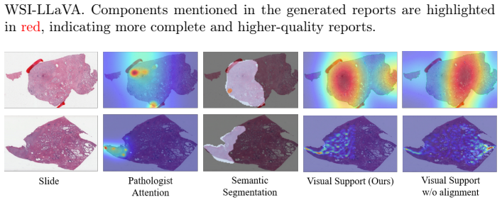
The paper's central claim is that aligning model attention to pathologist attention during training improves report generation. Concretely, they define a human saliency vector per image-token position by projecting recorded 20 Hz viewport-center scanpaths onto the patch grid and Gaussian-blurring them, and add a KL-divergence loss between softmax of this human vector and softmax of the model's last-layer attention over image tokens. Across HistGen (cross-attention) and WSI-LLaVA (self-attention), this yields consistent gains in BLEU, ROUGE-L, METEOR and in exact-match accuracy for Gleason pattern, PNI, EPE, and surgical margin, and produces attention heatmaps that correlate better with human
Load-bearing premise
The load-bearing premise is that the human saliency vector built from viewport trajectories is a faithful, token-level target distribution; if the recorded gaze is noisy, miscalibrated, or mis-synchronized with the verbal descriptions, the KL loss trains the model toward a flawed proxy rather than genuine diagnostic attention.
Editorial extensions
If this is right
- Experts' gaze can serve as a plug-and-play auxiliary supervision for pathology report generation, with no extra inputs or overhead at inference.
- Training with attention alignment prevents overfitting in low-data fine-tuning: WSI-LLaVA without alignment drops below zero-shot, while alignment lifts it above.
- The attention maps produced at inference are more human-aligned and more consistent with Gleason-region ground truth, strengthening interpretability of generated reports.
- The modest annotation load (about 7 hours total) suggests the approach can transfer to other cancer types and report-generation models.
Reading between the lines
- Extrapolating from the paper's mechanism, the same alignment loss could regularize any token-based vision-language model, and might reduce hallucinated findings by suppressing attention to regions the pathologist never examined—a testable extension using negative cases.
- The paper leaves the multi-scale saliency merge weights and Gaussian blur bandwidth unspecified; these choices determine the target distribution, so the method's sensitivity to them is an open empirical question.
- If human gaze is a strong prior for diagnostic relevance, the collected maps could serve as weak labels for weakly-supervised detection or grading of Gleason patterns, beyond report generation.
- The reported 10.9%/19.3% gains are averages over components; the per-component spread (8%-34% accuracy gains) suggests that gaze alignment helps most on components with clear visual signatures, a pattern the paper does not analyze.
Editorial analysis
A structured set of objections, weighed in public.
Referee Report
Summary. The paper proposes an auxiliary attention-alignment loss for pathology report generation. The authors collect a multimodal pathologist-attention dataset on 121 TCGA-PRAD prostate WSIs, with synchronized viewport trajectories and verbal descriptions. Viewport centers are converted into multi-scale, Gaussian-smoothed saliency maps, and a KL-divergence loss (Eq. 4) regularizes model attention over image tokens to match these human attention maps. The loss is added to the next-token objective for two models with different fusion mechanisms: HistGen (cross-attention) and WSI-LLaVA (self-attention). Experiments report gains in NLP-based metrics, component-level accuracy extracted by an LLM, and better agreement between inference-time model attention and pathologist attention. The central claim is that aligning model attention to expert gaze improves both report quality and interpretable visual grounding.
Significance. If the empirical claims held, this would be a useful and low-cost contribution: expert gaze would act as an auxiliary supervision signal for pathology vision-language models, and inference-time attention maps would provide interpretable visual evidence. The collected dataset of synchronized viewport trajectories and verbal descriptions is a novel resource, and the proposed loss is simple and architecture-agnostic. The paper also evaluates two common fusion designs, which strengthens the generality of the proposed method. However, the current evidence is not yet statistically convincing: the WSI-LLaVA evaluation rests on 11 test examples, the LLM-based metric extractor is unvalidated, and the visual-support evaluation partly measures the same quantity optimized by the training loss. The underlying idea is plausible and the issues are addressable, but the strength of the claims must be revised to match the evidence.
major comments (5)
- [§4.1, Table 1] Report-quality results for WSI-LLaVA are computed on 11 test examples. In Table 1, ACC-G improves from 54.5% to 63.6%, ACC-PNI from 81.8% to 90.9%, and ACC-EPE from 45.5% to 63.6% — corresponding to 1, 1, and 2 corrected samples, respectively. The abstract's 19.3% average accuracy gain is thus driven by a handful of cases. No confidence intervals, bootstrap, or significance tests accompany any metric. This is insufficient support for the claim that attention alignment consistently improves report quality. Please either evaluate on a substantially larger test set, provide uncertainty estimates (e.g., binomial confidence intervals), or temper the claims accordingly.
- [§4.1 Metrics] The component exact-match scores (ACC-G, ACC-PNI, ACC-EPE, ACC-S) are extracted by ChatGPT-5.2 from generated and ground-truth reports, but no validation of this extractor is reported. Extraction errors propagate directly into the main quantitative results. Please validate the ChatGPT-5.2 extractor against manual or pathologist-annotated labels on a held-out subset, or report an agreement metric, and state the prompt/version used.
- [§2, §3.2 Eq. (4)] The human attention vector a_human is derived from 20 Hz viewport-center sampling, duplicate removal, positive-label filtering, Gaussian blur, and multi-magnification merging. The Gaussian sigma/kernel size and magnification-merge weights are not specified, and the transformation from scanpath to a faithful token-level target distribution is assumed rather than validated. Because Eq. (4) trains the model to match this distribution, any bias in the saliency proxy is directly learned. Please report the exact construction parameters and provide a sensitivity analysis (e.g., varying sigma and merge weights) on a validation criterion such as component accuracy.
- [§3.2, Table 2] The NSS and KL improvements in Table 2 measure agreement between model attention and the same pathologist-attention maps used to define L_KL. Consequently, the NSS/KL gains are, to a substantial degree, an expected consequence of the training objective rather than independent evidence of better visual grounding. The ROC-AUC against Gleason segmentation is the non-circular signal, but the observed gains (0.500→0.537 for HistGen; 0.598→0.615 for WSI-LLaVA) are small and unaccompanied by uncertainty estimates. Please present ROC-AUC as the primary visual-support evidence and add confidence intervals or significance tests.
- [Abstract, §4.1] The abstract and introduction claim gains 'across five clinically relevant report components,' but §4.1 states that intraductal carcinoma is omitted because it does not appear in ground-truth reports, and Table 1 does not report ACC-S for WSI-LLaVA. In fact, component accuracies are reported for four components on HistGen and three on WSI-LLaVA. Please restate the claims to match the components actually evaluated, or provide additional annotations to cover the missing components.
minor comments (5)
- [Table 3 / Table 4] Tables 3 and 4 are identical in content and title ('Ablation on attention-extraction layer'). Please remove the duplicate and refer to a single table.
- [Table 1] The column header 'ACC-EDE' is inconsistent with the text's 'ACC-EPE' (extraprostatic extension). Please correct.
- [§3.3] The parenthetical '(Section 3.2)' for the saliency smoothing procedure should refer to Section 2, where human attention map construction is described.
- [§4.1] The text does not state whether the dataset, code, or trained models will be released. Since the dataset is a central contribution, a data-availability statement would aid reproducibility.
- [§4.1 Metrics] The use of ChatGPT-5.2 as a metric extractor is unusual and appears without any reference or version identifier. If it is a proprietary model, please provide the exact model identifier and date of use.
Circularity Check
No significant circularity: the central report-quality and accuracy gains are scored against independent ground-truth reports, and the visual-support KL metric is evaluated on held-out test slides.
full rationale
The load-bearing claim is that adding the attention-alignment loss L_KL (Eq. 4) improves report generation. The evidence in Table 1 (BLEU, METEOR, ROUGE-L, and component accuracy ACC-G/PNI/EPE/S) is computed against official HistGen/WSI-LLaVA test-set ground-truth reports, which are external to the training objective. Component accuracy is extracted with ChatGPT-5.2 and compared to ground-truth reports; it is not derived from the human-attention annotations. The visual-support evaluation in Table 2 does use NSS and KL against pathologist attention, which is the same target that L_KL optimizes. However, the paper explicitly states that visual-support evaluation is performed on 20 HistGen test slides and 15 WSI-LLaVA test samples, i.e., held-out data. An improvement in KL/NSS on held-out slides is therefore a generalization result, not an identity forced by construction. The additional ROC-AUC against Gleason-region segmentation labels from an external dataset provides independent evidence. The point about the small WSI-LLaVA test set (11 examples) is a legitimate statistical concern but not a circularity: it affects confidence in the empirical gain, not whether the derivation reduces to its inputs. No self-citation chain, imported uniqueness theorem, or ansatz-citation is load-bearing; the GazeVLM citation for the KL formulation is external and only provides the loss design. The core derivation is therefore self-contained and empirically testable against independent report ground truth.
Assumptions & free parameters
free parameters (3)
- λ (alignment-loss weight) =
1.0
- Human saliency map construction parameters (Gaussian sigma, kernel size, magnification merge weights) =
not reported
- Viewport sampling rate and point-to-scanpath conversion =
20 Hz
assumptions (4)
- domain assumption Transformer attention weights over image tokens faithfully reflect the visual evidence used for token prediction (Eq. 1–3).
- domain assumption Synchronized viewport trajectories and verbal descriptions provide reliable soft component labels for attention targets.
- ad hoc to paper Gaussian blurring and multi-magnification merging of scanpath centers yields a valid token-level saliency distribution.
- domain assumption TCGA-PRAD ground-truth reports and ChatGPT-5.2 extraction are accurate enough to score clinical components.
Cite this review
Pith. "Pith review of Pathologist Attention-Aligned Report Generation for Prostate Histopathology." pith.science (2026). https://pith.science/paper/ZRHQIEZE
@misc{pith2026260719624,
  author       = {Pith},
  title        = {Pith review of: Pathologist Attention-Aligned Report Generation for Prostate Histopathology},
  year         = {2026},
  howpublished = {\url{https://pith.science/paper/ZRHQIEZE}},
  note         = {Machine review of arXiv:2607.19624}
}
read the original abstract
The allocation of visual attention by pathologists during cancer diagnosis is a highly selective process that critically shapes the information extracted from whole-slide images (WSIs). Human attention helps medical imaging tasks such as classification and segmentation, and becomes a strong semantic cue for identifying diagnostically informative regions for report generation. In this paper, we introduce human attention into the training of pathologist report generation models. To this end, we collected a multimodal human-attention dataset of 121 prostate WSIs annotated with pathologists' multi-scale viewport trajectories synchronized with the pathologists' verbal descriptions and cursor movements for five clinically relevant components (e.g., Gleason patterns). Using this dataset, we finetune two report generation models with an attention-alignment loss that regularizes the model attention over image patches to match the distribution of pathologist attention. We evaluate our approach on prostate cancer report generation and visual question answering using two models with different internal attention mechanisms (i.e., how image tokens are integrated into the language decoder). Experiments show average gains of 10.9% on NLP-based metrics and 19.3% in accuracy across five clinically relevant report components. Further, model attention maps extracted at inference time, with minimal computational overhead, align more closely with pathologist attention, providing stronger visual support for the generated reports by highlighting the regions that most influence the output.
Figures


Figures from the paper (1 more)
Reference graph
Works this paper leans on
-
[1]
In: Proceedings of the acl workshop on intrinsic and extrinsic evaluation measures for machine translation and/or summa- rization
Banerjee, S., Lavie, A.: Meteor: An automatic metric for mt evaluation with im- proved correlation with human judgments. In: Proceedings of the acl workshop on intrinsic and extrinsic evaluation measures for machine translation and/or summa- rization. pp. 65–72 (2005)
2005
-
[2]
In: Medical Imaging with Deep Learning (2024)
Bhattacharya, M., Prasanna, P.: Gazediff: A radiologist visual attention guided diffusion model for zero-shot disease classification. In: Medical Imaging with Deep Learning (2024)
2024
-
[3]
arXiv preprint arXiv:2410.00307 (2024)
Bhattacharya, M., Singh, G., Jain, S., Prasanna, P.: Radgazegen: Radiomics and gaze-guided medical image generation using diffusion models. arXiv preprint arXiv:2410.00307 (2024)
arXiv 2024
-
[4]
arXiv preprint arXiv:2508.09478 (2025) 10 Xue et al
Bhattacharya, M., Singh, G., Jain, S., Prasanna, P.: Gazelt: Visual attention- guided long-tailed disease classification in chest radiographs. arXiv preprint arXiv:2508.09478 (2025) 10 Xue et al
arXiv 2025
-
[5]
PloS one9(8), e103447 (2014)
Brunye, T.T., Carney, P.A., Allison, K.H., Shapiro, L.G., Weaver, D.L., Elmore, J.G.: Eye movements as an index of pathologist visual expertise: a pilot study. PloS one9(8), e103447 (2014)
2014
-
[6]
In: International Workshop on Medical Optical Imaging and Virtual Microscopy Image Analysis
Chakraborty, S., Gupta, R., Ma, K., Govind, D., Sarder, P., Choi, W.T., Mahmud, W., Yee, E., Allard, F., Knudsen, B., et al.: Predicting the visual attention of pathologists evaluating whole slide images of cancer. In: International Workshop on Medical Optical Imaging and Virtual Microscopy Image Analysis. pp. 11–21. Springer (2022)
2022
-
[7]
In: MICCAI
Chakraborty, S., Gupta, R., Yaskiv, O., Friedman, C., Sheuka, N., Perez, D., Fried- man, P., Zelinsky, G., Saltz, J., Samaras, D.: Decoding the visual attention of pathologists to reveal their level of expertise. In: MICCAI. pp. 120–130. Springer (2024)
2024
-
[8]
Medical Image Analysis p
Chakraborty, S., Xue, R., Gupta, R., Yaskiv, O., Friedman, C., Sheuka, N., Perez, D., Friedman, P., Choi, W.T., Mahmud, W., et al.: Measuring and predicting where and when pathologists focus their visual attention while grading whole slide images of cancer. Medical Image Analysis p. 103752 (2025)
2025
Show all 33 references
-
[9]
arXiv preprint arXiv:2506.20964 (2025)
Chen, C., Weishaupt, L.L., Williamson, D.F., Chen, R.J., Ding, T., Chen, B., Vaidya, A., Le, L.P., Jaume, G., Lu, M.Y., et al.: Evidence-based diagnostic reason- ing with multi-agent copilot for human pathology. arXiv preprint arXiv:2506.20964 (2025)
2025
-
[10]
In: MIC- CAI
Chen, P., Li, H., Zhu, C., Zheng, S., Shui, Z., Yang, L.: Wsicaption: Multiple instance generation of pathology reports for gigapixel whole-slide images. In: MIC- CAI. pp. 546–556. Springer (2024)
2024
-
[11]
Cognitive science36(7), 1204–1223 (2012)
Coco,M.I.,Keller,F.:Scanpatternspredictsentenceproductioninthecross-modal processing of visual scenes. Cognitive science36(7), 1204–1223 (2012)
2012
-
[12]
Hug- ging Face Datasets,https://huggingface.co/datasets/Codatta/ Refined-TCGA-PRAD-Prostate-Cancer-Pathology-Dataset, accessed: 2026- 02-24
Codatta: Refined-tcga-prad prostate cancer pathology dataset. Hug- ging Face Datasets,https://huggingface.co/datasets/Codatta/ Refined-TCGA-PRAD-Prostate-Cancer-Pathology-Dataset, accessed: 2026- 02-24
2026
-
[13]
IEEE Transactions on Circuits and Systems for Video Technology (2025)
Ge, R., Li, R., Wang, C., Liu, Y., Zhu, H., Coatrieux, J.L., Zhang, D., Lu, J., Chen, Y., Li, S., et al.: Adaptation follow human attention: Gaze-assisted medical segment anything model. IEEE Transactions on Circuits and Systems for Video Technology (2025)
2025
-
[14]
Psychological science 11(4), 274–279 (2000)
Griffin, Z.M., Bock, K.: What the eyes say about speaking. Psychological science 11(4), 274–279 (2000)
2000
-
[15]
In: MICCAI
Guo, Z., Ma, J., Xu, Y., Wang, Y., Wang, L., Chen, H.: Histgen: Histopathol- ogy report generation via local-global feature encoding and cross-modal context interaction. In: MICCAI. pp. 189–199. Springer (2024)
2024
-
[16]
Iclr1(2), 3 (2022)
Hu, E.J., Shen, Y., Wallis, P., Allen-Zhu, Z., Li, Y., Wang, S., Wang, L., Chen, W., et al.: Lora: Low-rank adaptation of large language models. Iclr1(2), 3 (2022)
2022
-
[17]
In: CVPR
Jiang, M., Huang, S., Duan, J., Zhao, Q.: Salicon: Saliency in context. In: CVPR. pp. 1072–1080 (2015)
2015
-
[18]
IEEE Access (2025)
Kim, K.A., Hong, S., Yoo, S., Kang, Y., Shim, H.S.: Enhancing structured pathol- ogy report generation with foundation model and modular design. IEEE Access (2025)
2025
-
[19]
In: CVPR
Liang, Y., Lyu, X., Chen, W., Ding, M., Zhang, J., He, X., Wu, S., Xing, X., Yang, S., Wang, X., et al.: Wsi-llava: A multimodal large language model for whole slide image. In: CVPR. pp. 22718–22727 (2025)
2025
-
[20]
In: Text sum- marization branches out
Lin, C.Y.: Rouge: A package for automatic evaluation of summaries. In: Text sum- marization branches out. pp. 74–81 (2004) Attention-Aligned Report Generation 11
2004
-
[21]
Nature Communications16(1), 5493 (2025)
Nan, T., Zheng, S., Qiao, S., Quan, H., Gao, X., Niu, J., Zheng, B., Guo, C., Zhang, Y., Wang, X., et al.: Deep learning quantifies pathologists’ visual patterns for whole slide image diagnosis. Nature Communications16(1), 5493 (2025)
2025
-
[22]
arXiv preprint arXiv:2510.21356 (2025)
Pani, A., Yang, Y.: Gaze-vlm: Bridging gaze and vlms through attention regular- ization for egocentric understanding. arXiv preprint arXiv:2510.21356 (2025)
2025
-
[23]
In: Proceedings of the 40th annual meeting of the Association for Computational Linguistics
Papineni, K., Roukos, S., Ward, T., Zhu, W.J.: Bleu: a method for automatic evaluation of machine translation. In: Proceedings of the 40th annual meeting of the Association for Computational Linguistics. pp. 311–318 (2002)
2002
-
[24]
Vision research45(18), 2397–2416 (2005)
Peters, R.J., Iyer, A., Itti, L., Koch, C.: Components of bottom-up gaze allocation in natural images. Vision research45(18), 2397–2416 (2005)
2005
-
[25]
Cancer research77(21), e79–e82 (2017)
Saltz, J., Sharma, A., Iyer, G., Bremer, E., Wang, F., Jasniewski, A., DiPrima, T., Almeida, J.S., Gao, Y., Zhao, T., et al.: A containerized software system for gen- eration, management, and exploration of features from whole slide tissue images. Cancer research77(21), e79–e82 (2017)
2017
-
[26]
arXiv preprint arXiv:2505.20510 (2025)
Sun, Y., Si, Y., Zhu, C., Zhang, K., Shui, Z., Ding, B., Lin, T., Yang, L.: Cpathagent: An agent-based foundation model for interpretable high-resolution pathology image analysis mimicking pathologists’ diagnostic logic. arXiv preprint arXiv:2505.20510 (2025)
2025
-
[27]
arXiv preprint arXiv:2510.24653 (2025)
Thai, V., Li, R., Ling, M., Jiang, S., Wolfe, J., Machiraju, R., Hu, Y., Li, Z., Par- wani, A., Chen, J.: Eye-tracking, mouse tracking, stimulus tracking, and decision- making datasets in digital pathology. arXiv preprint arXiv:2510.24653 (2025)
2025
-
[28]
Nature communications16(1), 4886 (2025)
Tran, M., Schmidle, P., Guo, R.R., Wagner, S.J., Koch, V., Lupperger, V., Novotny, B., Murphree, D.H., Hardway, H.D., D’Amato, M., et al.: Generating dermatopathology reports from gigapixel whole slide images with histogpt. Nature communications16(1), 4886 (2025)
2025
-
[29]
NeurIPS30(2017)
Vaswani,A.,Shazeer,N.,Parmar,N.,Uszkoreit,J.,Jones,L.,Gomez,A.N.,Kaiser, L., Polosukhin, I.: Attention is all you need. NeurIPS30(2017)
2017
-
[30]
arXiv preprint arXiv:2512.06662 (2025)
Xue, R., Le, H., Xu, J., Mondal, S., Leite, A., Zelinsky, G., Hoai, M., Sama- ras, D.: Personalized image descriptions from attention sequences. arXiv preprint arXiv:2512.06662 (2025)
2025
-
[31]
Zhang, L., Yun, B., Li, Q., Wang, Y.: Historical report guided bi-modal concurrent learningforpathologyreportgeneration.In:MICCAI.pp.343–352.Springer(2025)
2025
-
[32]
NeurIPS36, 46595–46623 (2023)
Zheng, L., Chiang, W.L., Sheng, Y., Zhuang, S., Wu, Z., Zhuang, Y., Lin, Z., Li, Z., Li, D., Xing, E., et al.: Judging llm-as-a-judge with mt-bench and chatbot arena. NeurIPS36, 46595–46623 (2023)
2023
-
[33]
Cancer Imaging Arch9(10.7937), K9 (2016)
Zuley, M.L., Jarosz, R., Drake, B.F., Rancilio, D., Klim, A., Rieger-Christ, K., Lemmerman, J.: Radiology data from the cancer genome atlas prostate adenocar- cinoma [tcga-prad] collection. Cancer Imaging Arch9(10.7937), K9 (2016)
2016
Reviewed August 1, 2026 · model on record in the stance chip above.
Discussion (0). Continue with ORCID to comment.