REVIEW 4 major objections 5 minor 50 references
Nuclear mechanics controls the temporal dynamics of cell unjamming
T0 review · 4 major / 5 minor · reviewed 2026-08-11 · deepseek-v4-flash
Pith's one-line read This paper claims that nuclear size and stiffness regulate the jamming-unjamming transition in dense cell sheets by constraining the range of cell shapes a tissue can adopt, so that cell shape is a phenomenological indicator while the…
desk verdict Solid modeling core, but the claimed experimental verification of a universal morpho-dynamic relation is in-sample fitting — worth peer review with major revision. read the letter →
The pith
A machine-rendered reading of the paper's core claim, the machinery that carries it, and where it could break.
The reading
What carries the argument
The load-bearing object is an extended cellular Potts model in which each cell contains an explicitly deformable nucleus, treated as a separate compartment with its own area-conservation term and an internal contact energy $J_{\mathrm{int}}$ that sets nuclear stiffness; a harmonic spring anchors the nucleus to the cell centre of mass, and an active Brownian force drives cytoplasmic migration. The argument runs on dimensionless shape indices $S = \langle P/\sqrt{A}\rangle$ for cells and nuclei, and on an empirical double-exponential law $D(S_N,\phi) = D_0(\phi)\left[e^{-a(\phi)(S_N - S_N^*(\phi))} + e^{-b(\phi)(S_N - S_N^*(\phi))}\right]^{-1}$, whose $\phi$-dependent parameters (exponential $D_0$, linear $a$ with geometric limit $\phi^* = 28^2/30^2$, constant $b/a$, cubic $S_N^*$) are fitted to simulations and then used to rescale experimental data onto one master curve. That collapsed curve is the order parameter the paper introduces for nuclear jamming.
What would settle it
Grow a confluent monolayer of MCF-10A cells, pharmacologically enlarge nuclei (for example by inhibiting nuclear export) without changing cell-cell adhesion, and measure bin-averaged $D_2^{\min}$ against nuclear area fraction $\phi$ and nuclear shape index $S_N$; if the data no longer fall on the paper's rescaled master curve, or if mobility stays constant while the cell shape index distribution is unchanged, the claim that nuclear density constrains cell shape to control unjamming fails.
Extended reading notes
Core claim
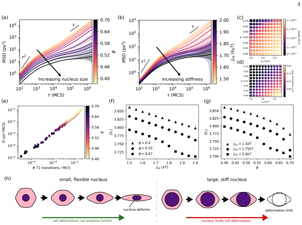
On the paper's own terms, the central discovery is that nuclear mechanics regulates tissue-scale jamming by constraining cell shape: larger nuclear area fraction $\phi$ and higher internal contact energy $J_{\mathrm{int}}$ make cells rounder, reduce T1 neighbour exchanges, and lower the long-time diffusion constant $D$, with combined changes lowering $D$ by nearly four orders of magnitude. The authors state the reconciliation directly: while cell shape determines jamming phenomenologically, nuclear density constrains possible cell shapes and thereby regulates the transition at the subcellular scale. Their simulations collapse onto a single cell-shape-diffusion master curve, and a rescaled nuclear-shape-diffusion law $D(S_N, \phi)$ is verified against MCF-10A and MDA-MB-436 monolayers, predicting measured $D_2^{\min}$ within 5 percent mean absolute percentage error. The paper therefore claims a universal morpho-dynamic link from statically measurable nuclear morphology to collective cell motion.
Load-bearing premise
The load-bearing premise is that the fitted functional forms—double-exponential shape-diffusion decay, exponential $D_0(\phi)$, linear $a(\phi)$, constant $b/a$, cubic $S_N^*(\phi)$, and the geometric upper limit $\phi^* = 28^2/30^2$—are the true universal relations and not just the best fits within this cellular Potts model family, since the paper gives no derivation of these forms from the model's Hamiltonian.
Editorial extensions
If this is right
- Increasing nuclear area fraction from $\phi=0.4$ to $\phi=0.7$ lowers the long-time diffusion constant by about one order of magnitude, and raising both nuclear size and stiffness together lowers it by almost four orders of magnitude.
- T1 neighbour-exchange rate and diffusion constant track each other, so nuclear mechanics controls jamming by throttling the rate at which cells can rearrange.
- All simulated state points fall on a single cell-shape-versus-diffusion master curve, making cell shape a universal indicator of tissue fluidity even though the nucleus sets the accessible shape range.
- Static nuclear shape and nuclear area fraction predict $D_2^{\min}$ in MCF-10A and MDA-MB-436 monolayers with a mean absolute percentage error below 5 percent, suggesting a physics-based prognostic marker readable from histological images.
Reading between the lines
- The paper does not test whether directly softening or shrinking nuclei is sufficient to unjam a monolayer while cell-cell adhesion is held fixed; an experiment with drugs targeting nuclear envelope components would isolate the nuclear contribution from the cytoskeletal one.
- Because the geometric bound $\phi^* = 28^2/30^2$ sets a formal limit at which $a(\phi)$ vanishes, the model predicts that mobility should drop to zero as the nucleus fills the cell; measuring whether diffusion vanishes near that packing fraction in experiments would test whether the bound is physical or only a fitting constraint.
- The double-exponential collapse is an empirical law fitted to cellular Potts model data, not derived from the model Hamiltonian; the same order parameter might survive with a different functional family in three dimensions or in monolayers with heterogeneous nuclear sizes, so the universality claim is only as strong as the persistence of these fitted forms.
Signed reviews
Editorial analysis
A structured set of objections, weighed in public.
Referee Report
Summary. The paper introduces a cellular Potts model with explicitly deformable nuclei to study unjamming in confluent monolayers. Simulations varying nuclear area fraction phi and nuclear stiffness J_int show that larger and stiffer nuclei reduce cell diffusivity, T1 transition rates, and cell shape index. The authors propose an empirical relation between nuclear shape index, nuclear area fraction, and collective diffusion, and report a collapse of simulation data onto a master curve. They further fit a piecewise version of this relation to binned experimental data from MCF-10A and MDA-MB-436 cell monolayers, reporting a mean absolute percentage error (MAPE) below 5% in both cell lines, and interpret the rescaled nuclear shape index as an order parameter for nuclear jamming.
Significance. If the quantitative claims were fully substantiated, the paper would offer a novel mechanistic bridge between nuclear mechanics and tissue-scale rigidity, with potential clinical relevance for cancer prognosis. The open-source simulation and analysis code, the transparency about the fitting procedure in Section IV D, and the qualitative consistency between simulations and two epithelial/mesenchymal cell lines are strengths. However, the central quantitative claims of a verified universal morpho-dynamic relation and a predictive order parameter are currently undermined by in-sample fitting and post-hoc exclusion of data points. The mechanistic narrative is plausible and qualitatively supported, but the evidence as presented does not establish the claimed universal relation.
major comments (4)
- [Section IV D 3, Eq. (20)] The reported MAPE values (4.42% for MCF-10A and 1.62% for MDA-MB-436, Fig. 3j,k) are in-sample training errors: Eq. (18) is fitted to the same binned data on which Eq. (20) is evaluated. The abstract's claim that the relation is 'verified experimentally with striking accuracy' is therefore not supported; the fit only shows that the piecewise-linear function with phi-dependent coefficients is flexible enough to approximate the training data. An out-of-sample evaluation (e.g., cross-validation, or fitting on one cell line and predicting the other) is needed to support the predictive claim.
- [Section IV D 1, Fig. 3g caption] The caption of Fig. 3g states that 'open symbols in g were excluded from the fit procedure to accomplish collapse.' Excluding data points that do not collapse, with the stated purpose of achieving collapse, is a post-hoc selection that invalidates the master-curve claim as a test of the proposed relation. The exclusion rule should be specified a priori, and the fit should be reported with and without the excluded points.
- [Section IV D 1, Eqs. (12)-(17)] The collapse in Fig. 3g is constructed by fitting phi-dependent parameters D0(phi), a(phi), b(phi), and S*_N(phi) to the simulation data. No independent test of these functional forms is provided, and no derivation from the CPM Hamiltonian is given. The 'order parameter' b(phi)(S_N - S*_N) is therefore a curve-fitting device rather than an independently validated physical quantity. To support universality, the authors should either derive the form from the model or validate it on data not used for fitting.
- [Section IV D 3, Eqs. (18)-(19)] The experimental fitting procedure changes the functional forms relative to the simulation relation: S*(phi) is taken to be linear rather than cubic (as in Eq. (17)), and a(phi) uses a free offset sigma in Eq. (19) rather than the geometric anchor phi* = 28^2/30^2 of Eq. (16). While the authors state that these changes are 'consistent with our previous rescaling method,' this means the experimental data do not test the simulation-derived relation; they test a more flexible variant with additional free parameters. The claim of a single universal relation is thereby weakened.
minor comments (5)
- [Introduction, page 2] The phrase 'immanent importance' should read 'imminent importance', and 'diagnosic tool' should be 'diagnostic tool'.
- [Fig. 2d caption] The caption contains the typo 'nuclera area fractions'; it should be 'nuclear area fractions'.
- [Table II and Section IV D 1] The fitted parameter values in Tables II and III are reported without uncertainties; confidence intervals should be provided, particularly for the parameters used in the collapse.
- [Section IV D 2] The experimental binning procedure uses 20 bins for phi and 40 bins for shape, with a cutoff of at least 100 observations per bin; the sensitivity of the fitted relation to the bin number and threshold should be discussed.
- [Section IV D 3] The manuscript does not report the number of experimental cells, tracks, or bins that enter the MAPE calculation; including these numbers would improve reproducibility.
Circularity Check
The quantitative 'universal morpho-dynamic relation' reduces to in-sample curve fitting: the simulation collapse is an algebraic restatement of the fitted double-exponential form, and the experimental <5% MAPE is training error on the same bins.
-
fitted input called prediction
[Section IV D 3 (Eqs. 18-20); Conclusion; Fig. 3j-k]
"Once the fit parameter in eq. (18) are determined, our model can be used to predict the expected D2_min at a given nuclear area fraction and shape index. We measure the accuracy of the predicted D2_min from our model using the mean absolute percentage error (MAPE), defined as MAPE = (1/N) Σ |y_i − ŷ_i|/y_i, (20) where y_i and ŷ_i are measured D2_min and the D2_min predicted by our model for the i-th bin, respectively, and N is the total number of bins. After fitting cell line specific parameters, our nuclear shape diffusion relation achieves a predictive MAPE below 5% for both cell lines."
Equation (18) contains free functions D0(φ), a(φ), b/a=γ, and S*(φ); equation (19) imposes a(φ)=ρ(φ−σ), and S*(φ) is taken to be linear. These parameters are fitted to the same binned experimental data whose bin-averaged D2_min values y_i are later inserted into Eq. (20). The MAPE therefore measures how well the fitted curve reproduces its own training data, i.e. in-sample goodness-of-fit, not out-of-sample predictive skill. A flexible piecewise function with a φ-dependent baseline, slope, and breakpoint can achieve <5% MAPE on the data used to fit it even if no universal morpho-dynamic relation exists. Calling this quantity 'predictive' and using it to claim experimental verification reduces the prediction to the fit.
-
self definitional
[Section IV D 1 (Eqs. 12-17); Results Section II C; Fig. 3g]
"An empirical law for D(S_N, φ) can be found by first fitting eq. (11) for each value of φ separately and then considering the fit parameters to be functions of φ, effectively assuming D(S_N, φ) = D0(φ) [ e^{−a(φ)(S−S*_N(φ))} + e^{−b(φ)(S−S*_N(φ))} ]^{−1}. (12) Remarkably, rescaling and shifting by nucleus-size-dependent parameters (see sections IV D 1 and IV C) collapses all nuclear-shape graphs onto single a curve (Fig. 3g-i)."
With the master-curve ordinate y=D/D0(φ) and abscissa x=b(φ)(S_N−S*_N(φ)), and with b/a=γ=const as imposed in Eq. (14), Eq. (12) becomes y=[e^{−x/γ}+e^{−x}]^{−1}, which is independent of φ. Hence the collapse in Fig. 3g is an algebraic restatement of the fitted form (12)-(17), not an independent test of that form. The fit parameters D0(φ), a(φ), b(φ), and S*_N(φ) are themselves determined from the simulation data, so the 'master curve' is constructed by definition from the same data. The open symbols excluded from the fit give a limited genuine check, but the displayed collapse is not evidence that the double-exponential family is the correct or unique morpho-dynamic law.
full rationale
The mechanistic simulation study (CPM with deformable nuclei; nuclear size/stiffness controlling accessible cell shapes, T1 rate, and diffusion) is self-contained and not circular: its qualitative trends follow from the model Hamiltonian and independent simulations, and no load-bearing self-citation chain was found. The circularity is confined to the quantitative 'universal morpho-dynamic relation' and its claimed experimental verification. First, the simulation collapse onto a master curve is a direct consequence of the fitted functional form (12), with γ=const making the rescaled expression independent of φ; it therefore does not independently validate the ansatz. Second, the experimental 'validation' fits Eq. (18) to binned data and evaluates Eq. (20) on those same bins, so the reported <5% MAPE is in-sample training error rather than a prediction. The qualitative agreement across MCF-10A and MDA-MB-436 and the open-symbol exclusion in Fig. 3g provide some supportive evidence, which prevents the score from being maximal, but the headline claim of a relation 'verified experimentally with striking accuracy' reduces to curve fitting as presented.
Assumptions & free parameters
free parameters (8)
- Simulation cell-shape fit parameters a, b, S*_C, D0 =
a=34.9, b=3.04, S*_C=3.75, D0=-5.75
- Simulation nuclear-shape fit parameters alpha, beta, gamma, rho, kappa, mu, nu =
alpha=11.1, beta=-12.8, gamma=4.57, rho=-287, kappa=3.21, mu=0.503, nu=4.63e-2
- Geometric upper limit phi* =
0.8711 (28^2/30^2)
- Experimental per-cell-line fit parameters for Eq. (18) =
not tabulated
- nucleus-cytoplasm spring constant k =
0.2 kBT/px^2
- active force magnitude F_active =
0.5 kBT/px
- polarity diffusion D_theta =
2e-3 MCS^-1
- area conservation strength lambda_area =
0.1 kBT/px^2
assumptions (6)
- domain assumption Confluent 2D tissue can be represented by a cellular Potts model with area conservation and contact energies.
- domain assumption The intracellular contact energy Jint controls nuclear stiffness, with higher Jint giving more compact, round nuclei.
- ad hoc to paper Nuclear shape index S_N is an order parameter for nuclear morphology, and nuclear area fraction phi captures density.
- ad hoc to paper The functional forms in Eqs. (11)-(17) describe the shape-diffusion relation universally.
- domain assumption The high intercellular contact energy keeps each nucleus inside its own cell at all times.
- domain assumption Binning cells by instantaneous phi and shape and averaging D2min per bin recovers the local relation between nuclear morphology and mobility.
invented entities (1)
-
Rescaled nuclear shape-diffusion order parameter b(phi)(S_N - S*_N)
Cite this review
Pith. "Pith review of Nuclear mechanics controls the temporal dynamics of cell unjamming." pith.science (2026). https://pith.science/paper/ORBJO5NQ
@misc{pith2026260807777,
  author       = {Pith},
  title        = {Pith review of: Nuclear mechanics controls the temporal dynamics of cell unjamming},
  year         = {2026},
  howpublished = {\url{https://pith.science/paper/ORBJO5NQ}},
  note         = {Machine review of arXiv:2608.07777}
}
read the original abstract
Cell unjamming in dense tissues is a complex but essential process in embryogenesis and cancer metastasis. Increasing evidence suggests that nuclear mechanics and density effects play a vital role in collective cell unjamming. However, state-of-the-art cell-shape-based theories fail to include nuclear and density effects, while computer models featuring rigid nuclei disagree with experimental observations of elongated nuclei promoting unjamming. Here, we introduce a computational model of confluent cells with explicitly deformable nuclei to study the dynamics of cell unjamming. Our simulations show an unjamming transition controlled by nuclear size and shape, reconciling conflicting theories of density-driven versus shape-driven mechanisms. We predict general relations connecting cellular and nuclear shape to collective cell motion, verified experimentally in distinct monolayers of MCF-10A and MDA-MB-436 breast cells, with striking accuracy. Our work establishes a rational connection between nuclear mechanics and tissue-scale rigidity transitions, highlighting the nucleus's key role in collective cell unjamming.
Figures


Figures from the paper (1 more)

Reference graph
Works this paper leans on
-
[1]
P. Friedl and D. Gilmour, Nature Reviews Molecular Cell Biology10, 445 (2009)
work page 2009
-
[2]
J.-A. Park, J. H. Kim, D. Bi, J. A. Mitchel, N. T. Qazvini, K. Tantisira, C. Y. Park, M. McGill, S.-H. Kim, B. Gweon, J. Notbohm, R. Steward Jr, S. Burger, S. H. Randell, A. T. Kho, D. T. Tambe, C. Hardin, S. A. Shore, E. Israel, D. A. Weitz, D. J. Tschumperlin, E. P. Henske, S. T. Weiss, M. L. Manning, J. P. Butler, J. M. Drazen, and J. J. Fredberg, Natu...
work page 2015
-
[3]
L. Atia, J. J. Fredberg, N. S. Gov, and A. F. Pegoraro, Cells & Development Quantitative Cell and Developmen- tal Biology,168, 203727 (2021)
work page 2021
-
[4]
J. A. Mitchel, A. Das, M. J. O’Sullivan, I. T. Stancil, S. J. DeCamp, S. Koehler, O. H. Oca˜ na, J. P. Butler, J. J. Fredberg, M. A. Nieto, D. Bi, and J.-A. Park, Nature Communications11, 5053 (2020)
work page 2020
-
[5]
D. Bi, J. H. Lopez, J. M. Schwarz, and M. L. Man- ning, Nature Physics11, 1074 (2015), publisher: Nature Publishing Group
work page 2015
-
[6]
T. E. Angelini, E. Hannezo, X. Trepat, M. Marquez, J. J. Fredberg, and D. A. Weitz, Proceedings of the National Academy of Sciences108, 4714 (2011), publisher: Pro- ceedings of the National Academy of Sciences
work page 2011
-
[7]
A. Francou and K. V. Anderson, Annual Review of Can- cer Biology4, 197 (2020)
work page 2020
-
[8]
R. Nishioka, S. Itoh, T. Gui, Z. Gai, K. Oikawa, M. Kawai, M. Tani, H. Yamaue, and Y. Muragaki, Ex- perimental and Molecular Pathology89, 149 (2010)
work page 2010
Show all 50 references
-
[9]
Oswald, S
L. Oswald, S. Grosser, D. M. Smith, and J. A. K¨ as, Journal of Physics D: Applied Physics50, 483001 (2017)
2017
-
[10]
Gottheil, J
P. Gottheil, J. Lippoldt, S. Grosser, F. Renner, M. Saibah, D. Tschodu, A.-K. Poߨ ogel, A.-S. Wegschei- der, B. Ulm, K. Friedrichs, C. Lindner, C. Engel, M. L¨ offler, B. Wolf, M. H¨ ockel, B. Aktas, H. Kubitschke, A. Niendorf, and J. A. K¨ as, Physical Review X13, 031003 (20...
2023
-
[11]
Grosser, J
S. Grosser, J. Lippoldt, L. Oswald, M. Merkel, D. M. Sussman, F. Renner, P. Gottheil, E. W. Morawetz, T. Fuhs, X. Xie, S. Pawlizak, A. W. Fritsch, B. Wolf, L.-C. Horn, S. Briest, B. Aktas, M. L. Manning, and J. A. K¨ as, Physical Review X11, 011033 (2021), pub- lisher: America...
2021
-
[12]
D. Bi, X. Yang, M. C. Marchetti, and M. L. Manning, Physical Review X6, 021011 (2016)
2016
-
[13]
K. Yu, A. J. Devanny, and L. J. Kaufman, PRX Life4, 033011 (2026)
2026
-
[14]
S. Kim, R. Amini, S.-T. Yen, P. Posp ´ ıˇ sil, A. Boutillon, I. A. Deniz, and O. Camp` as, Nature Materials , 1 (2024), publisher: Nature Publishing Group
2024
-
[15]
T. Fuhs, F. Wetzel, A. W. Fritsch, X. Li, R. Stange, S. Pawlizak, T. R. Kießling, E. Morawetz, S. Grosser, F. Sauer, J. Lippoldt, F. Renner, S. Friebe, M. Zink, K. Bendrat, J. Braun, M. H. Oktay, J. Condeelis, S. Briest, B. Wolf, L.-C. Horn, M. H¨ ockel, B. Aktas, M. C. Marche...
2022
-
[16]
Signatures of Jamming in the Cellular Potts Model,
A. J. Devanny, D. J. Lee, L. Kampman, and L. J. Kaufman, “Signatures of Jamming in the Cellular Potts Model,” (2023)
2023
-
[17]
J. Guck, S. Schinkinger, B. Lincoln, F. Wottawah, S. Ebert, M. Romeyke, D. Lenz, H. M. Erickson, R. Ananthakrishnan, D. Mitchell, J. K¨ as, S. Ulvick, and C. Bilby, Biophysical Journal88, 3689 (2005)
2005
-
[18]
X. Xie, F. Sauer, S. Grosser, J. Lippoldt, E. Warmt, A. Das, D. Bi, T. Fuhs, Alfons, and J. K¨ as, Soft Matter (2024), 10.1039/D3SM00630A, publisher: Royal Society of Chemistry
2024 doi
-
[19]
Biswas, O
A. Biswas, O. Mu˜ noz, K. Kim, C. Hoege, B. M. Lor- ton, R. Nikolay, M. L. Kraushar, D. Shechter, J. Guck, V. Zaburdaev, and S. Reber, Nature Communications 16, 7597 (2025)
2025
-
[20]
L. Atia, D. Bi, Y. Sharma, J. A. Mitchel, B. Gweon, S. A. Koehler, S. J. DeCamp, B. Lan, J. H. Kim, R. Hirsch, A. F. Pegoraro, K. H. Lee, J. R. Starr, D. A. Weitz, A. C. Martin, J.-A. Park, J. P. Butler, and J. J. Fredberg, Nature Physics14, 613 (2018), publisher: Na- ture Pub...
2018
-
[21]
Lammerding, Comprehensive Physiology1, 783 (2011)
J. Lammerding, Comprehensive Physiology1, 783 (2011)
2011
-
[22]
H. J. G. Bloom and W. W. Richardson, British Journal 8 of Cancer11, 359 (1957)
1957
-
[23]
Chiang and D
M. Chiang and D. Marenduzzo, Europhysics Letters116, 28009 (2016)
2016
-
[24]
Sadhukhan and S
S. Sadhukhan and S. K. Nandi, Physical Review E103, 062403 (2021)
2021
-
[25]
Scianna and L
M. Scianna and L. Preziosi, Axioms10, 32 (2021)
2021
-
[26]
Lecuit, P.-F
T. Lecuit, P.-F. Lenne, and E. Munro, Annual Review of Cell and Developmental Biology27, 157 (2011), pub- lisher: Annual Reviews
2011
-
[27]
E. Anon, X. Serra-Picamal, P. Hersen, N. C. Gauthier, M. P. Sheetz, X. Trepat, and B. Ladoux, Proceedings of the National Academy of Sciences109, 10891 (2012)
2012
-
[28]
Henkes, K
S. Henkes, K. Kostanjevec, J. M. Collinson, R. Sknepnek, and E. Bertin, Nature Communications11, 1405 (2020)
2020
-
[29]
A. L. McGregor, C.-R. Hsia, and J. Lammerding, Cur- rent Opinion in Cell Biology Cell nucleus,40, 32 (2016)
2016
-
[30]
K. Wolf, M. Te Lindert, M. Krause, S. Alexander, J. Te Riet, A. L. Willis, R. M. Hoffman, C. G. Figdor, S. J. Weiss, and P. Friedl, Journal of Cell Biology201, 1069 (2013)
2013
-
[31]
Magno, V
R. Magno, V. A. Grieneisen, and A. F. Mar´ ee, BMC Biophysics8, 8 (2015)
2015
-
[32]
Bechinger, R
C. Bechinger, R. Di Leonardo, H. L¨ owen, C. Reichhardt, G. Volpe, and G. Volpe, Reviews of Modern Physics88, 045006 (2016), arXiv:1602.00081 [cond-mat]
2016 arXiv
-
[33]
H. P. Jain, A. Voigt, and L. Angheluta, Scientific Re- ports13, 10096 (2023), publisher: Nature Publishing Group
2023
-
[34]
P. M. Davidson, C. Denais, M. C. Bakshi, and J. Lam- merding, Cellular and molecular bioengineering7, 293 (2014)
2014
-
[35]
L. P. Scianna, Marco, Mathematical Biosciences and En- gineering10, 235 (2013)
2013
-
[36]
Graner and J
F. Graner and J. A. Glazier, Physical Review Letters69, 2013 (1992), publisher: American Physical Society
1992
-
[37]
M. H. Swat, G. L. Thomas, J. M. Belmonte, A. Shirini- fard, D. Hmeljak, and J. A. Glazier, Methods in cell biology110, 325 (2012)
2012
-
[38]
Lippoldt,Jamming and Unjamming in Cancer Cells, Ph.D
J. Lippoldt,Jamming and Unjamming in Cancer Cells, Ph.D. thesis, Universit¨ at Leipzig (2021)
2021
-
[39]
Cellpose- SAM: superhuman generalization for cellular segmenta- tion,
M. Pachitariu, M. Rariden, and C. Stringer, “Cellpose- SAM: superhuman generalization for cellular segmenta- tion,” (2025), pages: 2025.04.28.651001 Section: New Results
2025
-
[40]
Utter and R
B. Utter and R. P. Behringer, Phys. Rev. Lett.100, 208302 (2008)
2008
-
[41]
Virtanen, R
P. Virtanen, R. Gommers, T. E. Oliphant, M. Haber- land, T. Reddy, D. Cournapeau, E. Burovski, P. Pe- terson, W. Weckesser, J. Bright, S. J. van der Walt, M. Brett, J. Wilson, K. J. Millman, N. Mayorov, A. R. J. Nelson, E. Jones, R. Kern, E. Larson, C. J. Carey, ˙I. Po- lat, Y...
2020
-
[42]
Di Tommaso, M
P. Di Tommaso, M. Chatzou, E. W. Floden, P. P. Barja, E. Palumbo, and C. Notredame, Nature Biotechnology 35, 316–319 (2017)
2017
-
[43]
M. P. Allen and D. J. Tildesley,Computer Simulation of Liquids, 2nd ed. (Oxford university press, Oxford, 2017)
2017
-
[44]
Stopper, A
D. Stopper, A. Thorneywork, R. Dullens, and R. Roth, The Journal of Chemical Physics148, 104501 (2018). IV. METHODS A. Computational model We employ an extended CPM [36] to simulate confluent layers of cells with deformable nuclei. Inspired by the compartmentalised description...
2018
-
[45]
Cells were seeded into 24-well plates with a flat microscopy bottom at a cell density marginally lower than the intended initial density of the experiment
Cell Culture & Microscopy MCF-10A cells and MDA-MB-436 cells were cultured according to standard protocol [38]. Cells were seeded into 24-well plates with a flat microscopy bottom at a cell density marginally lower than the intended initial density of the experiment. To enable...
-
[46]
Sub- sequently, the cell outlines are approximated via a watershed-based algorithm with the nuclei segmentations acting 10 as markers
Cell Tracking The nuclei in the microscopy images of the cell monolayer images are segmented withCellpose-SAM[39]. Sub- sequently, the cell outlines are approximated via a watershed-based algorithm with the nuclei segmentations acting 10 as markers. This yields two binary mask...
-
[47]
First, snapshots of our simulations are converted into undirected graphs, where edges connect neighbouring cells
T1 transition detection algorithm We detect T1 transitions with a simple, custom algorithm. First, snapshots of our simulations are converted into undirected graphs, where edges connect neighbouring cells. Each edge is represented by a sorted pair of cell IDs of neighbouring c...
-
[48]
Shape-diffusion relation As discussed in the results, the plot of the average cell shape against the average diffusion constant on semi- logarithmic axes exhibits two distinct slopes. For our simulation data, we find that fitting the functional form D(S) =D 0 e−a(S−S ∗ C) +e −...
-
[49]
Furthermore, while our simulations operate at a fixed nuclear packing density, packing density increases over time in our experiments with MCF-10A cells
Experimental data aggregation & shape diffusion relation In contrast to our simulations, there is natural variability in biological tissues. Furthermore, while our simulations operate at a fixed nuclear packing density, packing density increases over time in our experiments wi...
-
[50]
Active Matter Physics of Collective Metastasis
Experimental shape diffusion relation The averaged cellD 2 min per bin are then plotted against each bin’s average shape index, as described in section IV D 1. However, we find that eq. (11) does not yield a stable fit in our experiments. Therefore, consistent with the claim t...
2019
Reviewed August 11, 2026 · model on record in the stance chip above.
Discussion (0). Continue with ORCID to comment.