REVIEW 4 major objections 6 minor 27 references
PET/CT Radiogenomic Mutation Prediction in Non-Small Cell Lung Cancer Using Multi-Label Learning
T0 review · 4 major / 6 minor · reviewed 2026-08-11 · deepseek-v4-flash
Pith's one-line read Pairwise multi-label learning improves PET/CT mutation prediction only for certain NSCLC gene pairs.
desk verdict Useful cohort study, but the central pair-dependent benefit claim is not statistically established and the repeated-scan splitting is a patient-leakage risk. read the letter →
The pith
A machine-rendered reading of the paper's core claim, the machinery that carries it, and where it could break.
The reading
What carries the argument
The load-bearing mechanism is pairwise multi-label learning: one ResNet-18 backbone with two sigmoid outputs trained jointly, so the two mutation tasks share a learned image representation. The paper tests whether this shared representation produces positive transfer, negative transfer, or neither for each gene pair. The comparison is controlled by training single-gene models on the identical patient split and by weighting the binary cross-entropy loss per gene to counter class imbalance.
What would settle it
Recompute the KRAS/TP53 comparison with a strict patient-level split, placing all scans of each patient in the same set; if the joint-model AUC gains vanish or reverse, the conclusion that joint learning helps this pair is an artefact of scan-level leakage rather than shared imaging signal.
Extended reading notes
Core claim
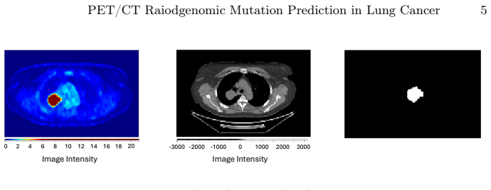
On its own terms, the paper's discovery is that pairwise multi-label learning transfers positively only between certain gene-mutation tasks. With a ResNet-18 taking the largest axial PET slice, the corresponding CT slice, and the tumour mask as input, joint KRAS/TP53 prediction increased test AUC from 0.575 to 0.641 for KRAS and from 0.689 to 0.712 for TP53 relative to single-gene baselines. For EGFR/KRAS, joint learning improved EGFR from 0.718 to 0.746 while KRAS fell from 0.632 to 0.557; the two mutations were mutually exclusive in this cohort. For EGFR/TP53, joint learning did not improve either task, with AUC dropping from 0.808 to 0.783 for EGFR and from 0.514 to 0.491 for TP53. The authors' conclusion is that benefit depends on the gene pair, so uniform adoption of multi-label learning is not warranted.
Load-bearing premise
Repeated PET/CT scans from the same patient were treated as independent samples and randomly split across training, validation, and test, so the model could memorise patient-specific appearance and inflate the reported AUC differences.
Editorial extensions
If this is right
- If KRAS and TP53 genuinely share imaging correlates, joint training is a cheap way to improve both predictions without new data.
- If the EGFR/KRAS asymmetry holds, a hybrid pipeline that predicts EGFR jointly with KRAS but reports KRAS from a single-gene model would dominate either uniform strategy.
- If EGFR and TP53 do not benefit from shared learning, future radiogenomic systems should treat task selection as an empirical per-pair question.
- If mutation-specific modelling is adopted, clinical workflows can match the model to the gene of interest rather than assuming one multi-label network fits all.
Reading between the lines
- The paper does not split by patient, so its AUC comparison may be optimistic; a patient-level split could shrink or reverse some pair gains, especially for patients with two scans.
- One testable extension is to measure gradient conflict or representation similarity between tasks; if KRAS/TP53 gradients align while EGFR/TP53 conflict, that would mechanistically explain the observed transfer pattern.
- Because only 2D single slices were used, 3D tumour-volume models might change which pairs benefit; the pair ordering found here should not be assumed to carry over to volumetric inputs.
- The conclusion that mutation-specific strategies are preferable would be strengthened or weakened by external multi-centre validation, which the paper lists as future work.
Editorial analysis
A structured set of objections, weighed in public.
Referee Report
Summary. The paper studies PET/CT-based radiogenomic prediction of EGFR, KRAS, and TP53 mutation status in non-small cell lung cancer using a ResNet-18 backbone, comparing single-gene classification with pairwise multi-label learning. It introduces a novel UK cohort (MIRAI, 263 patients / 293 PET/CT examinations) and reports that joint learning improves AUC for the KRAS/TP53 pair, improves only EGFR for the EGFR/KRAS pair, and provides no improvement for the EGFR/TP53 pair. The authors conclude that multi-label learning should not be applied uniformly and that mutation-specific modelling strategies may be preferable.
Significance. If the central comparison is statistically sound, the paper makes a useful contribution to PET/CT radiogenomics: it is among the first systematic evaluations of pairwise multi-label learning for mutation prediction in NSCLC, and it provides a new real-world UK cohort (MIRAI) and an openly described preprocessing pipeline, including an nnU-Net segmentation module validated on held-out manual segmentations (Table 3, Dice 0.78±0.12). The finding that multi-label benefit is gene-pair-dependent would also inform practical choices for radiogenomic modelling. However, the headline conclusion currently rests on AUC differences that fall within seed-to-seed variability, and the evaluation protocol admits patient-level leakage, so the significance of the empirical claim is not yet established.
major comments (4)
- [§3, Table 4] The central claim that multi-label learning improves KRAS/TP53 but not EGFR/TP53 is not supported by any statistical testing. All reported AUC differences are within one standard deviation of the five-seed means, e.g., KRAS 0.575±0.068 vs 0.641±0.050, TP53 0.689±0.100 vs 0.712±0.038, EGFR 0.718±0.057 vs 0.746±0.093, and TP53 0.514±0.107 vs 0.491±0.048. No paired significance test, confidence interval, or effect-size measure is reported. Since the same splits and seeds are used for single-gene and multi-label models, a paired statistical comparison (e.g., Wilcoxon signed-rank or bootstrap over patients) is feasible and should be provided; without it, the observed differences may be seed noise, and the 'no improvement' findings may be underpowered rather than evidence of absent benefit.
- [§2.4, §3] Repeated PET/CT examinations from the same patient are treated as independent samples and randomly assigned to training, validation, and test sets. If two scans from one patient appear in both training and test, the model can memorise patient-specific appearance, inflating AUC and distorting the single-gene versus multi-label comparison. The analysis should be repeated with a patient-level split (all scans from a given patient in the same fold), and the number of patients versus PET/CT examinations should be reported for each gene-pair cohort. This is a required robustness check for the quantitative conclusions.
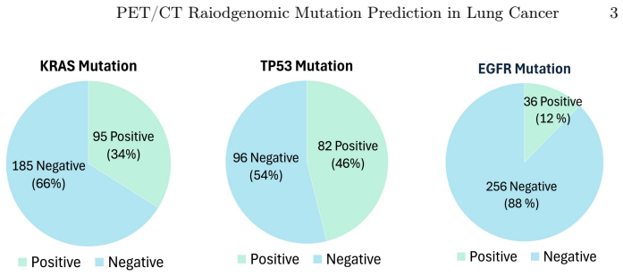
- [§2.4, Table 2] The exclusion of label combinations occurring fewer than six times removes all EGFR+/TP53+ cases (footnote a in Table 2), so the EGFR/TP53 multi-label model never sees the joint-positive class. The conclusion that 'no improvement was observed for the EGFR/TP53 pair' is therefore limited to the sub-cohort without dual-positive cases and should be stated as such. The analysis also changes the target distribution by exclusion; a sensitivity analysis reporting results on the full cohort, or an explicit discussion of why such exclusion is unavoidable, is needed before the pair-dependent benefit claim is accepted.
- [§2.4, Table 4] The evaluation reports mean±SD over five random seeds, which is a good start, but the spread in some cells is very large (e.g., EGFR AUC 0.783±0.357 in the joint EGFR/TP53 model). The paper should report the full distribution of results across seeds, not only the mean, and should indicate whether the qualitative pattern (improvement for KRAS/TP53, no improvement for EGFR/TP53) is stable across individual seeds. This is essential because the claim is about a consistent effect, not a single seed outcome.
minor comments (6)
- [Header, page 3] The running title contains a typo, 'Raiodgenomic' instead of 'Radiogenomic'; the same typo also appears in the header on page 5.
- [§2.4] The authors state that label combinations occurring fewer than six times were excluded, but they do not specify how many patients/scans were removed for each gene pair beyond the aggregate N reported in Table 2. Please state the number of excluded examinations and the number of affected patients for each pair.
- [§2.4, Table 2] The stratification is described as being based on target labels, but for multi-gene cohorts the target is a pair label while single-gene models use individual labels. It should be clarified whether the 'same patient split' means exactly the same assignment of examinations to folds for single-gene and multi-label models, and whether the stratification was recomputed for single-gene labels.
- [§3, Table 4] The bold formatting indicating the better-performing model per gene is visually clear, but it would be helpful to add a note that the values are means over five seeds and that the bold does not imply statistical significance.
- [§3] The discussion of negative transfer for KRAS in the EGFR/KRAS pair and the explanation based on class-imbalance weighting would be more convincing if accompanied by per-seed results or a simple ablation varying the positive weight; as written, the explanation is plausible but not directly supported by an experiment.
- [§2.1] The MIRAI dataset reference [2] is cited as 'In press'; if this is a companion paper, it would be helpful to state explicitly that the current work is based on a subset of MIRAI (108 of 130 segmented volumes with mutation labels) and to clarify the overlap with the dataset description paper.
Circularity Check
No circularity: held-out empirical AUC comparison, no self-citation load-bearing.
full rationale
This is an empirical benchmark study rather than a derivation chain: the central claim (pair-dependent benefit of multi-label learning) is obtained by comparing test-set AUCs of independently trained single-gene and pairwise multi-label ResNet-18 models on the same held-out stratified splits (Sections 2.4 and 3, Table 4). No predicted quantity is defined in terms of a fitted parameter, and no uniqueness theorem or ansatz is imported from prior work. The only self-citation is the authors' own MIRAI dataset [2], used as the cohort source; it is a data provenance citation and does not by itself determine the reported AUC comparisons. The segmentation backbone is independently validated against the external autoPET 2022 benchmark (Dice 0.78; Section 3, Table 3), which provides external support. The paper explicitly acknowledges limitations—class imbalance, single-centre data, absence of external validation, single 2D slices, and exclusion of EGFR+/TP53+ cases (Sections 3 and 4)—and the skeptic's concern that AUC differences lie within one standard deviation without significance testing is a statistical-robustness issue, not circularity. No equation or construction step reduces the result to an input; hence score 0.
Assumptions & free parameters
free parameters (1)
- per-gene positive weight in BCEWithLogitsLoss =
not reported
assumptions (4)
- domain assumption A single axial 2D slice at the largest cross-sectional area of the primary tumour captures enough imaging information for mutation prediction.
- domain assumption Manual and nnU-Net segmentations accurately delineate the primary tumour, with a mean Dice of 0.78 considered sufficient.
- domain assumption Mutation status from single-region biopsy within plus or minus 6 months of imaging is an adequate ground truth.
- domain assumption Repeated PET/CT examinations from the same patient are independent samples.
Cite this review
Pith. "Pith review of PET/CT Radiogenomic Mutation Prediction in Non-Small Cell Lung Cancer Using Multi-Label Learning." pith.science (2026). https://pith.science/paper/VXVKWAP2
@misc{pith2026260809721,
  author       = {Pith},
  title        = {Pith review of: PET/CT Radiogenomic Mutation Prediction in Non-Small Cell Lung Cancer Using Multi-Label Learning},
  year         = {2026},
  howpublished = {\url{https://pith.science/paper/VXVKWAP2}},
  note         = {Machine review of arXiv:2608.09721}
}
read the original abstract
Lung cancer remains one of the leading causes of cancer- related mortality worldwide. Although targeted therapies have improved outcomes for patients with non-small cell lung cancer (NSCLC), they rely on mutation profiling through tissue biopsy, an invasive procedure with several limitations. This study investigates PET/CT-based radio- genomic prediction of epidermal growth factor receptor (EGFR), tumour protein 53 (TP53), and Kirsten rat sarcoma viral oncogene (KRAS) mutations using deep learning. We further evaluate whether pairwise multi-label learning improves mutation prediction compared with conventional single-gene classification. To the best of our knowledge, this is among the first studies to systematically investigate multi-label learning for PET/CT radiogenomic mutation prediction in NSCLC. Experiments were conducted on a novel UK-based radiogenomics cohort. Joint pre- diction of KRAS and TP53 improved AUC from 0.58 to 0.64 for KRAS and from 0.69 to 0.71 for TP53. For the EGFR/KRAS pair, only EGFR benefited from joint learning, while no improvement was observed for the EGFR/TP53 pair. These findings demonstrate that the effectiveness of multi-label learning depends on the specific combination of gene mutations being modelled, suggesting that mutation-specific modelling strategies may be preferable for PET/CT radiogenomic prediction.
Figures
Reference graph
Works this paper leans on
-
[1]
CA: a cancer journal for clinicians74(3), 229–263 (2024)
Bray, F., et al.: Global cancer statistics 2022: Globocan estimates of incidence and mortality worldwide for 36 cancers in 185 countries. CA: a cancer journal for clinicians74(3), 229–263 (2024)
work page 2024
-
[2]
BMJ Digital Health & AI (In press)
Furukawa, M., et al.: Multimodal pet/ct imaging dataset for radiogenomic analysis in non-small cell lung cancer (mirai dataset): Description and initial results. BMJ Digital Health & AI (In press)
-
[3]
European Radiology36, 2157–2185 (2026)
Fuster-Matanzo, A., et al.: Prediction of oncogene mutation status in non-small cell lung cancer: a systematic review and meta-analysis with a special focus on artificial intelligence-based methods. European Radiology36, 2157–2185 (2026)
work page 2026
-
[4]
Gatidis, S., et al.: A whole-body FDG-PET/CT dataset with manually annotated tumor lesions. Scientific Data9(601) (2022)
work page 2022
-
[5]
Nature Machine Intelligence6, 1396– 1405 (2024)
Gatidis, S., et al.: Results from the autopet challenge on fully automated lesion segmentation in oncologic pet/ct imaging. Nature Machine Intelligence6, 1396– 1405 (2024)
work page 2024
-
[6]
Haixian, L., et al.: Machine learning approaches for EGFR mutation status pre- diction in NSCLC: an updated systematic review. Frontiers in Oncology15(2025)
work page 2025
-
[7]
In: Proceedings of the IEEE conference on computer vision and pattern recognition
He, K., et al.: Deep residual learning for image recognition. In: Proceedings of the IEEE conference on computer vision and pattern recognition. pp. 770–778 (2016) 10 M. Furukawa et al
work page 2016
-
[8]
Nature methods18, 203–211 (2021)
Isensee, F., et al.: nnu-net: a self-configuring method for deep learning-based biomedical image segmentation. Nature methods18, 203–211 (2021)
work page 2021
Show all 27 references
-
[9]
Lung 203(1) (2025)
Jeon, H., et al.: Update 2025: management of non-small-cell lung cancer. Lung 203(1) (2025)
2025
-
[10]
Translational Cancer Research8, S23–S47 (2019)
Karachaliou, N., et al.: EGFR first- and second-generation TKIs-there is still place for them in EGFR-mutant NSCLC patients. Translational Cancer Research8, S23–S47 (2019)
2019
-
[11]
Technology6(3-4), 79–100 (2019)
Krzyszczyk, P., et al.: The growing role of precision and personalized medicine for cancer treatment. Technology6(3-4), 79–100 (2019)
2019
-
[12]
Clinical and Transla- tional Oncology27, 1506–1515 (2025)
Li, S., et al.: Prediction of EGFR-TP53 genes co-mutations in patients with lung adenocarcinoma (LUAD) by 18F-FDG PET/CT radiomics. Clinical and Transla- tional Oncology27, 1506–1515 (2025)
2025
-
[13]
Cancer management and research pp
Lu, T., et al.: Trends in the incidence, treatment, and survival of patients with lung cancer in the last four decades. Cancer management and research pp. 943– 953 (2019)
2019
-
[14]
arXiv preprint arXiv:2505.23595 (2025)
Mohamed, Y., et al.: DeepChest: Dynamic gradient-free task weighting for effective multi-task learning in chest X-ray classification. arXiv preprint arXiv:2505.23595 (2025)
2025 arXiv
-
[15]
Scientific reports10(2020)
Pinheiro, G., et al.: Identifying relationships between imaging phenotypes and lung cancer-related mutation status: EGFR and KRAS. Scientific reports10(2020)
2020
-
[16]
PyTorch: Bcewithlogitsloss.https://docs.pytorch.org/docs/2.12/generated /torch.nn.BCEWithLogitsLoss.html, (Accessed: 30.06.2026)
2026
-
[17]
Applied Intelligence53, 25135–25153 (2023)
Read, J.: From multi-label learning to cross-domain transfer: a model-agnostic approach. Applied Intelligence53, 25135–25153 (2023)
2023
-
[18]
Cancer research77(14), 3922–3930 (2017)
Rios Velazquez, E., et al.: Somatic mutations drive distinct imaging phenotypes in lung cancer. Cancer research77(14), 3922–3930 (2017)
2017
-
[19]
European Journal of Radiology110, 148–155 (2019)
Rizzo, S., et al.: Genomics of non-small cell lung cancer (nsclc): Association be- tween ct-based imaging features and egfr and k-ras mutations in 122 patients— an external validation. European Journal of Radiology110, 148–155 (2019)
2019
-
[20]
Cancers14(2022)
Shao, J., et al.: Radiogenomic system for non-invasive identification of multiple actionable mutations and PD-L1 expression in non-small cell lung cancer based on CT images. Cancers14(2022)
2022
-
[21]
Frontiers in Oncology10(2021)
Shui, L., et al.: The era of radiogenomics in precision medicine: an emerging ap- proach to support diagnosis, treatment decisions, and prognostication in oncology. Frontiers in Oncology10(2021)
2021
-
[22]
Journal of Nuclear Medicine56(9), 1447–1452 (2015)
Teoh, E.J., et al.: Phantom and clinical evaluation of the bayesian penalized like- lihood reconstruction algorithm Q.clear on an LYSO PET/CT system. Journal of Nuclear Medicine56(9), 1447–1452 (2015)
2015
-
[23]
Frontiers in immunology13(2022)
Wang, C., et al.: Predicting egfr and pd-l1 status in nsclc patients using multitask ai system based on ct images. Frontiers in immunology13(2022)
2022
-
[24]
Radiology: Artificial Intelligence5(5) (2023)
Wasserthal, J., et al.: Totalsegmentator: robust segmentation of 104 anatomic structures in ct images. Radiology: Artificial Intelligence5(5) (2023)
2023
-
[25]
The Journal of Nuclear Medicine58(4), 569–576 (2017)
Yip, S.S., et al.: Associations between somatic mutations and metabolic imaging phenotypes in non-small cell lung cancer. The Journal of Nuclear Medicine58(4), 569–576 (2017)
2017
-
[26]
Neuroimage31(3), 1116–1128 (2006)
Yushkevich, P.A., et al.: User-guided 3D active contour segmentation of anatomi- cal structures: Significantly improved efficiency and reliability. Neuroimage31(3), 1116–1128 (2006)
2006
-
[27]
Cancers13(1814) (2021)
Zhang, T., et al.: Simultaneous identification of egfr, kras, erbb2, and tp53 mu- tations in patients with non-small cell lung cancer by machine learning-derived three-dimensional radiomics. Cancers13(1814) (2021)
2021
Reviewed August 11, 2026 · model on record in the stance chip above.
Discussion (0). Continue with ORCID to comment.