REVIEW 5 major objections 4 minor 47 references
Lesion-Aware Adaptive Fourier Neural Operator for CT-to-PSMA PET Synthesis in Prostate Cancer
T0 review · 5 major / 4 minor · reviewed 2026-08-15 · deepseek-v4-flash
Pith's one-line read The paper claims that conditioning a Fourier-neural-operator synthesis model on two CT-derived proxy channels and training with lesion-aware losses preserves lesion activity and tumor radiomics in synthetic PSMA-PET, where global…
desk verdict A well-motivated lesion-aware synthesis method whose headline TLA gains are likely optimistic because the loss weights and proxy scales were chosen on the test cohort. read the letter →
The pith
A machine-rendered reading of the paper's core claim, the machinery that carries it, and where it could break.
The reading
What carries the argument
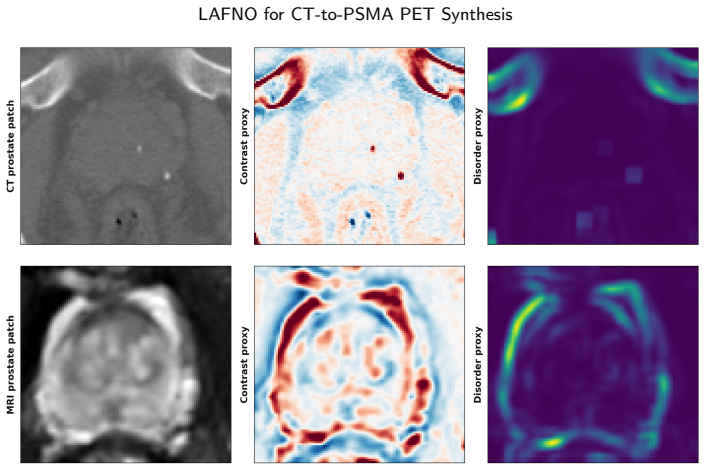
The central mechanism is the pair of differentiable CT proxy channels plus a lesion-aware loss. The contrast proxy is a Gaussian residual filter that captures the smooth density gradient from tumor core to periphery; the disorder proxy is a sliding-window local variance that captures peritumoral texture heterogeneity. They are computed from CT alone, so at inference no lesion segmentation is needed. They are pooled (average for contrast, max for disorder) and concatenated into the Adaptive Fourier Neural Operator bottleneck, which performs spectral channel mixing so the proxy-conditioned features influence long-range spatial structure. The lesion-wise total-activity loss uses a log-compressed, per-lesion-normalized summed-activity error so small lesions are not dominated by large ones; the tumor-contrast loss supervises the contrast operator on predicted versus ground-truth SUV within the tumor mask; and the peritumoral loss applies an exponential distance-decay weight in the 0–10 mm ring. Together these terms push the model to preserve lesion-level activity and tumor-adjacent structure rather than optimizing global similarity alone.
What would settle it
On an independent cohort of PSMA-PET/CT patients from different scanners, tracers, or institutions, train LAFNO and the CT-only AFNO-L1 baseline with the proxy scales fixed at $\sigma = 5$ mm and $w = 7.5$ mm; if per-patient TLA error and tumor-core radiomics reproducibility no longer improve over the baseline, the proxy conditioning claim is falsified. A cheaper check: replace the two proxy channels with identically shaped channels containing CT intensity or white noise, holding all losses fixed; if TLA error and ICC do not worsen, the proxies' specific encoding carries no weight.
Extended reading notes
Core claim
The paper's central claim is that a CT-to-PSMA-PET synthesis model conditioned on radiomics-motivated CT proxies and supervised at the lesion level preserves the clinically relevant PET signal that global L1 or MSE training discards. The contrast proxy $C(\mathbf{x}) = \mathrm{CT}(\mathbf{x}) - G_\sigma * \mathrm{CT}(\mathbf{x})$ with $\sigma = 5$ mm encodes the observed core-to-periphery CT attenuation gradient, and the disorder proxy $D(\mathbf{x})$ is local variance over a $7.5$ mm window, encoding tumor-adjacent texture heterogeneity; both are injected into the Adaptive Fourier Neural Operator bottleneck of a 3D U-Net. The loss combines whole-volume L1 with a per-lesion total-activity term, a tumor-core contrast term, and an exponentially distance-weighted peritumoral term. In the reported evaluation LAFNO attains SSIM 0.960 and 0.938 and per-patient TLA errors 48.3 and 64.0 percent for 18F- and 68Ga-PSMA, and the highest tumor-core radiomics ICCs across first-order, GLCM, GLRLM, and GLSZM classes for both tracers, while peritumoral reproducibility remains tracer-dependent. The authors interpret the remaining error as inherent one-to-many CT-to-PET mapping and tracer or SUV heterogeneity rather than a failure that purely architectural changes would fix.
Load-bearing premise
The load-bearing premise is that the two hand-coded CT filters—a 5 mm Gaussian residual and a 7.5 mm local variance—capture CT patterns that actually predict PSMA-PET uptake in and around lesions, and that the chosen scales generalize beyond the dataset where they were selected.
Editorial extensions
If this is right
- On 18F-PSMA, per-patient TLA error drops to 48.3 percent from 58.7 to 69.0 percent for the four baselines, so a trained model can estimate lesion activity from CT alone at inference.
- On 68Ga-PSMA, LAFNO reaches 64.0 percent per-patient TLA error and the highest fraction of lesions within 50 and 75 percent error, though cWDM is comparable on absolute lesion-level error.
- Tumor-core radiomics reproducibility is the highest of all compared models for every feature class on both tracers, indicating the synthetic volumes carry texture-level information rather than only pixel similarity.
- The ablation shows proxy conditioning alone cuts TLA error from 62.2 to 56.5 percent even without lesion masks or lesion-specific losses, so the CT-derived channels contribute independent information.
- Whole-volume SSIM (0.960 and 0.938) and PSNR remain competitive with baselines, so the lesion-aware objectives do not trade away global image quality.
Reading between the lines
- If the proxy scales were learned per scanner or tracer rather than fixed on the evaluation dataset, TLA gains might transfer more reliably across institutions; this is a testable extension not performed by the paper.
- Because the proxies need no lesion segmentation, coupling LAFNO with an automatic lesion detector would give a fully mask-free, CT-only pipeline for PSMA lesion burden.
- The 68Ga weakness and the peritumoral reproducibility peak at 3–5 mm suggest positron range and partial-volume effects are limiting; partial-volume correction before training or evaluation is a natural next test.
- The peritumoral term's effect is essentially confined to GLSZM in the 0–3 mm band, implying a single distance-weighted L1 penalty is too weak to shape the whole tumor-adjacent microenvironment; adversarial or multi-scale texture losses are worth trying.
Signed reviews
Editorial analysis
A structured set of objections, weighed in public.
Referee Report
Summary. The manuscript proposes LAFNO, a 3D U-Net with an Adaptive Fourier Neural Operator bottleneck for synthesizing PSMA-PET from CT in prostate cancer. The main novelties are two handcrafted CT-derived proxy channels (contrast and disorder) injected at the bottleneck, and a training objective that combines whole-volume L1 with lesion-wise total lesion activity (TLA), tumor-core contrast, and distance-weighted peritumoral losses. The authors evaluate on the TCIA PSMA-PET-CT-Lesions dataset with 18F- and 68Ga-PSMA cohorts against four baselines, reporting lesion-level and patient-level TLA error, fractions of lesions within error thresholds, and tumor-core/peritumoral radiomics ICC. They report that LAFNO remains competitive on SSIM/PSNR/MAE while improving TLA error and tumor-core radiomics reproducibility, with a cumulative ablation on the 18F cohort.
Significance. If the results are validated, LAFNO is a useful step toward clinically meaningful CT-to-PET synthesis because it targets lesion-level activity and radiomic structure rather than only global similarity, and the proxy-conditioning idea avoids per-inference radiomics extraction. Strengths include the cumulative ablation isolating each component, the use of lesion-level and radiomics evaluation beyond global metrics, and the explicit discussion of tracer-dependent limitations. However, the absence of a held-out validation set for hyperparameter selection, the lack of statistical inference, and an inconsistency in the TLA loss formulation mean the reported quantitative claims are not yet established. The work is therefore of interest to the medical image synthesis community, but the evidence requires strengthening before publication.
major comments (5)
- [§2.2, §5.1, §5.5] The proxy scales σ=5mm and w=7.5mm are described as chosen empirically, and the TLA loss formulation, λ_TLA=0.05, and λ_peri=0.05 are selected from curves and ablations computed on the same 18F test cohort used for the final evaluation (Figure 7, Table 5). With only N=47 test patients and no held-out validation or nested cross-validation, the reported improvements, e.g., patient-level TLA 48.3% vs. 62.2%, may be inflated by selection. Please either introduce a strict validation split for all hyperparameter choices or demonstrate that the conclusions are stable under repeated random splits.
- [Table 5] The cumulative ablation shows that adding the peritumoral term changes patient-level TLA error from 47.8% to 48.3%, lowers the ≤25% and ≤50% fractions from 25.2/54.9 to 24.0/50.9, and decreases every tumor-core ICC class (e.g., GLSZM from 0.760 to 0.709). The text states this term leaves the metrics 'essentially unchanged', which is contradicted by the table; it also raises the question of why the term is retained in the final model. This internal inconsistency, combined with the test-set tuning, undermines the ablation-based justification of the final configuration.
- [§2.4, Eq. (5); §3.2, Eq. (9)] The TLA loss is defined with ŝ_i and s_i as 'SUV values', but the model is trained on log-normalized PET values from Eq. (9). If the loss is computed on normalized outputs, then A_k and ^A_k are sums of nonlinearly transformed values and are not proportional to physical TLA; the inverse log transform in Eq. (9) is nonlinear, so the proportionality claim does not hold. Please state explicitly whether the inverse transform is applied before computing the TLA loss, or revise the loss definition and the interpretation of the reported TLA improvements accordingly.
- [§3.1, Table 3] For 68Ga-PSMA, the patient-level TLA error of LAFNO is 64.0% versus 63.9% for cWDM, so LAFNO does not reduce patient-level TLA error relative to the best baseline on that tracer. Since the 68Ga cohort is less affected by the tuning performed on 18F, this equality weakens the cross-tracer generalization claim and should be reported as a limitation rather than as a reduction.
- [§3.2, §3.3] The manuscript does not report confidence intervals, significance tests, or repeated-seed variability for the main TLA and ICC comparisons, and it does not specify the patient-level train/test split (e.g., whether all scans of a patient were confined to one split). These details are essential for assessing whether the reported differences are robust, and the split specification is needed to rule out leakage. Please add this information.
minor comments (4)
- [Section 4.3] The text refers to 'Figure 8' for peritumoral reproducibility by distance band, but the figure with that content is labeled Figure 6; the later ablation figure is Figure 8. Please fix the cross-reference.
- [Eq. (7)] The sentence 'with this value, the weight decreases to half of its boundary value from the tumor boundary' is inaccurate: with τ=5mm the half-value distance is τ ln 2 ≈ 3.47mm, not 5mm, where the weight is e^{-1} ≈ 0.368 of the boundary value.
- [Abstract] The abstract's phrase 'reducing per-patient TLA error to ... 64.0%' for 68Ga is misleading given that cWDM achieves 63.9% on the same metric; please qualify the claim.
- [Section 5.1] The paragraph describing the final choice of λ_TLA states that the overestimation rate is below 20% in the final model; the corresponding values (19.98% and 10.47%) appear only in the text and not in Table 3, making the table incomplete for this claim.
Circularity Check
Hyperparameters for the lesion-aware loss and proxy scales are selected on the same 18F-PSMA cohort used for the headline TLA evaluation, so the reported improvement is partly a selected result rather than an independent prediction; the derivation itself is not circular by construction.
-
fitted input called prediction
[Section 5.1 (loss-form and lambda_TLA choice, Figure 7) applied to the final model evaluated in Section 4.2 (Table 3); proxy scale w=7.5mm set in Section 2.2 on the same dataset.]
"We therefore adopt the lesion-wise L_TLA formulation. Its higher accuracy at large λ comes with an increased overestimation rate, which is why the final model uses a low weight of λTLA=0.05 and relies on the CT proxy channels and the tumor-contrast loss L_c to improve tumor-level performance. This keeps the overestimation rate below 20% in the final model: 19.98% for 18F-PSMA with 48.35% absolute TLA error ... as reported in Table 3."
The final TLA formulation, its weight, and the proxy window size are chosen by inspecting curves and ablations computed on the same 18F-PSMA cohort (N=47) that later supplies the headline per-patient TLA error of 48.3% in Table 3. lambda_TLA=0.05 was selected specifically to keep the overestimation rate below 20% on that cohort, so the reported 48.35% is not an out-of-sample prediction but a post-selection measurement on the tuning set. The 68Ga cohort (N=30) was not used for this selection, and there LAFNO does not beat cWDM on patient-level TLA (64.0% vs 63.9%), which is consistent with the 18F advantage being partly driven by test-set selection.
full rationale
LAFNO is an empirical deep-learning synthesis method, not a first-principles derivation, and most of its components (CT proxy channels, lesion-aware losses, radiomics evaluation) are independent of one another. I found no equation that is equivalent to its input by construction, and no load-bearing self-citation: the AFNO bottleneck is motivated by the authors' earlier work, but that citation only supports an architectural choice and is not invoked to forbid alternatives. The genuine circularity concern is evaluation-set selection: Section 5.1 tunes the per-lesion loss form and lambda_TLA on the same 18F-PSMA test cohort whose TLA error is then reported as the headline result, and Section 2.2 sets proxy scales empirically on the same dataset; Section 5.5 also retains L_peri after observing on that cohort that it slightly worsens TLA and tumor-core ICC while improving a narrow peritumoral GLSZM band. Thus the 18F improvement is partly a selected optimum rather than an independent prediction. The 68Ga results, which were not used for tuning, are weaker and do not show a patient-level TLA advantage over cWDM, reinforcing the interpretation that the 18F gain is inflated by selection. Because the central architecture and losses still have empirical content and the selection is disclosed in the ablation, the appropriate score is 4 rather than 6 or higher.
Assumptions & free parameters
free parameters (5)
- contrast proxy scale sigma =
5 mm
- disorder proxy window w =
7.5 mm
- peritumoral decay tau =
5 mm
- loss weights lambda_TLA, lambda_c, lambda_peri =
0.05, 0.02, 0.05
- SUVmax normalization ceiling =
cohort-dependent
assumptions (4)
- domain assumption Ground-truth PSMA-PET lesion masks and SUV values in the TCIA dataset are accurate enough for training and evaluation.
- domain assumption The CT-to-PET mapping is sufficiently learnable from paired CT/PET data despite being one-to-many.
- ad hoc to paper The two handcrafted proxies preserve the radiomics-observed trends outside the training cohort.
- standard math Standard deep learning training assumptions (batch norm, Adam, ReLU, etc.) work as expected.
invented entities (2)
-
Contrast proxy channel C(x)
-
Disorder proxy channel D(x)
Cite this review
Pith. "Pith review of Lesion-Aware Adaptive Fourier Neural Operator for CT-to-PSMA PET Synthesis in Prostate Cancer." pith.science (2026). https://pith.science/paper/5DPD7JP4
@misc{pith2026260810429,
  author       = {Pith},
  title        = {Pith review of: Lesion-Aware Adaptive Fourier Neural Operator for CT-to-PSMA PET Synthesis in Prostate Cancer},
  year         = {2026},
  howpublished = {\url{https://pith.science/paper/5DPD7JP4}},
  note         = {Machine review of arXiv:2608.10429}
}
read the original abstract
Deep learning models that synthesize PET from CT or MRI can reduce patient dose and scanner demand, but are typically optimized with global losses such as L1 or mean squared error (MSE) that treat all voxels similarly. In whole-body PSMA-PET, tumor voxels occupy only a small fraction of the volume, yet carry the clinically relevant activity signal; as a result, models can achieve high structural similarity index measure (SSIM) and peak signal-to-noise ratio (PSNR) while still underestimating lesion activity or failing to preserve tumor-specific structure. Radiomics provides biologically meaningful descriptors of tumor intensity and texture, but direct radiomics conditioning is time-consuming because it requires feature extraction from delineated lesion regions. We propose LAFNO, a Lesion-Aware Adaptive Fourier Neural Operator for CT-to-PSMA-PET synthesis that replaces high-dimensional radiomics conditioning with two efficient CT-derived proxy channels. Motivated by radiomics analysis of PSMA-avid tumor core and peritumoral regions, LAFNO uses a contrast proxy for local density variation and a disorder proxy for local texture heterogeneity, both injected into the model bottleneck. LAFNO combines whole-volume reconstruction with lesion-level total lesion activity (TLA), tumor-core contrast, and peritumoral supervision. We evaluated LAFNO against four baseline architectures on the TCIA PSMA-PET-CT-Lesions dataset. LAFNO remained competitive on whole-volume image quality, achieving SSIM of 0.960 and 0.938 for 18F- and 68Ga-PSMA, respectively, while reducing per-patient TLA error to 48.3% and 64.0% for 18F- and 68Ga-PSMA, respectively, and achieving the highest tumor-core radiomics reproducibility across all feature classes for both tracers. Peritumoral reproducibility remained tracer-dependent, indicating that biological fidelity in synthetic PSMA-PET remains challenging.
Figures


Figures from the paper (7 more)


Reference graph
Works this paper leans on
-
[1]
author Abdollahi, H. , author Harsini, S. , author Yousefirizi, F. , author Hatami, B. , author B \'e nard, F. , author Shariftabrizi, A. , author Alberts, I. , author Rahmim, A. , year 2026 . title Microenvironment at a distance: Multi-endocrine-organ radiomics to identify systemic signatures in psma-negative prostate cancer . journal Cancers volume 18 ,...
work page 2026
-
[2]
author Abtahi, M. , author Hong, C.M. , author Deveshwar, N. , author Nwihim, S. , author Larson, P.E. , author Hope, T.A. , year 2026 . title Fine-unetr for psma pet/ct lesion segmentation: Automated tumor quantification and overall survival stratification in prostate cancer . journal arXiv preprint arXiv:2606.17570
work page Pith review arXiv 2026
-
[3]
author Algohary, A. , author Shiradkar, R. , author Pahwa, S. , author Purysko, A. , author Verma, S. , author Moses, D. , author Shnier, R. , author Haynes, A.M. , author Delprado, W. , author Thompson, J. , et al., year 2020 . title Combination of peri-tumoral and intra-tumoral radiomic features on bi-parametric mri accurately stratifies prostate cancer...
work page 2020
-
[4]
author Almutairi, W.M. , author Gopaulchan, M. , author Bhaskara, R. , author Zheng, Q.H. , author Ocana, J.A. , author Langer, M. , author Perekattu Kuruvilla, T. , author Durm, G. , author Oderinde, O.M. , year 2026 . title Metabolic [18f]f-arag pet imaging of t cell activation: a functional complement to cell-specific immune tracers . journal EJNMMI Re...
-
[5]
author Almutairi, W.M. , author Zheng, Q.H. , author Langer, M. , author Oderinde, O.M. , year 2025 . title Application of immunopet imaging to enhance head and neck squamous cell carcinoma clinical management . journal Frontiers in Oncology volume 15 , pages 1644692 . :10.3389/fonc.2025.1644692
-
[6]
author Bhaskara, R. , author Oderinde, O. , year 2025 . title Enhancing synthetic pelvic ct generation from cbct using vision transformer with adaptive fourier neural operators . journal Biomedical Physics & Engineering Express volume 11 , pages 055013
work page 2025
-
[7]
author Boellaard, R. , author O'Doherty, M.J. , author Weber, W.A. , author Mottaghy, F.M. , author Lonsdale, M.N. , author Stroobants, S.G. , author Oyen, W.J.G. , author Kotzerke, J. , author Hoekstra, O.S. , author Pruim, J. , author Marsden, P.K. , year 2010 . title Fdg pet and pet/ct: Eanm procedure guidelines for tumour pet imaging: Version 1.0 . jo...
-
[8]
author Bouchelouche, K. , author Turkbey, B. , author Choyke, P.L. , year 2016 . title Psma pet and radionuclide therapy in prostate cancer , in: booktitle Seminars in nuclear medicine , organization Elsevier . pp. pages 522--535
work page 2016
Show all 47 references
-
[9]
, author De Risi, M
author Caldarella, C. , author De Risi, M. , author Massaccesi, M. , author Miccich \`e , F. , author Bussu, F. , author Galli, J. , author Rufini, V. , author Leccisotti, L. , year 2024 . title Role of 18f-fdg pet/ct in head and neck squamous cell carcinoma: current evidence ...
2024
-
[10]
, author Yao, X
author Chai, L. , author Yao, X. , author Yang, X. , author Na, R. , author Yan, W. , author Jiang, M. , author Zhu, H. , author Sun, C. , author Dai, Z. , author Yang, X. , year 2025 . title Synthesizing [18f] psma-1007 pet bone images from ct images with gan for early detect...
2025
-
[11]
, author Lombardi, A
author Danese, D. , author Lombardi, A. , author Attimonelli, M. , author Fasano, G. , author Di Noia, T. , year 2026 . title Flowlet: Conditional 3d brain mri synthesis using wavelet flow matching . journal Medical Image Analysis volume 113 , pages 104161 . http://dx.doi.org/...
2026
-
[12]
, author Islam, K.T
author Dayarathna, S. , author Islam, K.T. , author Uribe, S. , author Yang, G. , author Hayat, M. , author Chen, Z. , year 2024 . title Deep learning based synthesis of mri, ct and pet: Review and analysis . journal Medical image analysis volume 92 , pages 103046
2024
-
[13]
, author Gazzilli, M
author Dondi, F. , author Gazzilli, M. , author Albano, D. , author Rizzo, A. , author Treglia, G. , author Pisani, A.R. , author Palumbo, C. , author Rubini, D. , author Racca, M. , author Rubini, G. , et al., year 2024 . title Prognostic role of pre-and post-treatment [18f] ...
2024
-
[14]
, author Liu, Q
author Emami, H. , author Liu, Q. , author Dong, M. , year 2020 . title Frea-unet: frequency-aware u-net for modality transfer . journal arXiv preprint arXiv:2012.15397
2020 arXiv
-
[15]
, author Durrer, A
author Friedrich, P. , author Durrer, A. , author Wolleb, J. , author Cattin, P.C. , year 2024 . title cwdm: Conditional wavelet diffusion models for cross-modality 3d medical image synthesis . https://arxiv.org/abs/2411.17203, arXiv:2411.17203 http://arxiv.org/abs/2411.17203
2024 arXiv
-
[16]
, author Sheikhzadeh, P
author Ghafari, A. , author Sheikhzadeh, P. , author Seyyedi, N. , author Abbasi, M. , author Farzenefar, S. , author Yousefirizi, F. , author Ay, M.R. , author Rahmim, A. , year 2022 . title Generation of 18f-fdg pet standard scan images from short scans using cycle-consisten...
2022
-
[17]
, author Kinahan, P.E
author Gillies, R.J. , author Kinahan, P.E. , author Hricak, H. , year 2016 . title Radiomics: images are more than pictures, they are data . journal Radiology volume 278 , pages 563--577
2016
-
[18]
, author Mardani, M
author Guibas, J. , author Mardani, M. , author Li, Z. , author Tao, A. , author Anandkumar, A. , author Catanzaro, B. , year 2021 . title Adaptive fourier neural operators: Efficient token mixers for transformers . journal arXiv preprint arXiv:2111.13587
2021 arXiv
-
[19]
, author Shapiro, W.R
author Heppner, G.H. , author Shapiro, W.R. , author Rankin, J.K. , year 1981 . title Tumor heterogeneity . journal Pediatric Oncology 1: with a special section on Rare Primitive Neuroectodermal Tumors , pages 99--116
1981
-
[20]
, author Yamagata, K
author Ishibashi-Kanno, N. , author Yamagata, K. , author Hara, T. , author Takaoka, S. , author Fukuzawa, S. , author Uchida, F. , author Bukawa, H. , year 2023 . title Prognostic prediction using maximum standardized uptake value ratio of lymph node-to-primary tumor in preop...
2023
-
[21]
, author Zhu, J.Y
author Isola, P. , author Zhu, J.Y. , author Zhou, T. , author Efros, A.A. , year 2017 . title Image-to-image translation with conditional adversarial networks , in: booktitle Proceedings of the IEEE conference on computer vision and pattern recognition , pp. pages 1125--1134
2017
-
[22]
u stner, T. , author Gatidis, S. , author Fr \
author Jeblick, K. , author Schachtner, B. , author Mittermeier, A. , author Dexl, J. , author Wesp, P. , author K \"u stner, T. , author Gatidis, S. , author Fr \"u h, M. , author Fabritius, M.P. , author Herr, F. , et al., year 2026 . title A whole-body psma-pet/ct dataset w...
2026
-
[23]
, year 2026
author Jemal, A. , year 2026 . title Cancer statistics, 2026 . journal CA: A Cancer Journal for Clinicians volume 76 , pages e70043
2026
-
[24]
, author Kong, E
author Jung, E. , author Kong, E. , author Yu, D. , author Yang, H. , author Chicontwe, P. , author Park, S.H. , author Jeon, I. , year 2024 . title Generation of synthetic pet/mr fusion images from mr images using a combination of generative adversarial networks and condition...
2024
-
[25]
, author Na, I
author Kim, J. , author Na, I. , author Ko, E.S. , author Park, H. , year 2025 . title Tumor synthesis conditioned on radiomics , in: booktitle 2025 IEEE/CVF Winter Conference on Applications of Computer Vision (WACV) , organization IEEE . pp. pages 3635--3646
2025
-
[26]
, author Seifert, R
author Kim, M. , author Seifert, R. , author Fragemann, J. , author Kersting, D. , author Murray, J. , author Jonske, F. , author Pomykala, K.L. , author Egger, J. , author Fendler, W.P. , author Herrmann, K. , et al., year 2023 . title Evaluation of thresholding methods for t...
2023
-
[27]
, author Abedi-Ardekani, B
author Koca, D. , author Abedi-Ardekani, B. , author LeMaoult, J. , author Guyon, L. , year 2024 . title Peritumoral tissue (ptt): increasing need for naming convention . journal British Journal of Cancer volume 131 , pages 1111--1115
2024
-
[28]
, author Rios-Velazquez, E
author Lambin, P. , author Rios-Velazquez, E. , author Leijenaar, R. , author Carvalho, S. , author Van Stiphout, R.G. , author Granton, P. , author Zegers, C.M. , author Gillies, R. , author Boellard, R. , author Dekker, A. , et al., year 2012 . title Radiomics: extracting mo...
2012
-
[29]
, author Chen, J
author Liu, J. , author Chen, J. , author Qiu, L. , author Li, R. , author Li, Y. , author Li, T. , author Leng, X. , year 2025 . title The value of intratumoral and peritumoral ultrasound radiomics model constructed using multiple machine learning algorithms for non-mass brea...
2025
-
[30]
, author Al-Shalabi, M
author Mahdi, M.A. , author Al-Shalabi, M. , author Elbarougy, R. , author Alnfrawy, E.T. , author Hadi, M.U. , author Ali, R.F. , year 2026 . title Ct-to-pet synthesis in the head--neck and thoracic region via conditional 3d latent diffusion modeling . journal Bioengineering ...
2026
-
[31]
, author Eiber, M
author Maurer, T. , author Eiber, M. , author Schwaiger, M. , author Gschwend, J.E. , year 2016 . title Current use of psma--pet in prostate cancer management . journal Nature Reviews Urology volume 13 , pages 226--235
2016
-
[32]
, author Robinson, M.K
author Reddy, S. , author Robinson, M.K. , year 2010 . title Immuno-positron emission tomography in cancer models , in: booktitle Seminars in nuclear medicine , organization Elsevier . pp. pages 182--189
2010
-
[33]
, author Fischer, P
author Ronneberger, O. , author Fischer, P. , author Brox, T. , year 2015 . title U-net: Convolutional networks for biomedical image segmentation , in: booktitle International Conference on Medical image computing and computer-assisted intervention , organization Springer . pp...
2015
-
[34]
, author Udovicich, C
author Siva, S. , author Udovicich, C. , author Tran, B. , author Zargar, H. , author Murphy, D.G. , author Hofman, M.S. , year 2020 . title Expanding the role of small-molecule psma ligands beyond pet staging of prostate cancer . journal Nature Reviews Urology volume 17 , pag...
2020
-
[35]
, author Harper, M
author Smith, T. , author Harper, M. , year 2025 . title Prostate-specific membrane antigen (psma) pet-ct: Revolutionizing staging, restaging, and treatment response assessment . journal Ann. Urol. Oncol volume 8 , pages 200--210
2025
-
[36]
, author Martinez, J
author Subramanian, K. , author Martinez, J. , author Huicochea Castellanos, S. , author Ivanidze, J. , author Nagar, H. , author Nicholson, S. , author Youn, T. , author Nauseef, J.T. , author Tagawa, S. , author Osborne, J.R. , year 2023 . title Complex implementation factor...
2023
-
[37]
, author Finn, S
author Tonry, C. , author Finn, S. , author Armstrong, J. , author Pennington, S.R. , year 2020 . title Clinical proteomics for prostate cancer: understanding prostate cancer pathology and protein biomarkers for improved disease management . journal Clinical Proteomics volume ...
2020
-
[38]
, author Wee, L
author Traverso, A. , author Wee, L. , author Dekker, A. , author Gillies, R. , year 2018 . title Repeatability and reproducibility of radiomic features: a systematic review . journal International Journal of Radiation Oncology* Biology* Physics volume 102 , pages 1143--1158
2018
-
[39]
, author Vrachimis, A
author Tsechelidis, I. , author Vrachimis, A. , year 2022 . title Psma pet in imaging prostate cancer . journal Frontiers in Oncology volume 12 , pages 831429
2022
-
[40]
, year 2019
author Ulaner, G.A. , year 2019 . title Fundamentals of Oncologic PET/CT . publisher Elsevier . :10.1016/C2016-0-05304-4
2019 doi
-
[41]
, author Zhang, M
author Wang, G. , author Zhang, M. , author Cheng, M. , author Wang, X. , author Li, K. , author Chen, J. , author Chen, Z. , author Chen, S. , author Chen, J. , author Xiong, G. , et al., year 2021 . title Tumor microenvironment in head and neck squamous cell carcinoma: Funct...
2021
-
[42]
, author Yang, Y
author Wang, Y. , author Yang, Y. , author Li, J. , author Cheng, D. , author Xu, H. , author Huang, J. , year 2025 . title Dynamic fdg pet/ct imaging: Quantitative assessment, advantages and application in the diagnosis of malignant solid tumors . journal Frontiers in Oncolog...
2025
-
[43]
, author Korn, R.L
author Weiss, G.J. , author Korn, R.L. , year 2012 . title Interpretation of pet scans: Do not take suvs at face value . journal Journal of Thoracic Oncology volume 7 , pages 1744--1746 . :10.1097/JTO.0b013e31827450ae
2012 doi
-
[44]
, author Stockert, J.A
author Yadav, S.S. , author Stockert, J.A. , author Hackert, V. , author Yadav, K.K. , author Tewari, A.K. , year 2018 . title Intratumor heterogeneity in prostate cancer , in: booktitle Urologic Oncology: Seminars and Original Investigations , organization Elsevier . pp. page...
2018
-
[45]
, author Regan, K
author Zhang, S. , author Regan, K. , author Najera, J. , author Grinstaff, M.W. , author Datta, M. , author Nia, H.T. , year 2023 . title The peritumor microenvironment: physics and immunity . journal Trends in cancer volume 9 , pages 609--623
2023
-
[46]
, author Xie, M
author Zhou, H. , author Xie, M. , author Shi, H. , author Shou, C. , author Tang, M. , author Zhang, Y. , author Hu, Y. , author Liu, X. , year 2025 . title Integrating multimodal imaging and peritumoral features for enhanced prostate cancer diagnosis: a machine learning appr...
2025
-
[47]
, author Leger, S
author Zwanenburg, A. , author Leger, S. , author Valli \`e res, M. , author L \"o ck, S. , year 2016 . title Image biomarker standardisation initiative . journal arXiv preprint arXiv:1612.07003
2016 arXiv
Reviewed August 15, 2026 · model on record in the stance chip above.
Discussion (0). Continue with ORCID to comment.