REVIEW 4 major objections 5 minor 35 references
Multi-Task Diffusion Approach For Prediction of Glioma Tumor Progression
T0 review · 4 major / 5 minor · reviewed 2026-08-15 · deepseek-v4-flash
Pith's one-line read Two prior MRI scans are enough for a diffusion model to predict a glioma's future shape and a pixel-level risk map at any chosen day.
desk verdict A genuinely new multitask diffusion architecture for time-agnostic glioma progression prediction, but the calibrated-uncertainty claim rests on hand-built soft labels and a 7-patient test set, so the numbers are less solid than they look. read the letter →
The pith
A machine-rendered reading of the paper's core claim, the machinery that carries it, and where it could break.
The reading
What carries the argument
The load-bearing object is the logistic-normalized signed distance field (SDF) probability map, $P_t(x)=\sigma(\phi(x),\beta=0.1,\mu=15)$, which converts a binary Gross Tumor Volume mask into a smooth pixel-wise likelihood of tumor presence. This single representation does three jobs: it is the regression target for the predictive branch, the input that the deformation module warps between timepoints, and the reference against which uncertainty metrics (ECE, BCE, KL) are computed. The second mechanism is the pre-trained tumor evolution module, a U-Net-style network that takes two SDF maps and a time gap and returns a dense 2D displacement field via a spatial transformer, giving the diffusion model a learned prior for how the tumor moves and grows. Around these sit a sinusoidal temporal embedding injected through feature-wise modulation layers, a two-stage diffusion augmentation pipeline (full-sequence synthesis plus missing-modality imputation), and a radiotherapy-weighted focal loss that up-weights pixels inside high-dose regions.
What would settle it
Compare GliomaDiff's predicted probability maps and expected calibration error against independently produced clinical ground truth, for example expert neuroradiologist delineations of future GTV or biopsy-verified recurrence regions, rather than against the logistic-normalized SDF. If the ECE computed against those independent labels is substantially worse than the reported 0.0100, the calibrated-uncertainty claim is not supported. A simpler check: recompute all metrics on the seven public test patients using hard tumor masks only and see whether the reported Dice gains and calibration improvement persist.
Extended reading notes
Core claim
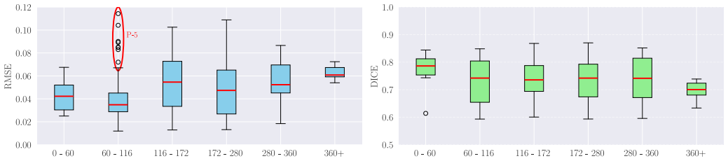
On its own terms, the paper establishes that glioma progression can be framed as a conditional generation task: given scans $S_{t_1}$ and $S_{t_2}$ with their acquisition dates, GliomaDiff synthesizes the future FLAIR (a fluid-sensitive MRI sequence) at a requested date $d_{t_3}$ and simultaneously regresses a probability map $P_{t_3}$ of tumor presence. The key move is to replace hard tumor masks with logistic-normalized signed distance fields: each pixel's distance to the tumor boundary is pushed through a logistic function ($\beta=0.1$, $\mu=15$ mm), so the core stays above 0.8 probability, the edge decays to 0.5 at 15 mm, and beyond that tumor presence is considered unlikely. A pre-trained U-Net deformation module predicts a dense displacement field between the two input SDF maps, conditioned on the time gap, and supplies this as a deformation prior to a shared-encoder, two-decoder diffusion network; a radiotherapy-weighted focal loss biases learning toward high-dose regions. The authors report that the full system beats a probabilistic-mask diffusion baseline on RMSE, KL divergence, and Dice, keeps calibration error low (ECE 0.0100), and remains accurate for intervals up to about a year, with Dice falling from 0.781 at 0–60 days to 0.699 beyond 365 days.
Load-bearing premise
The soft probability maps used as ground truth are generated by the authors' own logistic-normalized signed distance transform with hand-set parameters ($\beta=0.1$, $\mu=15$), and every calibration or uncertainty metric is measured against these synthetic labels; if that transform does not match real spatial uncertainty of tumor spread, the claim of calibrated uncertainty is only self-consistency.
Editorial extensions
If this is right
- A clinician with two routine follow-up scans could query the model at any future date and receive a risk map, enabling adaptive surveillance intervals, for example shorter scans when the predicted probability is high.
- The soft-label SDF target is a reusable recipe for other tumors or organs where boundary uncertainty matters more than crisp segmentation.
- Synthetic sequence augmentation helps only up to a point: 25 synthetic follow-ups improve Dice from 0.714 to 0.736 on the public test set, while 50 or 100 degrade it, so augmentation ratios must be tuned rather than maximized.
- The temporal behavior of the model gives a practical operating envelope: reliable up to roughly a year, with Dice above 0.73 through 365 days and declining beyond.
Reading between the lines
- The deformation module's SDF displacement field could be reused as a registration or shape-evolution prior for other longitudinal imaging tasks, since it is trained purely on tumor-distance maps and is time-gap conditioned.
- The paper's uncertainty claim is only as strong as the SDF assumption; a natural next step is to validate $P_{t_3}$ against expert consensus or recurrence maps, and if that fails, the method may still generate good FLAIR images while its risk maps remain self-referential.
- Because the model works on 2D axial slices and discards small-tumor slices, extending to full 3D volumes might change the error profile; testing on isotropic acquisitions would reveal whether the reported Dice holds in three dimensions.
- The radiotherapy-weighted focal loss already uses dose maps, so the same architecture could be extended to predict response to a specific planned treatment, such as simulating growth under a proposed dose distribution, rather than only unconditional progression.
Signed reviews
Editorial analysis
A structured set of objections, weighed in public.
Referee Report
Summary. The paper proposes GliomaDiff, a multi-task diffusion framework that takes two prior longitudinal MRI scans (S_t1, S_t2) with acquisition dates and predicts a future FLAIR scan S_t3 at an arbitrary target timepoint, together with a pixel-wise probability map of tumor evolution. Tumor labels are converted to soft probability maps through a logistic-normalized signed distance field (Eq. 5) with hand-set parameters β=0.1, μ=15, and the framework also integrates a learned deformation module, a diffusion-based data augmentation pipeline, and a radiotherapy-weighted focal loss. The method is trained on the public BGPD cohort (105 patients) and tested on 7 BGPD patients plus a 5-patient private dataset, reporting RMSE, BCE, KL divergence, ECE, Dice, and generative image quality metrics. The central claims are accurate time-agnostic prediction of glioma progression, calibrated uncertainty estimates, and generalization to an independent private set.
Significance. If the claims hold, the paper addresses a clinically relevant and technically challenging problem: forecasting tumor progression from sparse, irregularly collected longitudinal MRI. The multi-task formulation, the use of deformation fields as temporal priors, the augmentation pipeline for data-scarce settings, and the external private evaluation are all valuable contributions. The public BGPD training with a held-out private test set is a genuine strength, and the ablations in Table 2 give a reasonable picture of which components help. However, the current evaluation protocol does not yet support the calibrated-uncertainty claim, because all uncertainty metrics are computed against soft labels manufactured by the authors' own hand-set transform, and the augmentation budget is selected on the same test cohort that produces the headline numbers. These issues are load-bearing for the central claims, so the paper needs a revised validation protocol before it can be accepted.
major comments (4)
- [Section 4.1, Eq. (5); Tables 1, 2, 4; Fig. 4] The ECE, BCE, and KL metrics are all computed against the logistic-normalized signed distance field defined in Eq. (5) with hand-set β=0.1 and μ=15, justified only by 'empirical observations' with no quantitative fit. Because this transform is used as the probabilistic ground truth, the reported calibration improvement (ECE dropping from 0.0163 to 0.0100) measures agreement with a self-defined target rather than with any independent clinical probability. Under this protocol, a model that simply emits a distance-decayed function of the input mask would appear well calibrated. Please recompute ECE and reliability diagrams against binary GTV masks or another independently defined target, and report sensitivity of the uncertainty metrics to β and μ.
- [Section 5.4, Table 2] The best synthetic-augmentation budget (25 sequences) is selected on the same 7-patient test cohort used to report the headline results (RMSE 0.048, DSC 0.736, ECE 0.010). This post-hoc selection on the test set inflates the reported gains and means the improvements in Table 2 are not a valid estimate of performance on unseen data. Please move augmentation-budget selection to a held-out validation split or use nested cross-validation, and report final test-set results only for a configuration fixed before looking at the test cohort.
- [Section 5.4, Table 4] The generalization claim rests on 7 test patients from BGPD and 5 private patients, and the paper provides no re-implemented baselines trained and evaluated under the same protocol; the only external comparison to [19] is cross-dataset and uses a different number of input scans. Please add at least one re-implemented baseline (for example, a standard conditional diffusion model without the proposed deformation, augmentation, and RT-weighted modules) on the same splits, and report per-patient or bootstrapped confidence intervals for the aggregate metrics instead of treating all evaluation slices as independent samples.
- [Section 5.2 and Table 1] The binary baseline is scored against hard GTV masks while the probabilistic model is scored against its own logistic-normalized soft labels, so the ECE and CE improvements in Table 1 conflate a change in evaluation target with a change in model quality. Reporting DSC at a threshold P>0.8 chosen to mirror Eq. (5) does not resolve this, because the threshold is derived from the same transform being evaluated. Please report both models on both targets, or at least report DSC at a threshold that is not selected from the soft-label definition.
minor comments (5)
- [Abstract vs. Conclusion] The abstract and Section 5.4 claim forecasting 'up to 12 months ahead,' while the Conclusion states 'reliable forecasts up to six months ahead'; Table 3 shows DSC=0.699 for the >365-day bin. Please align these statements with the actual evidence.
- [Throughout] There are several typographical errors and inconsistent notations, e.g., 'Comarison' in Table 1, 'boundery' in Eq. (4), 'descrease' in Section 5.4, and 'the the' in Section 4.4. Please proofread carefully.
- [Section 5.4, Table 4] The table with per-patient results is referenced as 'Table 5.4' in the text but appears to be Table 4; please fix the cross-reference. Also, Figure 9 duplicates Figure 6.
- [Section 4.4 and 5.3] The augmentation pipeline relies on two diffusion networks from prior work, but no quantitative evaluation of the synthetic sequences is provided; please add at least a small quantitative or qualitative validation of the synthesized follow-ups.
- [Eq. (6)] The deformation loss notation is unclear: the similarity term is written as a sum over φ1,2 with no explicit per-pixel definition, and λ_smooth appears only in the second term while the first term is unweighted. Please clarify the exact loss expression.
Circularity Check
Probability-map supervision and all uncertainty metrics are defined by the authors' own Eq. (5) logistic SDF transform; the calibrated-uncertainty claim is self-referential, though hard-mask DSC and the private set provide independent grounding.
-
self definitional
[Eq. (5), Section 4.1; evaluation definitions in Section 5.2 and Figure 4]
"To achieve this, we convert each binary GTV mask into a continuous-valued probability map Pt using signed distance functions (SDFs). At each pixel x, Pt(x) represent the likelihood of the tumor presence. ... we define the probability function ... Pt(x)=σ(ϕ(x),β,μ)= 1/(1+e β(x−μ)) ... Based on empirical observations from our dataset and clinical practice, we configure this function so that the tumor core maintains a high probability (i.e, Pt >0.8), with the probability decaying toward 0.5 at approximately 15 mm from the boundary (β=0.1, μ=15)."
The 'ground-truth' probability maps are a hand-set logistic transform of the binary GTV mask, with no independent clinical probability source. Every uncertainty result in the paper (ECE, BCE, KL, and the reliability diagrams in Figure 4) is computed against these synthetic soft labels, so the 'observed frequencies' in the calibration curves are generated by Eq. (5), not by independent outcomes. A model that simply reproduces the same distance-decayed transform of its predicted mask would appear perfectly calibrated under this protocol. The headline ECE improvement (0.0163 vs 0.0100) also changes the evaluation target between the binary baseline and the proposed method, so part of the gain is an artifact of scoring against a self-defined target rather than improved calibration.
full rationale
The central generative and overlap claims are not circular: the model is trained on real BGPD images and binary GTV masks, and the private five-patient set provides independent evaluation with manually/nnU-Net-derived GTVs. The tumor-evolution and augmentation modules, while partly built on the authors' prior work ([5,30,34]), are used as engineering components rather than as the evidence for the paper's conclusions. The one load-bearing circularity is in the construction and evaluation of the probability maps. Eq. (5) defines P_t as a logistic-normalized signed distance field with hand-set beta=0.1 and mu=15, justified only by 'empirical observations and clinical practice'; all uncertainty metrics then measure agreement with this same deterministic transform. Consequently, the claimed 'calibrated uncertainty' and the 38% ECE reduction are self-referential: they certify fit to the authors' own label-generating function, not calibration to clinical tumor-presence frequencies. The DSC metric, computed at the P>0.8 threshold that mirrors Eq. (5), partly inherits this issue but still tests spatial overlap with real masks. I therefore assign a partial-circularity score of 4: the uncertainty quantification claim is partially circular by construction, while the image synthesis, hard-mask overlap, and cross-dataset generalization retain independent content.
Assumptions & free parameters
free parameters (6)
- Logistic decay sharpness beta =
0.1
- Logistic center mu =
15 mm
- RT-loss balance lambda_RT =
0.1
- Focal exponent gamma =
not specified
- Synthetic augmentation count =
25
- Dice threshold =
P_t > 0.8
assumptions (6)
- standard math Denoising diffusion probabilistic model equations (Eqs. 1-3)
- domain assumption Logistic-normalized SDF represents true tumor presence probability
- domain assumption nnU-Net auto-segmentation (Dice 0.76) is accurate enough to serve as ground truth for missing GTVs
- domain assumption Deformation fields between SDF maps capture tumor evolution sufficiently
- domain assumption Synthetic sequences from the authors' own diffusion models improve real-data training
- domain assumption Two input scans, FLAIR/T1CE, imputed T1/T2, and RTDOSE are sufficient to predict future progression
Cite this review
Pith. "Pith review of Multi-Task Diffusion Approach For Prediction of Glioma Tumor Progression." pith.science (2026). https://pith.science/paper/26SOKVNG
@misc{pith2026250910824,
  author       = {Pith},
  title        = {Pith review of: Multi-Task Diffusion Approach For Prediction of Glioma Tumor Progression},
  year         = {2026},
  howpublished = {\url{https://pith.science/paper/26SOKVNG}},
  note         = {Machine review of arXiv:2509.10824}
}
read the original abstract
Glioma, an aggressive brain malignancy characterized by rapid progression and its poor prognosis, poses significant challenges for accurate evolution prediction. These challenges are exacerbated by sparse, irregularly acquired longitudinal MRI data in clinical practice, where incomplete follow-up sequences create data imbalances and make reliable modeling difficult. In this paper, we present a multitask diffusion framework for time-agnostic, pixel-wise prediction of glioma progression. The model simultaneously generates future FLAIR sequences at any chosen time point and estimates spatial probabilistic tumor evolution maps derived using signed distance fields (SDFs), allowing uncertainty quantification. To capture temporal dynamics of tumor evolution across arbitrary intervals, we integrate a pretrained deformation module that models inter-scan changes using deformation fields. Regarding the common clinical limitation of data scarcity, we implement a targeted augmentation pipeline that synthesizes complete sequences of three follow-up scans and imputes missing MRI modalities from available patient studies, improving the stability and accuracy of predictive models. Based on merely two follow-up scans at earlier timepoints, our framework produces flexible time-depending probability maps, enabling clinicians to interrogate tumor progression risks at any future temporal milestone. We further introduce a radiotherapy-weighted focal loss term that leverages radiation dose maps, as these highlight regions of greater clinical importance during model training. The proposed method was trained on a public dataset and evaluated on an internal private dataset, achieving promising results in both cases
Figures


Figures from the paper (5 more)



Reference graph
Works this paper leans on
-
[19]
Qinghui Liu, Elies Fuster-Garcia, Ivar Thokle Hovden, Bradley J MacIntosh, Edvard OS Grødem, Petter Bran- dal, Carles Lopez-Mateu, Donatas Sedereviˇcius, Karoline Skogen, Till Schellhorn, et al. Treatment-aware diffusion probabilistic model for longitudinal mri generation and diffuse glioma growth prediction.IEEE Transactions on Medical Imaging, 2025
work page 2025
-
[1]
Birkan Tunç, David A Hormuth II, George Biros, and Thomas E Yankeelov. Modeling of glioma growth with mass effect by longitudinal magnetic resonance imag- ing.IEEE Transactions on Biomedical Engineering, 68(12):3713–3724, 2021
work page 2021
-
[2]
Roger Stupp, Warren P Mason, Martin J Van Den Bent, Michael Weller, Barbara Fisher, Martin JB Taphoorn, Karl Belanger, Alba A Brandes, Christine Marosi, Ulrich Bog- dahn, et al. Radiotherapy plus concomitant and adjuvant temozolomide for glioblastoma.New England journal of medicine, 352(10):987–996, 2005
work page 2005
-
[3]
Michael R Chicoine and Daniel L Silbergeld. Assessment of brain tumor cell motility in vivo and in vitro.Journal of neurosurgery, 82(4):615–622, 1995
work page 1995
-
[4]
Tongxue Zhou, Alexandra Noeuveglise, Romain Modzelewski, Fethi Ghazouani, Sébastien Thureau, Maxime Fontanilles, and Su Ruan. Prediction of brain tu- mor recurrence location based on multi-modal fusion and nonlinear correlation learning.Computerized Medical Imaging and Graphics, 106:102218, 2023
work page 2023
-
[5]
Aghiles Kebaili, Jérôme Lapuyade-Lahorgue, Pierre Vera, and Su Ruan. Multi-modal mri synthesis with condi- tional latent diffusion models for data augmentation in tu- mor segmentation.Computerized Medical Imaging and Graphics, page 102532, 2025
work page 2025
-
[6]
Lishan Cai, Doenja MJ Lambregts, Geerard L Beets, Monique Mass, Eduardo HP Pooch, Corentin Guéren- del, Regina GH Beets-Tan, and Sean Benson. An auto- mated deep learning pipeline for emvi classification and response prediction of rectal cancer using baseline mri: a multi-centre study.npj Precision Oncology, 8(1):17, 2024
work page 2024
-
[7]
Aghiles Kebaili, Jérôme Lapuyade-Lahorgue, and Su Ruan. Deep learning approaches for data augmenta- tion in medical imaging: A review.Journal of Imaging, 9(4):81, 2023
work page 2023
Show all 35 references
-
[8]
From passive diffusion to active cellu- lar migration in mathematical models of tumour invasion
Philippe Tracqui. From passive diffusion to active cellu- lar migration in mathematical models of tumour invasion. Acta biotheoretica, 43(4):443–464, 1995
1995
-
[9]
A mathematical model of glioma growth: the effect of extent of surgical resection
DE i wsp Woodward, J Cook, P Tracqui, GC Cruywagen, JD Murray, and EC Alvord Jr. A mathematical model of glioma growth: the effect of extent of surgical resection. Cell proliferation, 29(6):269–288, 1996
1996
-
[10]
Tumor heterogeneity: preclinical models, emerging technologies, and future applications.Frontiers in Oncology, 13:1164535, 2023
Marco Proietto, Martina Crippa, Chiara Damiani, Valentina Pasquale, Elena Sacco, Marco Vanoni, and Mara Gilardi. Tumor heterogeneity: preclinical models, emerging technologies, and future applications.Frontiers in Oncology, 13:1164535, 2023
2023
-
[11]
Radiomics: extracting more information from medical images using advanced feature analysis.European journal of cancer, 48(4):441– 446, 2012
Philippe Lambin, Emmanuel Rios-Velazquez, Ralph Lei- jenaar, Sara Carvalho, Ruud GPM Van Stiphout, Patrick Granton, Catharina ML Zegers, Robert Gillies, Ronald Boellard, André Dekker, et al. Radiomics: extracting more information from medical images using advanced feature anal...
2012
-
[12]
Simulation of glioblastoma growth using a 3d mul- tispecies tumor model with mass effect.Journal of math- ematical biology, 79:941–967, 2019
Shashank Subramanian, Amir Gholami, and George Biros. Simulation of glioblastoma growth using a 3d mul- tispecies tumor model with mass effect.Journal of math- ematical biology, 79:941–967, 2019
2019
-
[13]
Realistic simulation of the 3-d growth of brain tumors in mr images coupling diffusion with biomechanical deformation.IEEE transactions on med- ical imaging, 24(10):1334–1346, 2005
Olivier Clatz, Maxime Sermesant, P-Y Bondiau, Hervé Delingette, Simon K Warfield, Grégoire Malandain, and Nicholas Ayache. Realistic simulation of the 3-d growth of brain tumors in mr images coupling diffusion with biomechanical deformation.IEEE transactions on med- ical imagi...
2005
-
[14]
Tumor growth prediction with reaction- diffusion and hyperelastic biomechanical model by phys- iological data fusion.Medical Image Analysis, 25(1):72– 85, 2015
Ken CL Wong, Ronald M Summers, Electron Kebebew, and Jianhua Yao. Tumor growth prediction with reaction- diffusion and hyperelastic biomechanical model by phys- iological data fusion.Medical Image Analysis, 25(1):72– 85, 2015
2015
-
[15]
Nathan Gaw, Andrea Hawkins-Daarud, Leland S Hu, Hyunsoo Yoon, Lujia Wang, Yanzhe Xu, Pamela R Jack- son, Kyle W Singleton, Leslie C Baxter, Jennifer Es- chbacher, et al. Integration of machine learning and mech- anistic models accurately predicts variation in cell density of g...
2019
-
[16]
Gp-gan: Brain tumor growth prediction using stacked 3d generative adversarial networks from longitudinal mr images.Neural Networks, 132:321–332, 2020
Ahmed Elazab, Changmiao Wang, Syed Jamal Safdar Gardezi, Hongmin Bai, Qingmao Hu, Tianfu Wang, Chunqi Chang, and Baiying Lei. Gp-gan: Brain tumor growth prediction using stacked 3d generative adversarial networks from longitudinal mr images.Neural Networks, 132:321–332, 2020
2020
-
[17]
Continuous-time deep glioma growth models
Jens Petersen, Fabian Isensee, Gregor Köhler, Paul F Jäger, David Zimmerer, Ulf Neuberger, Wolfgang Wick, Jürgen Debus, Sabine Heiland, Martin Bendszus, et al. Continuous-time deep glioma growth models. InMedical Image Computing and Computer Assisted Intervention– MICCAI 2021:...
2021
-
[18]
Deep probabilistic modeling of glioma growth
Jens Petersen, Paul F Jäger, Fabian Isensee, Simon AA Kohl, Ulf Neuberger, Wolfgang Wick, Jürgen Debus, Sabine Heiland, Martin Bendszus, Philipp Kickingereder, et al. Deep probabilistic modeling of glioma growth. InMedical Image Computing and Computer Assisted Intervention–MIC...
2019
-
[20]
Denoising diffusion probabilistic models.Advances in neural infor- mation processing systems, 33:6840–6851, 2020
Jonathan Ho, Ajay Jain, and Pieter Abbeel. Denoising diffusion probabilistic models.Advances in neural infor- mation processing systems, 33:6840–6851, 2020
2020
-
[21]
Auto-encoding variational bayes.arXiv preprint arXiv:1312.6114, 2013
Diederik P Kingma and Max Welling. Auto-encoding variational bayes.arXiv preprint arXiv:1312.6114, 2013
2013 arXiv
-
[22]
Using soft labels to model uncertainty in medical image segmen- tation
João Lourenço-Silva and Arlindo L Oliveira. Using soft labels to model uncertainty in medical image segmen- tation. InInternational MICCAI brainlesion workshop, pages 585–596. Springer, 2021
2021
-
[23]
Clinically relevant modeling of tumor growth and treatment response.Science transla- tional medicine, 5(187):187ps9–187ps9, 2013
Thomas E Yankeelov, Nkiruka Atuegwu, David Hormuth, Jared A Weis, Stephanie L Barnes, Michael I Miga, Erin C Rericha, and Vito Quaranta. Clinically relevant modeling of tumor growth and treatment response.Science transla- tional medicine, 5(187):187ps9–187ps9, 2013
2013
-
[24]
Mathematical models of tumor cell prolifer- ation: A review of the literature.Expert review of anti- cancer therapy, 18(12):1271–1286, 2018
Angela M Jarrett, Ernesto ABF Lima, David A Hormuth, Matthew T McKenna, Xinzeng Feng, David A Ekrut, Anna Claudia M Resende, Amy Brock, and Thomas E Yankeelov. Mathematical models of tumor cell prolifer- ation: A review of the literature.Expert review of anti- cancer therapy, ...
2018
-
[25]
Estro-eano guideline on tar- get delineation and radiotherapy for idh-mutant who cns grade 2 and 3 diffuse glioma.Radiotherapy and Oncol- ogy, 202:110594, 2025
Brigitta G Baumert, Jaap PM Jaspers, Vera C Keil, Nor- bert Galldiks, Ewa Izycka-Swieszewska, Beate Timmer- mann, Anca L Grosu, Giuseppe Minniti, Umberto Ricardi, Frédéric Dhermain, et al. Estro-eano guideline on tar- get delineation and radiotherapy for idh-mutant who cns gra...
2025
-
[26]
Impact of clinical target volume margin reduction in glioblastoma patients treated with concurrent chemoradi- ation.Neuro-Oncology Practice, 11(3):249–254, 2024
Dario Di Perri, David Hofstede, Dianne Hartgerink, Karin Terhaag, Ruud Houben, Alida A Postma, Ann Hoeben, Monique Anten, Linda Ackermans, Inge Compter, et al. Impact of clinical target volume margin reduction in glioblastoma patients treated with concurrent chemoradi- ation.N...
2024
-
[27]
V oxelmorph: a learn- ing framework for deformable medical image registration
Guha Balakrishnan, Amy Zhao, Mert R Sabuncu, John Guttag, and Adrian V Dalca. V oxelmorph: a learn- ing framework for deformable medical image registration. IEEE transactions on medical imaging, 38(8):1788–1800, 2019
2019
-
[28]
Film: Visual reasoning with a general conditioning layer
Ethan Perez, Florian Strub, Harm De Vries, Vincent Du- moulin, and Aaron Courville. Film: Visual reasoning with a general conditioning layer. InProceedings of the AAAI conference on artificial intelligence, volume 32, 2018
2018
-
[29]
Focal loss for dense object detection
Tsung-Yi Lin, Priya Goyal, Ross Girshick, Kaiming He, and Piotr Dollár. Focal loss for dense object detection. InProceedings of the IEEE international conference on computer vision, pages 2980–2988, 2017
2017
-
[30]
Amm-diff: Adaptive multi-modality dif- fusion network for missing modality imputation.arXiv preprint arXiv:2501.12840, 2025
Aghiles Kebaili, Jérôme Lapuyade-Lahorgue, Pierre Vera, and Su Ruan. Amm-diff: Adaptive multi-modality dif- fusion network for missing modality imputation.arXiv preprint arXiv:2501.12840, 2025
2025 arXiv
-
[31]
Ai models col- lapse when trained on recursively generated data.Nature, 631(8022):755–759, 2024
Ilia Shumailov, Zakhar Shumaylov, Yiren Zhao, Nicolas Papernot, Ross Anderson, and Yarin Gal. Ai models col- lapse when trained on recursively generated data.Nature, 631(8022):755–759, 2024
2024
-
[32]
Burdenko’s glioblastoma progression dataset (Burdenko- GBM-Progression), 2023
Svetlana V Zolotova, Andrey V Golanov, Igor N Pronin, Alexandra V Dalechina, Anna A Nikolaeva, Alexandra S Belyashova, Dmitry Y Usachev, Ekaterina A Kondrat- eva, Polina V Druzhinina, Boris N Shirokikh, Talgat N Saparov, Mikhail G Belyaev, and Anvar I Kurmukov. Burdenko’s glio...
2023
-
[33]
nnu-net: Self-adapting framework for u-net- based medical image segmentation.arXiv preprint arXiv:1809.10486, 2018
Fabian Isensee, Jens Petersen, Andre Klein, David Zim- merer, Paul F Jaeger, Simon Kohl, Jakob Wasserthal, Gregor Koehler, Tobias Norajitra, Sebastian Wirkert, et al. nnu-net: Self-adapting framework for u-net- based medical image segmentation.arXiv preprint arXiv:1809.10486, 2018
2018 arXiv
-
[34]
Amm-diff: Adaptive multi-modality dif- fusion network for missing modality imputation
Aghiles Kebaili, Jérôme Lapuyade-Lahorgue, Pierre Vera, and Su Ruan. Amm-diff: Adaptive multi-modality dif- fusion network for missing modality imputation. In 2025 IEEE 22nd International Symposium on Biomedical Imaging (ISBI), pages 1–4, 2025
2025
-
[35]
Discriminative hamiltonian variational au- toencoder for accurate tumor segmentation in data-scarce regimes.Neurocomputing, 606:128360, 2024
Aghiles Kebaili, Jérôme Lapuyade-Lahorgue, Pierre Vera, and Su Ruan. Discriminative hamiltonian variational au- toencoder for accurate tumor segmentation in data-scarce regimes.Neurocomputing, 606:128360, 2024. 12
2024
Reviewed August 15, 2026 · model on record in the stance chip above.
Discussion (0). Continue with ORCID to comment.