REVIEW 3 major objections 6 minor 26 references
Deep Neural Encoder-Decoder Model to Relate fMRI Brain Activity with Naturalistic Stimuli
T0 review · 3 major / 6 minor · reviewed 2026-08-06 · deepseek-v4-flash
Pith's one-line read The paper claims that an end-to-end CNN can predict visual-cortex fMRI responses to movie frames and reconstruct the watched frame from that predicted activity, including for a held-out film.
desk verdict The decoding result is an autoencoder of movie frames through the encoder's predicted voxels, not a readout of measured brain activity; the encoding half is plausible but the central reconstruction claim is unsupported. read the letter →
The pith
A machine-rendered reading of the paper's core claim, the machinery that carries it, and where it could break.
The reading
What carries the argument
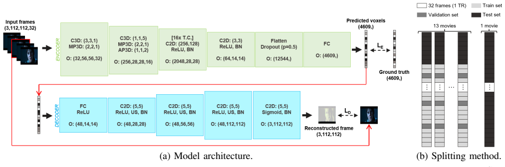
The machinery is the temporal encoder-decoder architecture: the encoder stacks temporal convolutional layers over 32-frame chunks of a film and outputs a 4609-dimensional voxel vector, while the decoder is a CNN that maps that voxel vector to a single target frame, defined as the chunk's middle frame. The encoder loss is $\mathcal{L}_E = \mathrm{MSE}(\mathbf{v}, \hat{\mathbf{v}}) + 0.5 \cdot \cos\text{-distance}(\mathbf{v}, \hat{\mathbf{v}})$, and the decoder loss is $\mathcal{L}_D = 0.35\,\mathcal{L}_{psim} + 0.35\,\mathcal{L}_{ssim} + 0.30\,\mathcal{L}_{tv}$, with a combined loss $\mathcal{L}_{ED} = 0.5\,\mathcal{L}_E + 0.5\,\mathcal{L}_D$. Temporal convolution is what lets the model bridge the mismatch between 32 frames per second of film and one fMRI volume every 1.3 seconds, and the decoder-as-regularizer is what ties reconstruction quality back to encoding quality. The saliency maps are produced by backpropagating SSIM gradients through the decoder and summing absolute saliencies across all test reconstructions.
What would settle it
Feed the trained decoder the actual measured, subject-averaged fMRI volumes from the test set, bypassing the encoder, and compare the SSIM of the resulting reconstructions with the reported 0.320; if scores fall to near the shuffled baseline, the decoder was reading the encoder's latent code rather than neural activity.
Extended reading notes
Core claim
The central claim is that an end-to-end trained encoder-decoder CNN, with temporal convolutional layers in the encoder and a fully convolutional decoder, predicts subject-averaged fMRI activity in visual cortex from chunks of 32 consecutive movie frames and reconstructs the middle frame of each chunk from the encoder's predicted activity, including for a film completely excluded from training. The decoder is fed the encoder's predicted voxel values rather than measured fMRI, and its loss combines perceptual similarity, SSIM, and total variation; the combined encoder-decoder loss uses the decoder as a regularizer that improves encoder predictions over an encoder trained alone. Saliency analysis by backpropagating SSIM gradients locates the most influential voxels in the middle-superior occipital area, fusiform gyrus, and calcarine cortex, which the authors connect to shape, face, and basic-feature processing respectively. The paper presents this as evidence that a deep model can serve as a proxy for studying visual representation in naturalistic movie watching.
Load-bearing premise
The decoder is trained and evaluated on the model's own predicted brain signals, not on measured brain signals, so the reconstruction-from-brain-activity claim assumes those predicted signals faithfully stand in for real measurements.
Editorial extensions
If this is right
- The same model can predict visual-cortex fMRI from movie frames and reconstruct a representative frame from that predicted activity, including on a held-out film never seen during training.
- Temporal convolutions over 32-frame chunks offer a way to handle the frame-rate-to-TR resolution gap without averaging away the movie's fast visual dynamics.
- Adding the decoder as a regularizer improves the encoder's voxel predictions compared with training the encoder alone, so reconstruction quality can be used to sharpen encoding.
- Decoder saliency maps implicate shape-, face-, and edge-selective regions, suggesting the reconstructions are driven by the same broad visual features the human visual system uses.
- A successful encoder-decoder pair of this kind provides a proxy through which deep network behavior can be used to probe visual processing in naturalistic stimuli.
Reading between the lines
- A direct test of the reconstruction claim would be to run the decoder on measured, subject-averaged test volumes rather than encoder-predicted activity, since the reported reconstructions never bypass the encoder.
- Because all fMRI was averaged across 30 subjects, the reconstruction likely reflects shared and stereotyped responses rather than an individual viewer's brain activity; subject-level decoding is a natural next step that would require handling lower signal-to-noise ratios.
- The saliency-region story could be tested directly by lesioning the top-saliency voxels in the predicted fMRI volumes and measuring how much SSIM degrades, which would connect causal perturbation to the reported correlational saliency maps.
- The same temporal-convolution design could transfer to other naturalistic modalities or to datasets with different preprocessing, although the authors note that cross-dataset transfer is not guaranteed.
Signed reviews
Editorial analysis
A structured set of objections, weighed in public.
Referee Report
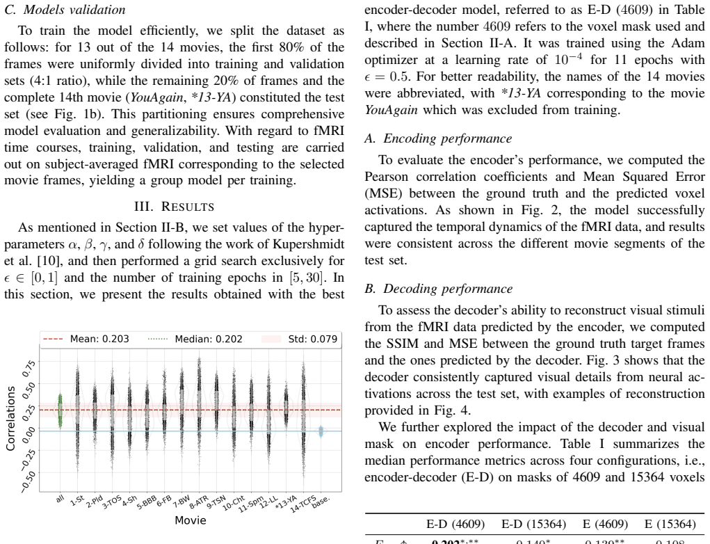
Summary. The paper proposes an end-to-end deep encoder-decoder model for naturalistic movie fMRI. The encoder is a temporal CNN that predicts subject-averaged fMRI voxel activity from chunks of 32 film frames; the decoder is a CNN that reconstructs the middle frame from the encoder's predicted fMRI, with a combined loss that includes perceptual, SSIM, and total-variation terms. Using the Emo-FilM dataset, the authors split 13 movies into training/validation and hold out the 14th movie plus the last 20% of the remaining movies as test. They report an average encoding correlation of 0.203 and an average decoding SSIM of 0.320 on the test set, and they use saliency maps to identify middle occipital, fusiform, and calcarine regions as most contributing to reconstruction. The central claims are that the model predicts visual cortex activity and reconstructs the visual stimulus from neural activity, and that the saliency results reflect the brain regions underlying the reconstructed visual features.
Significance. The paper addresses a timely problem—linking naturalistic movie stimuli to fMRI responses—and its encoder-decoder architecture with temporal convolutions is a reasonable design choice. Strengths include the use of the public Emo-FilM dataset, a held-out movie test protocol, null distributions for the encoding and decoding metrics, and non-parametric statistical comparisons between model variants. The saliency analysis yields falsifiable region-level predictions (middle occipital, fusiform, calcarine) that are consistent with established visual cortex functional organization. However, these strengths do not compensate for the fact that the central reconstruction claim is not tested against measured fMRI, and the evaluation hinges on subject-averaged data with a single split. If the authors add a ground-truth fMRI decoding experiment and cross-validated voxel selection, the work could make a solid contribution to naturalistic fMRI encoding/decoding.
major comments (3)
- [Section III-B, Eq. (2)-(3)] The abstract claims that the model performs 'reconstruction of corresponding visual inputs from neural activity,' but the decoder's input is always the encoder's predicted fMRI, not measured fMRI. In the end-to-end setup, the encoder output is a deterministic function of the movie frames, so the decoder can learn to invert that movie-derived latent representation without reading a brain signal. The reported SSIM of 0.320 (Fig. 3) therefore characterizes the encoder-decoder as an autoencoder of movie frames, not as a neural decoding pipeline. The paper does not report any experiment in which ground-truth fMRI (even subject-averaged) is fed to the decoder at train or test time. This point is load-bearing: either the authors must train/evaluate a decoder on measured fMRI, or the decoding claim must be reframed as 'reconstruction from encoder-predicted fMRI,' removing the neural-decoding implications from the abstract and discussion.
- [Section II-A (voxel selection)] The SNR-based voxel selection is described as a preprocessing step applied to the whole dataset, including the held-out movie and the last 20% of volumes from the remaining movies, before the train/validation/test split (Fig. 1b). Because the top 30% SNR voxels are chosen using test-set signal statistics, the encoding correlations in Fig. 2 and the comparisons in Table I are optimistically biased. The voxel selection must be performed on the training set only, or the entire pipeline (including voxel selection) must be cross-validated, to support the claimed generalizability.
- [Section II-A, Section III-A] All results are on subject-averaged fMRI with a single train/validation/test split. The Mann-Whitney U tests in Table I compare distributions over voxels or frames within that one split; they do not provide evidence about stability across subjects or cross-validation folds. The paper's contribution (3) claims robustness based on the splitting method, but without reporting per-subject encoding performance or repeated cross-validation, the generalization claim is not substantiated. Please provide error bars across subjects (at least for the encoder correlation) or use leave-one-movie-out cross-validation for the central encoding and decoding metrics.
minor comments (6)
- [Section II-A] The sentence 'We exclusively considered the visual and divided the films into N chunks of 32 RGB frames' is incomplete; 'visual' should be 'visual modality' or 'visual frames.'
- [Fig. 3] The caption for Fig. 3 states 'SSIM scores between true and decoder-predicted movie frames' but does not mention that the decoder input is encoder-predicted fMRI, which is essential for interpreting the result.
- [Table I] The entries 'Dssim' and 'Dmse' for the encoder-only models are given as '/', but no footnote explains that no decoder was trained; add a note.
- [Eq. (1)] The notation 'cos(̸ (v, ˆv))' is unusual and potentially a typo; please define whether it is cosine distance or cosine similarity and use standard notation such as '1 - cos_sim' if distance is meant.
- [Fig. 2 caption] The caption reports an 'average correlation score of 0.203' while Table I reports a median Ecorr of 0.202; clarify whether the stated value is the mean or the median and ensure consistency.
- [Reference [10]] Kupershmidt et al. is cited as an arXiv preprint; please check whether a peer-reviewed version is available and cite that version if so.
Circularity Check
Decoder is trained and evaluated on encoder-predicted fMRI, so the reconstruction claim reduces to autoencoding movie frames rather than decoding measured brain activity; voxel selection also leaks test information.
-
self definitional
[Section II-B (end-to-end loss, Eq. 3); Section III-B (Decoder performance)]
"In this setup, the decoder reconstructs movie frames using the fMRI predicted by the encoder, serving as an additional regularization module to enhance encoding performance."
The encoder's output is a deterministic function of the input movie frames, so the decoder is trained and evaluated on a movie-derived latent code, not on measured brain activity. The path frames -> encoder -> decoder -> frames is an autoencoder, and the reported SSIM (0.320), MSE, and saliency maps describe inversion of the encoder's own representation. The paper reports no experiment, at train or test time, in which ground-truth fMRI is fed to the decoder. Therefore the abstract's claim of reconstructing visual inputs 'from neural activity' reduces by construction to reconstructing from encoder predictions.
-
other
[Section II-A (SNR voxel selection) relative to Section II-C (dataset split)]
"we further refined the parameter space of our models by selecting the top 30% voxels with the highest signal-to-noise ratio (SNR). This resulted in 4609 remaining voxels."
The SNR statistic is computed over the entire dataset before the train/validation/test split described in Section II-C. Consequently, the held-out movie and the final 20% of volumes contribute to choosing the voxel mask on which all reported test metrics are computed. This leaks test information into feature selection, making the held-out evaluation partly self-referential, although it is not the main definitional issue.
full rationale
The encoding half of the paper is self-contained: the encoder is trained on movie frames and evaluated against measured fMRI on a fully held-out movie (YouAgain) and the last 20% of the remaining movies, so the Pearson correlation of 0.203 is not fitted to test data. However, the decoding half is circular in the precise sense that the decoder's input is the encoder's prediction, a deterministic transform of the frames to be reconstructed, rather than measured brain activity. Equation (3) and Section III-B make this explicit, so the 'reconstruction from neural activity' claim reduces to autoencoding. In addition, the SNR-based voxel mask is computed on the whole dataset before splitting, leaking test information into feature selection. These two issues make the central decoding claim partially circular, even though the encoder results provide independent content.
Assumptions & free parameters
free parameters (8)
- alpha =
0.5
- beta =
0.35
- gamma =
0.35
- delta =
0.30
- epsilon =
0.5
- number of training epochs =
11
- SNR voxel selection quantile =
30%
- HRF delay =
4 TRs
assumptions (5)
- domain assumption BOLD signal can be aligned to the stimulus by a fixed 4-TR delay without deconvolution of the hemodynamic response.
- domain assumption Averaging fMRI across subjects preserves the shared neural representation of interest and removes subject-specific variability.
- domain assumption The Schaefer 1000 parcellation correctly identifies visual cortex voxels.
- ad hoc to paper Top 30% SNR voxels are the most informative for both encoding and decoding.
- domain assumption VGG16 perceptual similarity approximates human visual similarity for reconstruction.
Cite this review
Pith. "Pith review of Deep Neural Encoder-Decoder Model to Relate fMRI Brain Activity with Naturalistic Stimuli." pith.science (2026). https://pith.science/paper/27JOETCH
@misc{pith2026250712009,
  author       = {Pith},
  title        = {Pith review of: Deep Neural Encoder-Decoder Model to Relate fMRI Brain Activity with Naturalistic Stimuli},
  year         = {2026},
  howpublished = {\url{https://pith.science/paper/27JOETCH}},
  note         = {Machine review of arXiv:2507.12009}
}
read the original abstract
We propose an end-to-end deep neural encoder-decoder model to encode and decode brain activity in response to naturalistic stimuli using functional magnetic resonance imaging (fMRI) data. Leveraging temporally correlated input from consecutive film frames, we employ temporal convolutional layers in our architecture, which effectively allows to bridge the temporal resolution gap between natural movie stimuli and fMRI acquisitions. Our model predicts activity of voxels in and around the visual cortex and performs reconstruction of corresponding visual inputs from neural activity. Finally, we investigate brain regions contributing to visual decoding through saliency maps. We find that the most contributing regions are the middle occipital area, the fusiform area, and the calcarine, respectively employed in shape perception, complex recognition (in particular face perception), and basic visual features such as edges and contrasts. These functions being strongly solicited are in line with the decoder's capability to reconstruct edges, faces, and contrasts. All in all, this suggests the possibility to probe our understanding of visual processing in films using as a proxy the behaviour of deep learning models such as the one proposed in this paper.
Figures

Figures from the paper (1 more)

Reference graph
Works this paper leans on
-
[1]
High-resolution image reconstruction with latent diffusion models from human brain activity,
Y . Takagi and S. Nishimoto, “High-resolution image reconstruction with latent diffusion models from human brain activity,” Cold Spring Harbor Laboratory , 2022
work page 2022
-
[2]
End- to-end deep image reconstruction from human brain activity,
G. Shen, K. Dwivedi, K. Majima, T. Horikawa, and Y . Kamitani, “End- to-end deep image reconstruction from human brain activity,”Frontiers in Computational Neuroscience , vol. 13, 2019
work page 2019
-
[3]
From voxels to pixels and back: Self-supervision in natural-image reconstruction from fmri,
R. Beliy, G. Gaziv, A. Hoogi, F. Strappini, T. Golan, and M. Irani, “From voxels to pixels and back: Self-supervision in natural-image reconstruction from fmri,” in Proceedings of the Conference on Neural Information Processing Systems (NeurIPS) , vol. 32, 2019
work page 2019
-
[4]
G. Gaziv, R. Beliy, N. Granot, A. Hoogi, F. Strappini, T. Golan, and M. Irani, “Self-supervised natural image reconstruction and large-scale semantic classification from brain activity,” NeuroImage, vol. 254, p. 119121, 2022
work page 2022
-
[5]
Very deep convolutional networks for large-scale image recognition,
K. Simonyan and A. Zisserman, “Very deep convolutional networks for large-scale image recognition,” arXiv, 2014
2014
-
[6]
Reconstructing visual experiences from brain activity evoked by natural movies,
S. Nishimoto, A. T. Vu, T. Naselaris, Y . Benjamini, B. Yu, and J. L. Gallant, “Reconstructing visual experiences from brain activity evoked by natural movies,” Current Biology, vol. 21, no. 19, pp. 1641–1646, 2011
work page 2011
-
[7]
K. Han, H. Wen, J. Shi, K.-H. Lu, Y . Zhang, D. Fu, and Z. Liu, “Varia- tional autoencoder: An unsupervised model for encoding and decoding fmri activity in visual cortex,” NeuroImage, vol. 198, pp. 125–136, 2019
work page 2019
-
[8]
Cortical response to naturalistic stimuli is largely predictable with deep neural networks,
M. Khosla, G. H. Ngo, K. Jamison, A. Kuceyeski, and M. R. Sabuncu, “Cortical response to naturalistic stimuli is largely predictable with deep neural networks,” Science Advances , vol. 7, no. 22, 2021
work page 2021
Show all 26 references
-
[9]
Neural encoding and decoding with deep learning for dynamic natural vision,
H. Wen, J. Shi, Y . Zhang, K.-H. Lu, J. Cao, and Z. Liu, “Neural encoding and decoding with deep learning for dynamic natural vision,” Cerebral Cortex, vol. 28, no. 12, pp. 4136–4160, 2017
2017
-
[10]
A penny for your (visual) thoughts: Self-supervised reconstruction of natural movies from brain activity,
G. Kupershmidt, R. Beliy, G. Gaziv, and M. Irani, “A penny for your (visual) thoughts: Self-supervised reconstruction of natural movies from brain activity,” arXiv, vol. 2206.03544, 2022
2022 arXiv
-
[11]
Brain2pix: Fully convolutional naturalistic video frame reconstruction from brain activity,
L. Le, L. Ambrogioni, K. Seeliger, Y . G ¨uc ¸l¨ut¨urk, M. van Gerven, and U. G ¨uc ¸l¨u, “Brain2pix: Fully convolutional naturalistic video frame reconstruction from brain activity,” Frontiers in Neuroscience, vol. 16, 2022
2022
-
[12]
Spurious reconstruction from brain activity,
K. Shirakawa, Y . Nagano, M. Tanaka, S. C. Aoki, K. Majima, Y . Muraki, and Y . Kamitani, “Spurious reconstruction from brain activity,” arXiv, 2024
2024
-
[13]
Using goal-driven deep learning models to understand sensory cortex,
D. L. K. Yamins and J. J. DiCarlo, “Using goal-driven deep learning models to understand sensory cortex,” Nature Neuroscience, vol. 19, no. 3, pp. 356–365, 2016
2016
-
[14]
Emo-film: A multimodal dataset for affective neuroscience using naturalistic stimuli,
E. Morgenroth, S. Moia, L. Vilaclara, R. Fournier, M. Muszynski, M. Ploumitsakou, M. Almato-Bellavista, P. Vuilleumier, and D. V . D. Ville, “Emo-film: A multimodal dataset for affective neuroscience using naturalistic stimuli,” preprint, Neuroscience, February 2024
2024
-
[15]
Local-global parcellation of the human cerebral cortex from intrinsic functional connectivity mri,
A. Schaefer, R. Kong, E. M. Gordon, T. O. Laumann, X.-N. Zuo, A. J. Holmes, S. B. Eickhoff, and B. T. T. Yeo, “Local-global parcellation of the human cerebral cortex from intrinsic functional connectivity mri,” Cerebral Cortex, vol. 28, no. 9, pp. 3095–3114, 2017
2017
-
[16]
Automated anatomical labelling atlas 3,
E. T. Rolls, C.-C. Huang, C.-P. Lin, J. Feng, and M. Joliot, “Automated anatomical labelling atlas 3,” NeuroImage, vol. 206, p. 116189, 2020
2020
-
[17]
Separate visual pathways for perception and action,
M. A. Goodale and A. D. Milner, “Separate visual pathways for perception and action,” Trends in neurosciences, vol. 15, no. 1, pp. 20– 25, 1992
1992
-
[18]
The fusiform face area: a cortical region specialized for the perception of faces,
N. Kanwisher and G. Yovel, “The fusiform face area: a cortical region specialized for the perception of faces,” Philosophical Transactions of the Royal Society B: Biological Sciences , vol. 361, no. 1476, pp. 2109– 2128, 2006
2006
-
[19]
Meadows, Calcarine Cortex , pp
M.-E. Meadows, Calcarine Cortex , pp. 472–472. New York, NY: Springer New York, 2011
2011
-
[20]
Examining the impact of fmri preprocessing steps on machine learning-based classification of autism spectrum disorder,
R. C. Sotero, J. M. Sanchez-Bornot, I. Shaharabi-Farahani, and Y . Iturria-Medina, “Examining the impact of fmri preprocessing steps on machine learning-based classification of autism spectrum disorder,” in Proceedings of the 2023 7th International Conference on Medical and He...
2023
-
[21]
Revealing the multidimensional mental representations of natural objects underlying human similarity judgements,
M. N. Hebart, C. Y . Zheng, F. Pereira, and C. I. Baker, “Revealing the multidimensional mental representations of natural objects underlying human similarity judgements,” Nature human behaviour, vol. 4, no. 11, pp. 1173–1185, 2020
2020
-
[22]
Concept-based explainability for an eeg transformer model,
A. Gjølbye, T. Lehn-Schiøler, ´A. J ´onsd´ottir, B. Arnard ´ottir, and L. K. Hansen, “Concept-based explainability for an eeg transformer model,” arXiv, 2023
2023
-
[23]
Concept-based ai interpretability in physiological time-series data: Example of abnormality detection in electroencephalography,
A. Brenner, F. Knispel, F. P. Fischer, P. Rossmanith, Y . Weber, H. Koch, and E. Kutafina, “Concept-based ai interpretability in physiological time-series data: Example of abnormality detection in electroencephalography,” Computer Methods and Programs in Biomedicine, vol. 257,...
2024
-
[24]
An improved saliency model of visual attention dependent on image content,
S. Novin, A. Fallah, S. Rashidi, and M. R. Daliri, “An improved saliency model of visual attention dependent on image content,” Frontiers in Human Neuroscience , vol. 16, p. 862588, 2023
2023
-
[25]
Sanity checks for saliency maps,
J. Adebayo, J. Gilmer, M. Muelly, I. Goodfellow, M. Hardt, and B. Kim, “Sanity checks for saliency maps,” in Advances in Neural In- formation Processing Systems (S. Bengio, H. Wallach, H. Larochelle, K. Grauman, N. Cesa-Bianchi, and R. Garnett, eds.), vol. 31, Curran Associate...
2018
-
[26]
Interpretability beyond feature attribution: Quantitative testing with concept activation vectors (tcav),
B. Kim, M. Wattenberg, J. Gilmer, C. Cai, J. Wexler, F. Viegas, and R. Sayres, “Interpretability beyond feature attribution: Quantitative testing with concept activation vectors (tcav),” arXiv, 2017
2017
Reviewed August 6, 2026 · model on record in the stance chip above.
Discussion (0). Continue with ORCID to comment.