REVIEW 4 major objections 4 minor 60 references
Microwave focusing with temporal interference for non-invasive deep brain stimulation
T0 review · 4 major / 4 minor · reviewed 2026-08-03 · deepseek-v4-flash
Pith's one-line read Microwave temporal interference can focus low-frequency stimulation envelopes at deep brain targets in a realistic head model.
desk verdict A useful proof-of-principle for microwave TI focusing, but the headline claim runs ahead of what the simulations actually show—especially since the optimized 'envelope' is a norm-based upper bound that ignores field polarization. read the letter →
The pith
A machine-rendered reading of the paper's core claim, the machinery that carries it, and where it could break.
The reading
What carries the argument
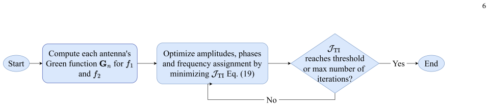
The load-bearing object is the temporal-interference envelope amplitude E_AM(r) = 2 min(||E_f1(r)||, ||E_f2(r)||), which the paper treats as the quantity neurons respond to. The two-stage pipeline is the mechanism: iterative time-reversal (a virtual source at the target, back-propagated fields to rank candidate antenna positions, with Bayesian updates of source parameters and an attenuation-compensation exponent) supplies positions, orientations, and initial excitations; then a genetic algorithm over the Green's-function representation E_fi = sum(G_ni * m_ni * exp(j*alpha_ni)) refines amplitudes, phases, and binary frequency assignment to minimize J_TI = (background mean E_AM)/(focal mean E_
What would settle it
An in-vitro or in-vivo neuron preparation exposed to two 700 MHz fields with a 100 Hz offset, at the amplitudes the optimized array would produce at a deep target: if action potentials do not phase-lock to the 100 Hz envelope, or if the required amplitudes exceed head SAR limits, the central claim fails.
Extended reading notes
Core claim
The paper's core discovery is that adding iterative time reversal (iTR) as an initializer makes temporal-interference (TI) optimization practical and precise in a heterogeneous, lossy head. After iTR fixes the array geometry and initial steering parameters, a genetic algorithm minimizes the ratio of background-to-focal envelope amplitude E_AM = 2 min(|E_f1|,|E_f2|) by assigning each element to one of two carriers, 700 MHz and 700.0001 MHz, and adjusting amplitude and phase. The resulting normalized maps show a confined envelope at the intended location, with noticeable buildup only in superficial non-neuronal tissues, and the side-focus case shows a deliberate asymmetry in frequency assignme
Load-bearing premise
The whole method rests on the temporal-interference hypothesis at microwave carriers: neurons will demodulate two 700 MHz fields separated by 100 Hz and respond to the envelope E_AM = 2 min(|E_f1|, |E_f2|), at field strengths this array can deliver without exceeding safety limits.
Editorial extensions
If this is right
- If the central claim is correct, non-invasive DBS becomes a numerical optimization problem over an external array: positions, orientations, frequency split, amplitudes, and phases can all be solved jointly.
- The frequency-split asymmetry for off-center targets—putting more f1 elements near the target hemisphere and f2 elements opposite—provides a general steering principle for TI arrays.
- Because results are normalized, the immediate next step is to determine the absolute envelope amplitudes required for neuron activation and compare them against SAR and thermal limits; the paper identifies this as an open issue.
- The idealized magnetic-point-dipole sources imply that the reported focusing is a lower bound; physical directive antennas should perform at least as well, motivating dedicated antenna design.
- The framework extends naturally to multi-objective optimization, such as balancing stimulation envelope against energy absorption along a Pareto front, as the paper suggests.
Reading between the lines
- A testable extension suggested by the side-focus result: because E_AM uses min(|E_f1|,|E_f2|), asymmetric tissue losses must be compensated by asymmetric element counts, so one could verify that the optimal solution indeed balances the two carrier amplitudes near the focal point.
- The paper does not model the neuron itself; feeding the simulated field maps into single-compartment or cable neuron models at 700 MHz carriers would test whether a 100 Hz envelope actually drives spiking at usable amplitudes.
- The safety argument treats off-target envelope amplitude as benign because skin and skull lack neurons; an inference beyond the paper is that patient-specific variation in skull thickness and tissue layering will alter the envelope maps, so clinical translation would likely require per-patient optimization.
- The 100 Hz difference frequency sits in the classic DBS range, and the same framework could in principle deliver multiple simultaneous targets by adding more carriers or different difference frequencies, though the paper does not explore this.
Signed reviews
Editorial analysis
A structured set of objections, weighed in public.
Referee Report
Summary. The manuscript proposes a computational proof-of-principle for non-invasive microwave deep brain stimulation using an external array of magnetic point dipoles. A two-stage pipeline is presented: iterative time reversal (iTR) selects antenna positions and orientations, and a genetic algorithm optimizes amplitudes, phases, and frequency assignment to minimize the background-to-focal ratio of the temporal-interference envelope E_AM defined in Eq. (17). The method is evaluated in a 77-tissue voxel model of the Duke head for a center focus and a side focus at 700 MHz and Δf = 100 Hz. The central claim is that amplitude-modulated microwave fields can be effectively focused at deep brain targets, with normalized field plots as the main evidence.
Significance. If the central claim holds, the work would be a useful computational framework combining iTR and TI in a realistic head model, with potential value as a benchmark for non-invasive DBS. Strengths include the use of the IT'IS Duke model, a large number of tissue types, full-wave COMSOL simulations, and a clearly described optimization pipeline. The method description is sufficiently detailed to allow reproduction in principle. However, the quantitative support is currently incomplete, and the TI envelope metric in Eq. (17) is polarization-blind, which materially weakens the link between the reported field plots and the actual neuronal stimulation that the paper claims to demonstrate. These issues significantly reduce the current support for the central conclusion.
major comments (4)
- [§II-B, Eq. (17)] The TI envelope is defined as E_AM = 2 min(||E_f1||, ||E_f2||), which depends only on field magnitudes. For a neuron with axis u, the slow envelope of the projection u·(E_f1 e^{jω1t} + E_f2 e^{jω2t}) is 2 min(|u·E_f1|, |u·E_f2|), not the norm-based quantity. Since ||E_fi|| ≥ |u·E_fi|, Eq. (17) is an upper bound over neuron orientations; for orthogonal equal-magnitude fields at the focus it reports 2A while the best achievable projection envelope is √2 A. The objective J_TI in Eq. (19) and all E_AM maps (Figs. 13–14) therefore optimize and report an upper bound, not the low-frequency envelope that would actually drive a neuron. This is an internal consistency problem, not merely a biological extrapolation. The authors acknowledge in Sec. V-A2 that orientation effects are left to future work, but that acknowledgment does not rescue the current central claim. The objective should be revised
- [Abstract and §IV, §VI] The abstract states that 'Systematic numerical studies, including perturbation analysis and statistical evaluation, demonstrate consistent spatial localization and robustness across all reported configurations' and that 'safety is quantified using specific absorption rate (SAR), ensuring compliance with exposure limits.' The full text contains no perturbation analysis, no statistical evaluation, no numeric SAR values, and no comparison with IEEE or ICNIRP limits. The results section reports only normalized field plots. These are load-bearing claims in the abstract and must either be added to the manuscript or removed from the abstract. As written, the abstract overstates the content of the paper.
- [§IV, Eqs. (11), (19)] The focal maxima shown in Figs. 10–14 are the fitted optima of objective functions that explicitly reward concentration of the optimized quantity in the focal region: J_iTR combines HIR and FIQ in Eq. (11), and J_TI minimizes the background-to-focal envelope ratio in Eq. (19). Normalized plots alone therefore provide no independent evidence of focusing performance. The authors should report the numerical values of HIR, FIQ, and J_TI for both targets, compare them with a non-optimized or single-frequency baseline, and state the achieved background/focal suppression. Without these quantitative results, the claim that the method 'effectively focuses' is largely a restatement of the optimization objective rather than a measured outcome.
- [§VI] The medical safety section is entirely qualitative. It asserts that SAR and thermal effects can be kept within limits and that therapeutic activation occurs below heating thresholds, but no actual SAR or temperature calculation is presented anywhere in the paper. Since the abstract explicitly promises safety quantification through SAR, this is a missing load-bearing analysis. The authors should compute and report SAR distributions for the optimized arrays and assess them against the ICNIRP/IEEE limits for head tissues, including off-target regions.
minor comments (4)
- [Throughout] Typos and formatting issues: 'V oxelized' appears in several places; 'V .' is used before reference numbers in the bibliography; the caption of Fig. 9 says 'dipole direction' but the displayed quantity is not described in the text.
- [§II-B] The statement that 'GA outperforms PSO in both runtime and final objective values' is given without any supporting comparison data. Either report the comparison or remove the claim.
- [§II-A and §II-B] The values of the iTR weights c1 and c2, the attenuation-compensation exponent b, and the GA settings (population size, number of generations, termination criteria) are not specified. These parameters are needed for reproducibility and to understand the sensitivity of the results.
- [§IV] The units of the field magnitudes in Figs. 10–14 are labelled '(V/m)' but the fields are normalized and the absolute scaling is not defined in the text. This is confusing; either provide absolute values or remove the unit labelling.
Circularity Check
No significant circularity: the optimized E_AM maps are the explicit objective of the GA, and the paper does not present them as independent predictions; the load-bearing biological TI premise is an external assumption.
full rationale
The paper's derivation chain is an optimization study, not a prediction-from-first-principles chain. The TI envelope E_AM in Eq. (17) is adopted from [44] as a definition, and the objective JTI in Eq. (19) directly minimizes the background-to-focal ratio of that same quantity. The reported focal E_AM maps are therefore the output of the optimizer, not an independently predicted outcome. This is transparent in the text: the algorithm 'optimize[s] amplitudes, phases, and frequency assignment ... by minimizing the objective function JTI'. Since the paper does not frame the optimized field as an out-of-sample prediction, this does not meet the 'fitted input called prediction' or 'self-definitional' circularity patterns. The biological premise that neurons respond to the low-frequency envelope of microwave carriers is explicitly labeled a hypothesis ('Under this TI hypothesis, neuronal activation is expected...') and is supported by external citations [29], [45]-[47]; it is an assumption about neurophysiology, not a result derived from this paper's equations. The polarization-blindness of Eq. (17) is a real validity limitation, and the paper acknowledges it in Sec. V-A2 ('A natural extension for future studies is to incorporate the orientations of nerve tracts ... the electric fields could be projected along the known nerve directions'); this affects whether the norm-based E_AM is the physiologically relevant stimulus, but it is not a circular derivation. There are no load-bearing self-citations: the cited iTR and hyperthermia works [22], [24], [54] involve H. Dobsicek Trefna, who is acknowledged for discussions but is not an author of this paper. For completeness, the abstract promises 'perturbation analysis and statistical evaluation' that do not appear in the full text, and Sec. V-B asserts that TI alone can focus without showing supporting results; these are reporting/validation gaps, not circularity. Overall, the central numerical demonstration is equivalent to reporting that the GA successfully minimized its stated objective in a realistic head model, which is an in-sample optimization result rather than a circularly derived prediction.
Assumptions & free parameters
free parameters (8)
- b (attenuation compensation exponent)
- c1, c2 (iTR objective weights)
- sigma (Gaussian sampling width) =
0.8/3 * d_min ≈ 1.6 cm (given d_min=6 cm)
- d_min (minimum inter-element spacing) =
6 cm
- N_max (maximum number of antenna elements) =
35
- Operating frequencies f1, delta_f =
700 MHz, 100 Hz
- Matching layer permittivity epsilon_ml =
40
- Focal region radius =
1 cm
assumptions (7)
- domain assumption Temporal interference hypothesis: neurons respond to the low-frequency envelope of two high-frequency fields, and the relevant stimulus measure is E_AM = 2 min(|E_f1|, |E_f2|) (Eq. 17).
- standard math Huygens' principle and time-reversal theory: fields inside a volume can be reconstructed from sources on an enclosing surface.
- domain assumption The straight-line path attenuation integral (Eqs. 7-8) is an adequate compensation model in heterogeneous tissue.
- domain assumption The IT'IS Duke voxel model with 77 isotropic tissues and Cole-Cole parameters is a faithful representation of the human head at 400 MHz-1 GHz.
- domain assumption The lossless matching layer (epsilon=40) and the scattering boundary condition approximate the clinical environment.
- domain assumption Magnetic point dipoles are sufficient as antenna models for optimization benchmarking.
- domain assumption The genetic algorithm converges to a near-global optimum of J_TI with the (unreported) solver settings.
Cite this review
Pith. "Pith review of Microwave focusing with temporal interference for non-invasive deep brain stimulation." pith.science (2026). https://pith.science/paper/4WHAV5T5
@misc{pith2026260218477,
  author       = {Pith},
  title        = {Pith review of: Microwave focusing with temporal interference for non-invasive deep brain stimulation},
  year         = {2026},
  howpublished = {\url{https://pith.science/paper/4WHAV5T5}},
  note         = {Machine review of arXiv:2602.18477}
}
read the original abstract
Deep Brain Stimulation (DBS) is an effective treatment for neurological disorders but requires invasive surgery. This work presents a method for non-invasive DBS, based on microwave focusing of amplitude-modulated electric fields using an external antenna array of magnetic point dipoles. The proposed method combines iterative time reversal (iTR) and temporal interference (TI) optimization to jointly address electromagnetic field focusing and physiologically relevant neural stimulation. Antenna element positions, orientations, frequencies, amplitudes, and phases are optimized to localize stimulation within a target region. The method is evaluated in an anatomically realistic voxel head model with heterogeneous and lossy tissue properties. Systematic numerical studies, including perturbation analysis and statistical evaluation, demonstrate consistent spatial localization and robustness across all reported configurations. Safety is quantified using specific absorption rate (SAR), ensuring compliance with exposure limits. The study further provides insight into the influence of key parameters on field behavior and the associated trade-offs between focality, penetration, and safety in physiologically relevant stimulation. To the authors knowledge, this is the first study to combine iTR and TI optimization for microwave-based DBS in a realistic voxel head model, establishing a promising framework for safe non-invasive deep brain stimulation.
Figures


Figures from the paper (10 more)



Reference graph
Works this paper leans on
-
[1]
P. A. Iaizzo,Introduction to Neurophysiology. Cham: Springer Inter- national Publishing, 2020, pp. 1–64
2020
-
[2]
Deep brain stimulation: current challenges and future directions,
A. M. Lozano, N. Lipsman, H. Bergman, P. Brown, S. Chabardes, J. W. Chang, K. Matthews, C. C. McIntyre, T. E. Schlaepfer, M. Schulder, Y . Temel, J. V olkmann, and J. K. Krauss, “Deep brain stimulation: current challenges and future directions,”Nat. Rev. Neurol., vol. 15, no. 3, pp. 148–160, 2019
2019
-
[3]
Deep brain stimulation for epilepsy,
V . Salanova, “Deep brain stimulation for epilepsy,”Epilepsy Behav., vol. 88, p. 21–24, Nov. 2018
2018
-
[4]
Deep brain stimulation in treatment-resistant depression: A systematic review and meta-analysis on efficacy and safety,
Y . Wu, J. Mo, L. Sui, J. Zhang, W. Hu, C. Zhang, Y . Wang, C. Liu, B. Zhao, X. Wang, K. Zhang, and X. Xie, “Deep brain stimulation in treatment-resistant depression: A systematic review and meta-analysis on efficacy and safety,”Front. Neurosci., vol. 15, Apr. 2021
2021
-
[5]
Technology of deep brain stimulation: current status and future directions,
J. K. Krauss, N. Lipsman, T. Aziz, A. Boutet, P. Brown, J. W. Chang, B. Davidson, W. M. Grill, M. I. Hariz, A. Horn, M. Schulder, A. Mammis, P. A. Tass, J. V olkmann, and A. M. Lozano, “Technology of deep brain stimulation: current status and future directions,”Nat. Rev. Neurol., vol. 17, p. 75–87, 2020
2020
-
[6]
Krames, P
E. Krames, P. H. Peckham, and A. R. Rezai,Neuromodulation, 1st ed. London: Academic Press, 2009
2009
-
[7]
Neural synchrony in brain disorders: Relevance for cognitive dysfunctions and pathophysiology,
P. J. Uhlhaas and W. Singer, “Neural synchrony in brain disorders: Relevance for cognitive dysfunctions and pathophysiology,”Neuron, vol. 52, no. 1, pp. 155–168, 2006
2006
-
[8]
Basal ganglia,
R. Erzurumlu, G. Sengul, and E. Ulupinar, “Basal ganglia,” inHuman Neuroanatomy, R. Erzurumlu, G. Sengul, and E. Ulupinar, Eds. Aca- demic Press, 2024, pp. 79–93
2024
Show all 60 references
-
[9]
Gunduz,Deep Brain Stimulation: Emerging Technologies and Appli- cations
A. Gunduz,Deep Brain Stimulation: Emerging Technologies and Appli- cations. Cham: Springer International Publishing, 2020, pp. 223–243
2020
-
[10]
Frequency-dependent effects of subthalamic deep brain stimulation on motor symptoms in parkinson’s disease: a meta- analysis of controlled trials,
D. Su, H. Chen, W. Hu, Y . Liu, Z. Wang, X. Wang, G. Liu, H. Ma, J. Zhou, and T. Feng, “Frequency-dependent effects of subthalamic deep brain stimulation on motor symptoms in parkinson’s disease: a meta- analysis of controlled trials,”Sci. Rep., vol. 8, no. 1, p. 14456, 2018
2018
-
[11]
Cerebral vascular dysfunctions detected in human small vessel disease and implications for preclinical studies,
J. M. Wardlaw, H. Benveniste, and A. Williams, “Cerebral vascular dysfunctions detected in human small vessel disease and implications for preclinical studies,”Annu. Rev. Physiol., vol. 84, no. 1, p. 409–434, Feb. 2022
2022
-
[12]
Non-invasive brain stimulation: from field modeling to neuronal activation,
D. Klooster, E. Raaijmakers, M. van Rossum, M. van Beurden, P. Boon, and R. Mestrom, “Non-invasive brain stimulation: from field modeling to neuronal activation,” in2020 50th Eur. Microwave Conf. (EuMC), 2021, pp. 1059–1062
2021
-
[13]
L. M. Koponen and A. V . Peterchev,Transcranial Magnetic Stimulation: Principles and Applications. Cham: Springer International Publishing, 2020, pp. 245–270
2020
-
[14]
D. Q. Truong, N. Khadka, and M. Bikson,Transcranial Electrical Stimulation. Cham: Springer International Publishing, 2020, pp. 271– 292
2020
-
[15]
A method for non-destructive microwave focusing for deep brain and tissue stimulation,
V . Harid, H. Kim, B.-Z. Li, and T. Lei, “A method for non-destructive microwave focusing for deep brain and tissue stimulation,”PLoS ONE, vol. 18, no. 2, p. e0278765, 2023
2023
-
[16]
On the opportunities and challenges in microwave medical sensing and imaging,
R. Chandra, H. Zhou, I. Balasingham, and R. M. Narayanan, “On the opportunities and challenges in microwave medical sensing and imaging,”IEEE Trans. Biomed. Eng., vol. 62, no. 7, pp. 1667–1682, 2015
2015
-
[17]
Microwave tomography for brain imaging: Feasibility assessment for stroke detection,
S. Semenov and D. Corfield, “Microwave tomography for brain imaging: Feasibility assessment for stroke detection,”Int. J. Antennas Propag., vol. 2008, p. 254830, 2008
2008
-
[18]
Stroke detection and diagnosis with a microwave helmet,
A. Fhager and M. Persson, “Stroke detection and diagnosis with a microwave helmet,” in2012 6th Eur. Conf. Antennas Propag. (EuCAP), 2012, pp. 1796–1798
2012
-
[19]
Microwave systems for head imaging: Challenges and recent developments,
A. Abbosh, “Microwave systems for head imaging: Challenges and recent developments,” in2013 IEEE MTT-S Int. Microwave Workshop Ser. RF Wireless Technol. Biomed. Healthcare Appl. (IMWS-BIO), 2013, pp. 1–3. 12
2013
-
[20]
Design and experimental evaluation of a non-invasive microwave head imaging system for intracranial haemorrhage detection,
A. T. Mobashsher, K. S. Bialkowski, A. M. Abbosh, and S. Crozier, “Design and experimental evaluation of a non-invasive microwave head imaging system for intracranial haemorrhage detection,”PLoS One, vol. 11, no. 4, p. e0152351, 04 2016
2016
-
[21]
Sensor-based microwave brain imaging system (smbis): An experimental six-layered tissue based human head phantom model for brain tumor diagnosis using electromagnetic signals,
A. Hossain, M. Tariqul Islam, A. Hoque, S. Kamal Abdul Rahim, A. S. Alshammari, M. E. Chowdhury, and M. S. Soliman, “Sensor-based microwave brain imaging system (smbis): An experimental six-layered tissue based human head phantom model for brain tumor diagnosis using electroma...
2023
-
[22]
Iterative time-reversal for multi- frequency hyperthermia,
M. Zanoli and H. Dobsicek Trefna, “Iterative time-reversal for multi- frequency hyperthermia,”Phys. Med. Biol., vol. 66, no. 4, p. 045015, 2021
2021
-
[23]
Feasibility of heating brain tumors using a 915 MHz annular phased-array,
D. B. Rodrigues, J. Ellsworth, and P. Turner, “Feasibility of heating brain tumors using a 915 MHz annular phased-array,”IEEE Antennas Wireless Propag. Lett., vol. 20, no. 4, p. 423–427, Apr. 2021
2021
-
[24]
Antenna arrangement in UWB helmet brain applicators for deep microwave hyperthermia,
M. Zanoli, E. Ek, and H. Dobsicek Trefna, “Antenna arrangement in UWB helmet brain applicators for deep microwave hyperthermia,” Cancers, vol. 15, no. 5, p. 1447, 2023
2023
-
[25]
Joint optimization of antenna system matching and specific absorption rate focusing in microwave hyperthermia cancer treatment,
M. Firuzalizadeh, R. Gaffoglio, G. Giordanengo, M. Righero, M. Zuc- chi, G. Musacchio Adorisio, A. Bellone, A. Vallan, G. Perrone, and G. Vecchi, “Joint optimization of antenna system matching and specific absorption rate focusing in microwave hyperthermia cancer treatment,” C...
2025
-
[26]
Advancements in non-invasive microwave brain stimulation: A comprehensive survey,
F. E. S. Pereira, S. K. Jagatheesaperumal, S. R. Benjamin, P. C. do Nasci- mento Filho, F. T. Duarte, and V . H. C. de Albuquerque, “Advancements in non-invasive microwave brain stimulation: A comprehensive survey,” Phys. Life Rev., vol. 48, pp. 132–161, 2024
2024
-
[27]
Resonance, oscillation and the intrinsic frequency preferences of neurons,
B. Hutcheon and Y . Yarom, “Resonance, oscillation and the intrinsic frequency preferences of neurons,”Trends Neurosci., vol. 23, no. 5, pp. 216–222, 2000
2000
-
[28]
The origin of extracellular fields and currents — EEG, ECoG, LFP and spikes,
G. Buzs ´aki, C. A. Anastassiou, and C. Koch, “The origin of extracellular fields and currents — EEG, ECoG, LFP and spikes,”Nat. Rev. Neurosci., vol. 13, no. 6, p. 407–420, May 2012
2012
-
[29]
Noninvasive deep brain stimulation via temporally interfering electric fields,
N. Grossman, D. Bono, N. Dedic, S. B. Kodandaramaiah, A. Rudenko, H.-J. Suk, A. M. Cassara, E. Neufeld, N. Kuster, L.-H. Tsai, A. Pascual- Leone, and E. S. Boyden, “Noninvasive deep brain stimulation via temporally interfering electric fields,”Cell, vol. 169, no. 6, pp. 1029– ...
2017
-
[30]
A system of hemispherical antenna arrays for noninvasive deep brain stimulation,
M. A. Safar and R. W. Newcomb, “A system of hemispherical antenna arrays for noninvasive deep brain stimulation,”Math. Probl. Eng., vol. 2023, p. 8019934, 2023
2023
-
[31]
Wireless beam- forming for electromagnetic field focusing in brain tissue,
W. Lee, P. Faeghi, A. D. Dorval, and J. S. Walling, “Wireless beam- forming for electromagnetic field focusing in brain tissue,” in2024 54th Eur. Microwave Conf. (EuMC), 2024, pp. 489–492
2024
-
[32]
Microwave beamforming for non-invasive brain stimulation,
A. Madannejad, S. Sadeghi, J. EbrahimiZadeh, F. Ravanbakhsh, M. D. Perez, and R. Augustine, “Microwave beamforming for non-invasive brain stimulation,” in2020 IEEE MTT-S Int. Microwave Biomed. Conf. (IMBioC). IEEE, Dec. 2020, p. 1–4
2020
-
[33]
Electro- magnetic phased arrays for regional hyperthermia: optimal frequency and antenna arrangement,
M. Seebass, R. Beck, J. Gellermann, J. Nadobny, and P. Wust, “Electro- magnetic phased arrays for regional hyperthermia: optimal frequency and antenna arrangement,”Int. J. Hyperthermia, vol. 17, no. 4, p. 321–336, Jul. 2001
2001
-
[34]
Efficient focusing of microwave hyperthermia for small deep-seated breast tumors treatment using particle swarm optimization,
H. Elkayal and N. Ismail, “Efficient focusing of microwave hyperthermia for small deep-seated breast tumors treatment using particle swarm optimization,”Comput. Methods Biomech. Biomed. Eng., vol. 24, no. 9, pp. 985–994, July 2021, epub 2021 Jun 16
2021
-
[35]
Implementation of thinned array synthesis in hyperthermia treatment planning of 434 MHz phased array breast applicator using genetic algorithm,
D. Baskaran and K. Arunachalam, “Implementation of thinned array synthesis in hyperthermia treatment planning of 434 MHz phased array breast applicator using genetic algorithm,”IEEE J. Electromagn., RF Microw. Med. Biol., vol. 7, no. 1, pp. 32–38, 2023
2023
-
[36]
Metasurface-based time-reversal focusing for brain tumor microwave hyperthermia,
M. J. Hajiahmadi, R. Faraji-Dana, and C. Caloz, “Metasurface-based time-reversal focusing for brain tumor microwave hyperthermia,”IEEE Trans. Antennas Propag., vol. 70, no. 12, p. 12237–12246, Dec. 2022
2022
-
[37]
Time reversal based microwave hyperthermia treatment of breast cancer,
B. Guo, L. Xu, and J. Li, “Time reversal based microwave hyperthermia treatment of breast cancer,”Microw. Opt. Technol. Lett., vol. 47, no. 4, p. 335–338, 2005
2005
-
[38]
Time reversal of electromagnetic waves,
J. de Rosny, G. Lerosey, A. Tourin, and M. Fink, “Time reversal of electromagnetic waves,” inModeling and Computations in Electromag- netics, H. Ammari, Ed. Berlin, Heidelberg: Springer Berlin Heidelberg, 2008, pp. 187–202
2008
-
[39]
Development of a new genera- tion of high-resolution anatomical models for medical device evaluation: the virtual population 3.0,
M.-C. Gosselin, E. Neufeld, H. Moser, E. Huber, S. Farcito, L. Ger- ber, M. Jedensj ¨o, I. Hilber, F. D. Gennaro, B. Lloyd, E. Cherubini, D. Szczerba, W. Kainz, and N. Kuster, “Development of a new genera- tion of high-resolution anatomical models for medical device evaluation...
2014
-
[40]
Comsol multiphysics,
COMSOL AB, “Comsol multiphysics,” Stockholm, Sweden, 2025. [Online]. Available: https://www.comsol.com
2025
-
[41]
[Online]
The MathWorks, Inc., “Matlab,” Natick, MA, USA, 2024. [Online]. Available: https://www.mathworks.com
2024
-
[42]
Theory of electromagnetic time- reversal mirrors,
J. de Rosny, G. Lerosey, and M. Fink, “Theory of electromagnetic time- reversal mirrors,”IEEE Trans. Antennas Propag., vol. 58, no. 10, pp. 3139–3149, 2010
2010
-
[43]
bayesopt (Bayesian Optimization),
The MathWorks, Inc., “bayesopt (Bayesian Optimization),” Natick, MA, USA, 2025, accessed: Jun. 6, 2025. [Online]. Available: https://www.mathworks.com/help/stats/bayesopt.html
2025
-
[44]
Emvelop stimulation: minimally invasive deep brain stimulation using temporally interfering electromagnetic waves,
F. Ahsan, T. Chi, R. Cho, S. A. Sheth, W. Goodman, and B. Aazhang, “Emvelop stimulation: minimally invasive deep brain stimulation using temporally interfering electromagnetic waves,”J. Neural Eng., vol. 19, no. 4, p. 046005, Jul. 2022
2022
-
[45]
Effects of modulated VHF fields on the central nervous system,
S. M. Bawin, L. K. Kaczmarek, and W. R. Adey, “Effects of modulated VHF fields on the central nervous system,”Ann. N. Y. Acad. Sci., vol. 247, no. 1, p. 74–81, Feb. 1975
1975
-
[46]
Responses of neurons to an amplitude modulated microwave stimulus,
R. C. Beason and P. Semm, “Responses of neurons to an amplitude modulated microwave stimulus,”Neurosci. Lett., vol. 333, no. 3, p. 175–178, Nov. 2002
2002
-
[47]
Biophysics of amplitude-modulated giga-hertz electromagnetic waves stimulation,
F. Ahsan, A. C. Govindaraju, R. M. Raphael, T. Chi, S. A. Sheth, W. Goodman, and B. Aazhang, “Biophysics of amplitude-modulated giga-hertz electromagnetic waves stimulation,” in2023 57th Asilomar Conf. Signals, Syst., Comput.IEEE, Oct. 2023, p. 1463–1468
2023
-
[48]
Genetic algorithm and direct search toolbox: GA Function,
The MathWorks, Inc., “Genetic algorithm and direct search toolbox: GA Function,” Natick, MA, USA, 2025, accessed: Jul. 22, 2025. [Online]. Available: https://se.mathworks.com/help/gads/ga.html
2025
-
[49]
Microwave hyperthermia of brain tumors: A 2d assessment parametric numerical study,
J. Redr, T. Pokorny, T. Drizdal, O. Fiser, M. Brunat, J. Vrba, and D. Vrba, “Microwave hyperthermia of brain tumors: A 2d assessment parametric numerical study,”Sensors, vol. 22, no. 16, p. 6115, 2022
2022
-
[50]
Differential evolution optimization of microwave focused hyperthermia phased array excitation for targeted breast cancer heating,
C. Lyu, W. Li, and B. Yang, “Differential evolution optimization of microwave focused hyperthermia phased array excitation for targeted breast cancer heating,”Sensors, vol. 23, no. 8, 2023
2023
-
[51]
Foundation for research on information technologies in society,
IT’IS Foundation, “Foundation for research on information technologies in society,” 1999, accessed: Aug. 2025. [Online]. Available: https: //itis.swiss/
1999
-
[52]
Tissue Properties Database,
——, “Tissue Properties Database,” 2018, version V4.0
2018
-
[53]
W. R. Hendee and E. R. Ritenour,Medical Imaging Physics. Wiley- Liss, 2002
2002
-
[54]
Radiobiological evaluation of com- bined gamma knife radiosurgery and hyperthermia for pediatric neuro- oncology,
M. Ghaderi Aram, M. Zanoli, H. Nordstr ¨om, I. Toma-Dasu, K. Blom- gren, and H. Dob ˇs´ıˇcek Trefn ´a, “Radiobiological evaluation of com- bined gamma knife radiosurgery and hyperthermia for pediatric neuro- oncology,”Cancers, vol. 13, no. 13, p. 3277, Jun. 2021
2021
-
[55]
Orientation selective DBS of entorhinal cortex and medial septal nucleus modulates activity of rat brain areas involved in memory and cognition,
L. Wu, A. Canna, O. Narvaez, J. Ma, S. Sang, L. J. Lehto, A. Sierra, H. Tanila, Y . Zhang, O. Gr ¨ohn, W. C. Low, P. Filip, S. Mangia, and S. Michaeli, “Orientation selective DBS of entorhinal cortex and medial septal nucleus modulates activity of rat brain areas involved in m...
2022
-
[56]
Multi-objective optimization using genetic algorithms: A tutorial,
A. Konak, D. W. Coit, and A. E. Smith, “Multi-objective optimization using genetic algorithms: A tutorial,”Reliab. Eng. Syst. Saf., vol. 91, no. 9, pp. 992–1007, 2006, special Issue - Genetic Algorithms and Reliability
2006
-
[57]
gamultiobj – find pareto front of multiple fitness func- tions using genetic algorithm,
MathWorks, “gamultiobj – find pareto front of multiple fitness func- tions using genetic algorithm,” https://se.mathworks.com/help/gads/ gamultiobj.html, 2025, accessed: 2025-08-14. [58]IEEE Standard for Safety Levels with Respect to Human Exposure to Electric, Magnetic, and E...
2025
-
[59]
Guidelines for limiting exposure to electromagnetic fields (100 kHz to 300 GHz),
International Commission on Non-Ionizing Radiation Protection, “Guidelines for limiting exposure to electromagnetic fields (100 kHz to 300 GHz),”Health Phys., vol. 118, no. 5, p. 483–524, May 2020
2020
-
[60]
A guide for water bolus temperature selection for semi- deep head and neck hyperthermia treatments using the hypercollar3d applicator,
T. Drizdal, G. C. van Rhoon, R. F. Verhaart, O. Fiser, and M. M. Paulides, “A guide for water bolus temperature selection for semi- deep head and neck hyperthermia treatments using the hypercollar3d applicator,”Cancers, vol. 13, no. 23, p. 6126, Dec. 2021
2021
-
[61]
Fast optimization of temperature focusing in hyperthermia treatment of sub-superficial tumors,
R. G. et al., “Fast optimization of temperature focusing in hyperthermia treatment of sub-superficial tumors,” 2020, arXiv:2006.13261
2020 arXiv
Reviewed August 3, 2026 · model on record in the stance chip above.
Discussion (0). Continue with ORCID to comment.