Forming Weakly Interacting Multi Layers of Graphene by using Atomic Force Microscope Tip Scanning and Evidence of Competition Between Inner and Outer Raman Scattering Processes Piloted by Structural Defects
Pith reviewed 2026-05-24 16:24 UTC · model grok-4.3
The pith
AFM tip scanning folds single-layer graphene into weakly interacting multi-layers while maintaining in-plane properties and linking defects to Raman scattering competition.
A machine-rendered reading of the paper's core claim, the machinery that carries it, and where it could break.
Core claim
We report an alternative route based on nanomechanical folding induced by AFM tip to obtain weakly interacting multi-layer graphene from CVD grown single-layer graphene. The tip first cuts, then pushes and folds graphene during zigzag movements. We show that the SLG in plane properties are maintained under the folding process and that a few tens of graphene layers are stacked, with a limited amount of structural defects. A blue shift of about 20 cm-1 of the 2D band is observed. The relative intensity of the 2D- and 2D+ bands have been related to structural defects, giving evidence of their role in the inner and outer processes at play close to the Dirac cone.
What carries the argument
AFM tip nanomechanical folding that cuts and stacks graphene layers, analyzed through Raman plots including A_D/A_G × E_L^4 versus Γ_G and A_2D-/A_2D+ versus A_2D/A_G to connect defects to scattering processes.
Load-bearing premise
The Raman spectral changes result directly from the AFM-induced folding and the structural defects it creates rather than from substrate interactions or measurement artifacts.
What would settle it
Measuring the Raman spectra on the folded graphene after transferring it to a different substrate and finding no blue shift or altered 2D- to 2D+ ratios would falsify the claim that defects from folding drive these changes.
Figures
read the original abstract
We report on an alternative route based on nanomechanical folding induced by AFM tip to obtain weakly interacting multi-layer graphene (wi-MLG) from a chemical vapor deposition (CVD) grown single-layer graphene (SLG). The tip first cuts, then pushes and folds graphene during zigzag movements. The pushed graphene has been analyzed using various Raman microscopy plots: $A_D /A_G \times E_L{}^4$ vs $\Gamma_G$, $\omega_{2D}$ vs $\Gamma_{2D}$, $\Gamma_{2D}$ vs $\Gamma_G$, $\omega_{2D+/-}$ vs $\Gamma_{2D+/-}$, and $A_{2D-}/A_{2D+}$ vs $A_{2D}/A_G$. We show that the SLG in plane properties are maintained under the folding process and that a few tens of graphene layers are stacked, with a limited amount of structural defects. A blue shift of about 20 cm-1 of the 2D band is observed. The relative intensity of the 2D$_-$ and 2D$_+$ bands have been related to structural defects, giving evidence of their role in the inner and outer processes at play close to the Dirac cone.
Editorial analysis
A structured set of objections, weighed in public.
Referee Report
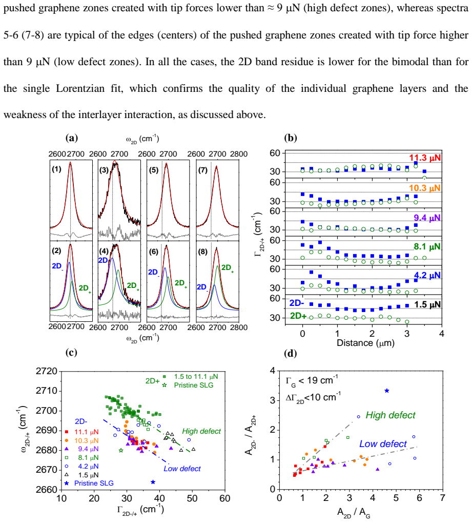
Summary. The manuscript reports an AFM tip-based nanomechanical folding method to convert CVD-grown single-layer graphene (SLG) into weakly interacting multi-layer graphene (wi-MLG). Analysis via multiple Raman correlation plots (A_D/A_G × E_L^4 vs Γ_G, ω_2D vs Γ_2D, Γ_2D vs Γ_G, ω_2D+/- vs Γ_2D+/-, and A_2D-/A_2D+ vs A_2D/A_G) leads to the claims that SLG in-plane properties are preserved, a few tens of layers are stacked with limited structural defects, a ~20 cm⁻¹ blue shift of the 2D band occurs, and the relative intensities of the 2D- and 2D+ bands demonstrate the role of defects in piloting inner and outer Raman scattering processes near the Dirac cone.
Significance. If the observed Raman shifts and intensity correlations can be isolated to the AFM folding and resulting defects, the work supplies an alternative fabrication route for wi-MLG and experimental evidence connecting structural defects to specific scattering channels in the Raman response of graphene. The use of several independent correlation plots constitutes a strength in the experimental design.
major comments (2)
- [Abstract] Abstract: The central attribution of the ~20 cm⁻¹ 2D-band blue shift, the A_2D-/A_2D+ vs A_2D/A_G correlation, and the maintenance of SLG-like properties specifically to AFM-induced folding and limited defects lacks reported before/after spectra on the same flake, Raman data from scanned but unfolded regions, or independent layer-count verification (AFM step height or optical contrast). Without these controls the interpretation cannot exclude substrate interactions, doping, or strain as alternative sources.
- [Abstract] Abstract: No raw spectra, error bars, number of sampled locations, or explicit exclusion criteria are supplied for any of the five listed Raman correlation plots, so the statistical reliability of the claimed defect-mediated inner/outer process distinction cannot be evaluated.
Simulated Author's Rebuttal
We thank the referee for the constructive comments on our manuscript. We address each major point below and have revised the manuscript accordingly to strengthen the experimental controls and statistical presentation.
read point-by-point responses
-
Referee: [Abstract] Abstract: The central attribution of the ~20 cm⁻¹ 2D-band blue shift, the A_2D-/A_2D+ vs A_2D/A_G correlation, and the maintenance of SLG-like properties specifically to AFM-induced folding and limited defects lacks reported before/after spectra on the same flake, Raman data from scanned but unfolded regions, or independent layer-count verification (AFM step height or optical contrast). Without these controls the interpretation cannot exclude substrate interactions, doping, or strain as alternative sources.
Authors: We agree that before/after spectra on the exact same flake location would be the strongest control but are not possible because the folding process irreversibly alters the flake. In the revised manuscript we have added Raman spectra from adjacent regions scanned by the AFM tip but left unfolded; these show SLG-like 2D-band position and width, supporting that the ~20 cm⁻¹ blue shift and intensity correlations arise from the folded wi-MLG rather than substrate, doping or strain. We have also included AFM step-height profiles confirming a few tens of layers. The existing multi-plot Raman correlations already discriminate against uniform doping/strain scenarios according to established literature. revision: partial
-
Referee: [Abstract] Abstract: No raw spectra, error bars, number of sampled locations, or explicit exclusion criteria are supplied for any of the five listed Raman correlation plots, so the statistical reliability of the claimed defect-mediated inner/outer process distinction cannot be evaluated.
Authors: We have revised the manuscript to include representative raw spectra for each correlation plot, error bars (standard deviation), the number of sampled locations (n = 12–18 per plot), and explicit exclusion criteria (spectra with SNR < 5 or obvious cosmic-ray artifacts were discarded). These additions allow direct assessment of the statistical robustness of the defect-piloted inner/outer scattering interpretation. revision: yes
Circularity Check
No circularity: purely experimental observations with no derivations or fitted predictions
full rationale
The paper reports AFM-induced folding of CVD graphene followed by Raman microscopy plots (A_D/A_G vs Γ_G, ω_2D vs Γ_2D, etc.) and direct band assignments. No equations, models, parameters fitted to subsets, or predictions are presented that could reduce to inputs by construction. All claims rest on observed spectral shifts and intensity ratios interpreted via standard Raman literature; no self-citation chains or ansatzes are invoked as load-bearing steps. This matches the default case of an experimental report with independent content.
Axiom & Free-Parameter Ledger
axioms (1)
- domain assumption Standard assignment of D, G, and 2D Raman bands in graphene and their established relations to defects, layer number, and electronic structure near the Dirac cone.
Reference graph
Works this paper leans on
-
[1]
Novoselov, K. S.; Geim, A. K.; Morozov, S. V.; Jiang, D.; Zhang, Y.; Dubonos, S. V.; Grigorieva, I. V.; Firsov, A. A., Electric field effect in atomically thin carbon films. Science 2004, 306 (5696), 666-669
work page 2004
-
[2]
Xu, J. H.; Wang, Y. Z.; Hu, S. S., Nanocomposites of graphene and graphene oxides: Synthesis, molecular functionalization and application in electrochemical sensors and biosensors. A review. Microchimica Acta 2017, 184 (1), 1-44
work page 2017
-
[3]
Meunier, V.; Souza, A. G.; Barros, E. B.; Dresselhaus, M. S., Physical properties of low- dimensional sp(2)-based carbon nanostructures. Reviews of Modern Physics 2016, 88 (2) 025005
work page 2016
-
[4]
Ni, Z. H.; Wang, Y. Y.; Yu, T.; You, Y. M.; Shen, Z. X., Reduction of Fermi velocity in folded graphene observed by resonance Raman spectroscopy. Physical Review B 2008, 77 (23) 235403
work page 2008
-
[5]
W.; Dominguez-Adame, F., Tuning the Fermi velocity in Dirac materials with an electric field
Diaz-Fernandez, A.; Chico, L.; Gonzalez, J. W.; Dominguez-Adame, F., Tuning the Fermi velocity in Dirac materials with an electric field. Scientific Reports 2017, 7, 8058
work page 2017
-
[6]
Kim, S.; Shin, S.; Kim, T.; Du, H.; Song, M.; Kim, K. S.; Cho, S.; Lee, S. W.; Seo, S., A reliable and controllable graphene doping method compatible with current CMOS technology and the demonstration of its device applications. Nanotechnology 2017, 28 (17) 175710
work page 2017
-
[7]
M.; Liu, F., Strain engineering of graphene: a review
Si, C.; Sun, Z. M.; Liu, F., Strain engineering of graphene: a review. Nanoscale 2016, 8 (6), 3207-3217
work page 2016
-
[8]
Chemical Society Reviews 2018, 47 (13), 4860-4908
Anichini, C.; Czepa, W.; Pakulski, D.; Aliprandi, A.; Ciesielski, A.; Samori, P., Chemical sensing with 2D materials. Chemical Society Reviews 2018, 47 (13), 4860-4908
work page 2018
- [9]
-
[10]
Baig, N.; Saleh, T. A., Electrodes modified with 3D graphene composites: a review on methods for preparation, properties and sensing applications. Microchimica Acta 2018, 185 (6) UNSP 283
work page 2018
-
[11]
Hazra, A.; Basu, S., Graphene Nanoribbon as Potential On-Chip Interconnect Material—A Review. C 2018, 4, 4
work page 2018
-
[12]
Di Bernardo, I.; Avvisati, G.; Chen, C. Y.; Avila, J.; Asensio, M. C.; Hu, K. L.; Ito, Y.; Hines, P.; Lipton-Duffin, J.; Rintoul, L.; Motta, N.; Mariani, C.; Betti, M. G., Topology and doping effects in three-dimensional nanoporous graphene. Carbon 2018, 131, 258-265
work page 2018
-
[13]
Yi, C. L.; Chen, X. M.; Zhang, L. Y.; Wang, X. Q.; Ke, C. H., Nanomechanical z-shape folding of graphene on flat substrate. Extreme Mechanics Letters 2016, 9, 84-90
work page 2016
-
[14]
Chang, J. S.; Kim, S.; J., S. H.; J., Y.; J., C. K.; Li, X.; Kim, S., Graphene Nanoribbons with Atomically Sharp Edges Produced by AFM Induced Self-Folding. Small 2018, 14 (47) 1803386
work page 2018
-
[15]
Blees, M. K.; Barnard, A. W.; Rose, P. A.; Roberts, S. P.; McGill, K. L.; Huang, P. Y.; Ruyack, A. R.; Kevek, J. W.; Kobrin, B.; Muller, D. A.; McEuen, P. L., Graphene kirigami. Nature 2015, 524 (7564), 204-+
work page 2015
-
[16]
H.; Zhang, F.; Yan, Z.; Ma, Q.; Li, X
Zhang, Y. H.; Zhang, F.; Yan, Z.; Ma, Q.; Li, X. L.; Huang, Y. G.; Rogers, J. A., Printing, folding and assembly methods for forming 3D mesostructures in advanced materials. Nature Reviews Materials 2017, 2 (4) 17019
work page 2017
-
[17]
Physical Review Letters 2006, 97 (3) 036803
Latil, S.; Henrard, L., Charge carriers in few-layer graphene films. Physical Review Letters 2006, 97 (3) 036803
work page 2006
-
[18]
Goutham, S.; Bykkam, S.; Sadasivuni, K. K.; Kumar, D. S.; Ahmadipour, M.; Ahmad, Z. A.; Rao, K. V., Room temperature LPG resistive sensor based on the use of a few-layer graphene/SnO2 nanocomposite. Microchimica Acta 2018, 185 (1) UNSP 69
work page 2018
-
[19]
Sanchez, V. C.; Jachak, A.; Hurt, R. H.; Kane, A. B., Biological Interactions of Graphene-Family Nanomaterials: An Interdisciplinary Review. Chemical Research in Toxicology 2012, 25 (1), 15-34. 22
work page 2012
-
[20]
T.; Ho, J.; Nezich, D.; Son, H
Reina, A.; Jia, X. T.; Ho, J.; Nezich, D.; Son, H. B.; Bulovic, V.; Dresselhaus, M. S.; Kong, J., Large Area, Few-Layer Graphene Films on Arbitrary Substrates by Chemical Vapor Deposition. Nano Letters 2009, 9 (1), 30-35
work page 2009
-
[21]
R.; Chauhan, N.; Sharma, S.; Mathur, R
Dhakate, S. R.; Chauhan, N.; Sharma, S.; Mathur, R. B., The production of multi-layer graphene nanoribbons from thermally reduced unzipped multi-walled carbon nanotubes. Carbon 2011, 49 (13), 4170-4178
work page 2011
-
[22]
Li, Y. T.; Wu, B.; Guo, W.; Wang, L. F.; Li, J. B.; Liu, Y. Q., Tailoring graphene layer-to-layer growth. Nanotechnology 2017, 28 (26), 265101
work page 2017
-
[23]
Merlen, A.; Buijnsters, J. G.; Pardanaud, C., A Guide to and Review of the Use of Multiwavelength Raman Spectroscopy for Characterizing Defective Aromatic Carbon Solids: from Graphene to Amorphous Carbons. Coatings 2017, 7 (10) 153
work page 2017
-
[24]
Ferrari, A. C.; Basko, D. M., Raman spectroscopy as a versatile tool for studying the properties of graphene. Nature Nanotechnology 2013, 8 (4), 235-246
work page 2013
-
[25]
Nano Letters 2007, 7 (2), 238-242
Graf, D.; Molitor, F.; Ensslin, K.; Stampfer, C.; Jungen, A.; Hierold, C.; Wirtz, L., Spatially resolved raman spectroscopy of single- and few-layer graphene. Nano Letters 2007, 7 (2), 238-242
work page 2007
-
[26]
Physical Review B 2011, 84 (3) 035433
Venezuela, P.; Lazzeri, M.; Mauri, F., Theory of double-resonant Raman spectra in graphene: Intensity and line shape of defect-induced and two-phonon bands. Physical Review B 2011, 84 (3) 035433
work page 2011
-
[27]
Berciaud, S.; Ryu, S.; Brus, L. E.; Heinz, T. F., Probing the Intrinsic Properties of Exfoliated Graphene: Raman Spectroscopy of Free-Standing Monolayers. Nano Letters 2009, 9 (1), 346-352
work page 2009
-
[28]
R.; Felten, A.; Bakaraki, A.; Landois, P.; Colomer, J
Bayle, M.; Reckinger, N.; Huntzinger, J. R.; Felten, A.; Bakaraki, A.; Landois, P.; Colomer, J. F.; Henrard, L.; Zahab, A. A.; Sauvajol, J. L.; Paillet, M., Dependence of the Raman spectrum characteristics on the number of layers and stacking orientation in few-layer graphene. Physica Status Solidi B-Basic Solid State Physics 2015, 252 (11), 2375-2379
work page 2015
-
[29]
Physical Review B 2010, 82 (20) 201409
Mohr, M.; Maultzsch, J.; Thomsen, C., Splitting of the Raman 2D band of graphene subjected to strain. Physical Review B 2010, 82 (20) 201409
work page 2010
-
[30]
Berciaud, S.; Li, X. L.; Htoon, H.; Brus, L. E.; Doorn, S. K.; Heinz, T. F., Intrinsic Line Shape of the Raman 2D-Mode in Freestanding Graphene Monolayers. Nano Letters 2013, 13 (8), 3517-3523
work page 2013
-
[31]
Wang, X. Y.; Christopher, J. W.; Swan, A. K., 2D Raman band splitting in graphene: Charge screening and lifting of the K-point Kohn anomaly. Scientific Reports 2017, 7, 13539
work page 2017
-
[32]
Physica Status Solidi B-Basic Solid State Physics 2016, 253 (12), 2326-2330
Neumann, C.; Banszerus, L.; Schmitz, M.; Reichardt, S.; Sonntag, J.; Taniguchi, T.; Watanabe, K.; Beschoten, B.; Stampfer, C., Line shape of the Raman 2D peak of graphene in van der Waals heterostructures. Physica Status Solidi B-Basic Solid State Physics 2016, 253 (12), 2326-2330
work page 2016
-
[33]
Physical Review B 2009, 80 (23) 233407
Casiraghi, C., Doping dependence of the Raman peaks intensity of graphene close to the Dirac point. Physical Review B 2009, 80 (23) 233407
work page 2009
-
[34]
Journal of Materials Science 2010, 45 (19), 5135-5149
Ferralis, N., Probing mechanical properties of graphene with Raman spectroscopy. Journal of Materials Science 2010, 45 (19), 5135-5149
work page 2010
-
[35]
Lee, J. E.; Ahn, G.; Shim, J.; Lee, Y. S.; Ryu, S., Optical separation of mechanical strain from charge doping in graphene. Nature Communications 2012, 3, 1024
work page 2012
-
[36]
J.; Heiranian, M.; Janicek, B.; Budrikis, Z.; Zapperi, S.; Huang, P
Zhang, Y. J.; Heiranian, M.; Janicek, B.; Budrikis, Z.; Zapperi, S.; Huang, P. Y.; Johnson, H. T.; Aluru, N. R.; Lyding, J. W.; Mason, N., Strain Modulation of Graphene by Nanoscale Substrate Curvatures: A Molecular View. Nano Letters 2018, 18 (3), 2098-2104
work page 2018
-
[37]
Mueller, N. S.; Heeg, S.; Alvarez, M. P.; Kusch, P.; Wasserroth, S.; Clark, N.; Schedin, F.; Parthenios, J.; Papagelis, K.; Galiotis, C.; Kalbac, M.; Vijayaraghavan, A.; Huebner, U.; Gorbachev, R.; Frank, O.; Reich, S., Evaluating arbitrary strain configurations and doping in graphene with Raman spectroscopy. 2D Materials 2018, 5 (1) 015016
work page 2018
-
[38]
G.; Novotny, L., Raman characterization of defects and dopants in graphene
Beams, R.; Cancado, L. G.; Novotny, L., Raman characterization of defects and dopants in graphene. Journal of Physics-Condensed Matter 2015, 27 (8) 083002
work page 2015
-
[39]
Cancado, L. G.; da Silva, M. G.; Ferreira, E. H. M.; Hof, F.; Kampioti, K.; Huang, K.; Penicaud, A.; Achete, C. A.; Capaz, R. B.; Jorio, A., Disentangling contributions of point and line defects in the Raman spectra of graphene-related materials. 2D Materials 2017, 4 (2) 025039. 23
work page 2017
-
[40]
G.; Takai, K.; Enoki, T.; Endo, M.; Kim, Y
Cancado, L. G.; Takai, K.; Enoki, T.; Endo, M.; Kim, Y. A.; Mizusaki, H.; Speziali, N. L.; Jorio, A.; Pimenta, M. A., Measuring the degree of stacking order in graphite by Raman spectroscopy. Carbon 2008, 46 (2), 272-275
work page 2008
-
[41]
Herziger, F.; Tyborski, C.; Ochedowski, O.; Schleberger, M.; Maultzsch, J., Tunable quantum interference in bilayer graphene in double-resonant Raman scattering. Carbon 2018, 133, 254-259
work page 2018
-
[42]
L., Effect of rotational stacking faults on the Raman spectra of folded graphene
Poncharal, P.; Ayari, A.; Michel, T.; Sauvajol, J. L., Effect of rotational stacking faults on the Raman spectra of folded graphene. Physical Review B 2009, 79 (19) 195417
work page 2009
-
[43]
Eliel, G. S. N.; Ribeiro, H. B.; Sato, K.; Saito, R.; Lu, C. C.; Chiu, P. W.; Fantini, C.; Righi, A.; Pimenta, M. A., Raman Excitation Profile of the G-band Enhancement in Twisted Bilayer Graphene. Brazilian Journal of Physics 2017, 47 (6), 589-593
work page 2017
-
[44]
Ferrari, A. C.; Meyer, J. C.; Scardaci, V.; Casiraghi, C.; Lazzeri, M.; Mauri, F.; Piscanec, S.; Jiang, D.; Novoselov, K. S.; Roth, S.; Geim, A. K., Raman spectrum of graphene and graphene layers. Physical Review Letters 2006, 97 (18) 187401
work page 2006
-
[45]
May, P.; Lazzeri, M.; Venezuela, P.; Herziger, F.; Callsen, G.; Reparaz, J. S.; Hoffmann, A.; Mauri, F.; Maultzsch, J., Signature of the two-dimensional phonon dispersion in graphene probed by double-resonant Raman scattering. Physical Review B 2013, 87 (7) 075402
work page 2013
-
[46]
A.; Clair, S.; Patrone, L.; Abel, M.; Porte, L.; Chuzel, O.; Parrain, J
Valyaev, D. A.; Clair, S.; Patrone, L.; Abel, M.; Porte, L.; Chuzel, O.; Parrain, J. L., Grafting a homogeneous transition metal catalyst onto a silicon AFM probe: a promising strategy for chemically constructive nanolithography. Chemical Science 2013, 4 (7), 2815-2821
work page 2013
-
[47]
A.; Francois, C.; Patrone, L.; Balaban, T
Mesquita, V.; Botton, J.; Valyaev, D. A.; Francois, C.; Patrone, L.; Balaban, T. S.; Abel, M.; Parrain, J. L.; Chuzel, O.; Clair, S., Catalytic Scanning Probe Nanolithography (cSPL): Control of the AFM Parameters in Order to Achieve Sub-100-nm Spatially Resolved Epoxidation of Alkenes Grafted onto a Surface. Langmuir 2016, 32 (16), 4034-4042
work page 2016
- [48]
-
[49]
Ramirez-Jimenez, R.; Alvarez-Fraga, L.; Jimenez-Villacorta, F.; Climent-Pascual, E.; Prieto, C.; de Andres, A., Interference enhanced Raman effect in graphene bubbles. Carbon 2016, 105, 556-565
work page 2016
-
[50]
Acs Applied Materials & Interfaces 2017, 9 (4), 4119-4125
Alvarez-Fraga, L.; Climent-Pascual, E.; Aguilar-Pujol, M.; Ramirez-Jimenez, R.; Jimenez- Villacorta, F.; Prieto, C.; de Andres, A., Efficient Heterostructures for Combined Interference and Plasmon Resonance Raman Amplification. Acs Applied Materials & Interfaces 2017, 9 (4), 4119-4125
work page 2017
-
[51]
Das, A.; Pisana, S.; Chakraborty, B.; Piscanec, S.; Saha, S. K.; Waghmare, U. V.; Novoselov, K. S.; Krishnamurthy, H. R.; Geim, A. K.; Ferrari, A. C.; Sood, A. K., Monitoring dopants by Raman scattering in an electrochemically top-gated graphene transistor. Nature Nanotechnology 2008, 3 (4), 210-215
work page 2008
-
[52]
K.; Ijas, M.; Yoon, D.; Sassi, U.; Ferrari, A
Bruna, M.; Ott, A. K.; Ijas, M.; Yoon, D.; Sassi, U.; Ferrari, A. C., Doping Dependence of the Raman Spectrum of Defected Graphene. Acs Nano 2014, 8 (7), 7432-7441
work page 2014
-
[53]
Physica Status Solidi B-Basic Solid State Physics 2016, 253 (12), 2355-2361
Bousa, M.; Anagnostopoulos, G.; del Corro, E.; Drogowska, K.; Pekarek, J.; Kavan, L.; Kalbac, M.; Parthenios, J.; Papagelis, K.; Galiotis, C.; Frank, O., Stress and charge transfer in uniaxially strained CVD graphene. Physica Status Solidi B-Basic Solid State Physics 2016, 253 (12), 2355-2361
work page 2016
-
[54]
Ji, J. T.; He, R.; Jie, Y. H.; Zhang, A. M.; Ma, X. L.; Pan, L. J.; Wang, L.; Zhang, L. Y.; Zhang, Q. M., Modification of electronic band structure in mL plus nL (m=1, 2; n=1-5) free-stacking graphene. Applied Physics Letters 2016, 109 (15) 153111
work page 2016
-
[55]
Carbon 1984, 22 (4-5), 375-385
Lespade, P.; Marchand, A.; Couzi, M.; Cruege, F., Characterization of carbon materials with raman microspectrometry. Carbon 1984, 22 (4-5), 375-385
work page 1984
-
[56]
M., Calculation of the Raman G peak intensity in monolayer graphene: role of Ward identities
Basko, D. M., Calculation of the Raman G peak intensity in monolayer graphene: role of Ward identities. New Journal of Physics 2009, 11, 095011
work page 2009
-
[57]
Chen, C. F.; Park, C. H.; Boudouris, B. W.; Horng, J.; Geng, B. S.; Girit, C.; Zettl, A.; Crommie, M. F.; Segalman, R. A.; Louie, S. G.; Wang, F., Controlling inelastic light scattering quantum pathways in graphene. Nature 2011, 471 (7340), 617-620. 24
work page 2011
-
[58]
Physical Review B 2017, 95 (19) 195422
Reichardt, S.; Wirtz, L., Ab initio calculation of the G peak intensity of graphene: Laser-energy and Fermi-energy dependence and importance of quantum interference effects. Physical Review B 2017, 95 (19) 195422
work page 2017
-
[59]
Hu, K. M.; Xue, Z. Y.; Liu, Y. Q.; Long, H.; Peng, B.; Yan, H.; Di, Z. F.; Wang, X.; Lin, L.; Zhang, W. M., Tension-Induced Raman Enhancement of Graphene Membranes in the Stretched State. Small 2019, 15 (2) 1804337
work page 2019
-
[60]
Bartolome, J.; Alvarez-Fraga, L.; Aguilar-Pujol, M. X.; Cortijo, S.; Cremades, A.; Prieto, C.; de Andres, A., Grain selective Cu oxidation and anomalous shift of graphene 2D Raman peak in the graphene-Cu system. 2D Materials 2019, 6 (1) 015023
work page 2019
discussion (0)
Sign in with ORCID, Apple, or X to comment. Anyone can read and Pith papers without signing in.