REVIEW 5 major objections 6 minor 34 references
Automated Measurement of Optic Nerve Sheath Diameter Using Ocular Ultrasound Video
T0 review · 5 major / 6 minor · reviewed 2026-08-07 · deepseek-v4-flash
Pith's one-line read A handcrafted video-analysis pipeline measures optic nerve sheath diameter automatically, reaching 0.04 mm mean error and 0.782 ICC against expert measurements.
desk verdict A plausible ONSD automation pipeline that is currently unreproducible: the center-finding loop can loop forever and the reported error metric doesn't match the paper's own formula. read the letter →
The pith
A machine-rendered reading of the paper's core claim, the machinery that carries it, and where it could break.
The reading
What carries the argument
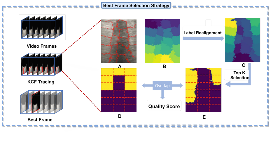
The central mechanism is the frame-quality score: the overlap $S = \frac{2|Y \cap \hat{Y}|}{|Y| + |\hat{Y}|}$ between SLIC-generated segmentation $\hat{Y}$ and a manually designed 3×6 binary template $Y$ of the optic nerve sheath's echo pattern, which selects the optimal frame for measurement. Measurement then proceeds through a GMM foreground mask to initialize a search on the column-sum signal $v(n)$, gradient-descent location of the nerve center, and a weighted KL-divergence signal $L(d)$ that pinpoints the left and right boundaries whose separation gives the ONSD.
What would settle it
Take a set of ocular ultrasound videos with varied probe angles or orbital pathologies (e.g., orbital fat atrophy, trauma, post-surgical changes), have two experts identify the frame with maximal ONSD in each video, and compare the frame chosen by the template-scoring method against the experts' choice via the final measured diameter. If the automatically selected frame yields ONSD values deviating more than about 0.05 mm from the expert-selected frames in a substantial fraction of cases, the template assumption is the point of failure.
Extended reading notes
Core claim
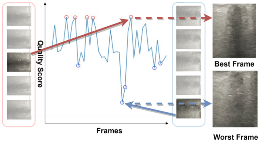
The authors establish that a fully handcrafted, annotation-free pipeline for ONSD measurement from ultrasound video achieves expert-level agreement. Frame selection is treated as template matching: SLIC superpixels are compared against a manually designed 3×6 binary echo-intensity template, and the frame with the highest Dice-like overlap score is chosen as optimal. After GMM-based foreground extraction and a column-sum signal analysis, the sheath boundaries are refined using a KL-divergence measure weighted by an anatomically derived Gaussian. On 40 videos, the automatic measurements differ from the averaged expert gold standard by 0.04 mm mean error (MSE 0.054), with ICC 0.782, close to the inter-expert ICC of 0.952.
Load-bearing premise
The frame-selection stage assumes that every good-quality optic nerve sheath ultrasound frame matches a fixed 3×6 template of hyperechoic retrobulbar fat on the left, right, and superior sides and hypoechoic sheath below; if real anatomy or probe orientation departs from that pattern, the selected "optimal" frame may be wrong.
Editorial extensions
If this is right
- Clinicians can obtain ONSD measurements directly from raw ultrasound video, eliminating manual frame selection and measurement, which reduces operator dependence and enables serial bedside monitoring of intracranial pressure.
- Because the method uses no annotated training data, it can be deployed in low-resource settings where deep-learning approaches requiring hundreds of labeled videos are infeasible.
- The frame-quality scoring approach could generalize to other ultrasound video analysis tasks that require picking a single best diagnostic frame from a sequence.
- The weighted KL-divergence boundary refinement technique could be reused for other low-contrast boundary detection problems in medical ultrasound.
Reading between the lines
- The frame-scoring stage could be validated independently of the full pipeline by checking whether the automatically selected frame actually contains the maximal ONSD across all video frames, rather than only comparing the final measurement to expert averages.
- The reported ICC of 0.782 sits below the expert-expert ICC of 0.952, suggesting the method may be better suited for tracking ICP trends over time than for binary clinical decisions at a fixed ONSD threshold.
- Because the template assumes hyperechoic retrobulbar fat on the left, right, and superior flanks of the sheath, the method may silently fail in patients with orbital trauma, post-surgical changes, or atypical anatomy; a confidence threshold on the frame-quality overlap could flag such cases.
Editorial analysis
A structured set of objections, weighed in public.
Referee Report
Summary. The manuscript proposes an automated pipeline for measuring optic nerve sheath diameter (ONSD) from ocular ultrasound video. The pipeline comprises three stages: optimal frame selection using KCF tracking followed by SLIC superpixel segmentation and template-overlap scoring; ONS localization using a row-sum signal and a Gaussian Mixture Model; and boundary refinement using a KL-divergence-based weighted search. The authors report a mean error of 0.04 mm, MSE of 0.054, and ICC of 0.782 against the average of two expert clinicians on 40 ultrasound videos, and argue that the method reduces operator dependence in ONSD measurement.
Significance. If the technical content were sound, the paper would address a real clinical need by offering a training-free, interpretable alternative to deep-learning-based ONSD measurement, which is relevant for non-invasive intracranial pressure monitoring. The pipeline is composed of classical image-processing components, which is a strength in resource-constrained settings. However, the central derivation contains multiple load-bearing errors, and the reported results cannot be reproduced from the method as written. There is no shared code or data, and the external validation claims in the Discussion lack methodological support. The potential clinical significance does not compensate for the current technical deficiencies.
major comments (5)
- [Section 3.2.3, Algorithm 1] Algorithm 1 cannot terminate on a discrete signal. The loop continues while Δv(n) ≠ 0; when Δv(n) < 0, d is incremented, and when Δv(n) > 0, d is decremented. At a strict local minimum n0, Δv(n0) < 0 moves to n0+1, and then Δv(n0+1) > 0 moves back to n0, producing an infinite oscillation. The pseudocode also uses n in the condition while updating d, so either n is never updated or the intended update rule is inconsistent. No plateau, tolerance, or maximum-iteration condition is provided. Consequently, dcenter is undefined, and the downstream boundary refinement and the diameter D in Eq. (9) cannot be computed. This invalidates the central measurement claim.
- [Section 3.2.2, Eq. (5)] Equation (5) is algebraically wrong: k(n) = (Σ_m M(n,m)) / (Σ_n Σ_m M(n,m)) is not identically equal to 1. The numerator is the foreground-pixel count in row n, while the denominator is the total foreground-pixel count; their ratio equals 1 only for a single row that contains all foreground pixels. The correct normalization would be Σ_n k(n) = 1. As written, the initialization condition k(d) = 0.5 has no solution, so the starting point for Algorithm 1 is undefined.
- [Section 3.3, Eq. (11) and Table 1] Equation (11) defines the mean error as a percentage (mean absolute percentage error, multiplied by 100), but Table 1 reports 'Mean Error' in millimeters (0.020, 0.043, 0.044, 0.040) and the surrounding text explicitly says 'mean errors (in millimeters, mm)'. These are different quantities. The manuscript must state which metric is actually used and align the formula with the reported values. Without this correction, the headline accuracy of 0.04 mm is not supported by the stated evaluation procedure.
- [Section 5, Discussion (external validation and synthetic noise)] The Discussion reports results on an external dataset (ONSD-Benchmark, n=32, ICC 0.74) and under synthetic noise (SNR = 15 dB, MAE 0.04–0.06 mm), but no methodological details are provided in Section 3: the source and annotation of the external dataset, the preprocessing steps, how the pixel-to-mm mapping system was adapted, or how synthetic noise and motion artifacts were generated and applied. These claims are therefore unverifiable and should either be fully described or clearly labeled as preliminary observations.
- [Section 3.2.1, optimal frame selection] The 'optimal frame' is defined as the frame whose SLIC segmentation maximally overlaps a manually designed binary template (Fig. 2D). This is a hand-crafted quality prior rather than an independent reference, so the selection is inherently self-consistent with that template. The Discussion reports a validation against expert-selected frames, but the protocol is not specified (number of experts, selection criteria, statistical analysis). This is a major concern for the central claim that the method automatically identifies the clinically optimal frame, and it needs a more rigorous and transparent evaluation.
minor comments (6)
- [Abstract and Section 3.1] The dataset description in Section 3.1 contains an incomplete sentence: '8) underwent optic nerve sheath diameter (ONSD) ultrasound.' This appears to be a residual fragment and should be corrected.
- [Figure captions] Figure 2 caption contains 'groud truth' and Figure 4 caption contains 'sigmal'; these typos should be fixed.
- [Section 3.3, Eq. (12)] Equation (12) uses '1/nu' where 'n' is presumably the number of images; the notation should be defined and made consistent with Eq. (11).
- [Section 3.2.4, Eq. (8)] The Gaussian weighting parameters (μ, σ) in Eq. (8) are derived from the same localization output (dcenter and dleft) that is being refined, so the prior is not independent of the data. The authors should discuss this potential bias or provide a sensitivity analysis.
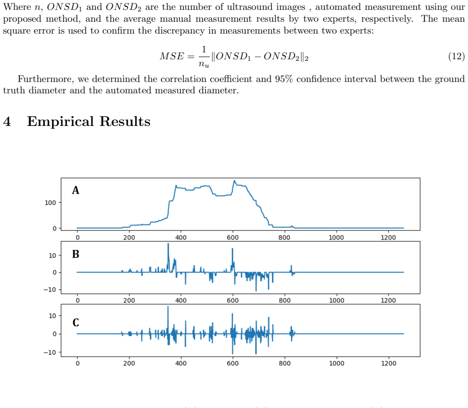
- [Section 3.2.3 and Figure 5] The information-entropy-based keyframe extraction described around Figure 5 is not integrated into the method or evaluated; it appears as an orphaned component.
- [References] Reference [26] is cited for KCF tracking, but the cited article describes a dense spatio-temporal context tracker rather than a kernel correlation filter; the citation should be corrected or the method should be renamed.
Circularity Check
No significant circularity: the accuracy claim is grounded in external expert measurements, and the frame-selection template does not determine the reported ONSD values.
full rationale
The central claim (mean error 0.04 mm, MSE 0.054, ICC 0.782) is evaluated against the average of two expert clinicians (Table 1), which is an external ground truth not produced by the algorithm. The optimal-frame stage uses a manually designed binary template Y (Fig. 1D) and a Dice-style overlap score (Eq. 1), but this only selects a frame; the diameter D is subsequently computed from v(n) row sums, GMM foreground extraction, and KL-divergence boundary refinement (Eqs. 3-9), so the template is not equivalent to the measurement output. The KL weighting parameters mu and sigma in Eq. 8 are derived from the coarse localization interval, but the boundary condition Delta Ltilde(dtilde_left) = 0 is not equal by construction to mu or to the coarse edges; it is a regularized optimization condition on a data-dependent divergence signal. No load-bearing result is justified by self-citation, and no fitted parameter is renamed as a prediction. The paper's Algorithm 1 termination issue and the Eq. (11) metric-unit inconsistency are correctness and quality concerns, not circular reasoning.
Assumptions & free parameters
free parameters (6)
- Template mask Y (3x6 pattern) =
Binary pattern with r1,r2,r3,r4,r6,r7,r9 set to 255
- Gaussian weighting parameters (mu, sigma) =
mu = (dcenter - dleft)/2, sigma = (dcenter - dleft)/6 per image
- SLIC superpixel parameters =
unspecified
- Binarization threshold k (number of top-intensity superpixels) =
unspecified
- GMM component count =
unspecified
- Pixel-to-mm mapping system =
unspecified
assumptions (5)
- ad hoc to paper Optic nerve sheath appears hypoechoic and retrobulbar fat hyperechoic in B-mode ultrasound, so the fixed 3x6 template pattern is a valid representation of a good frame.
- domain assumption The signal v(n) (column-summed pixel intensity) has the optic nerve sheath center at a local minimum flanked by two local maxima.
- domain assumption GMM with unspecified component count separates retrobulbar fat from other tissue in the binary mask M(n,m).
- ad hoc to paper Algorithm 1 converges to the trough center by following the gradient sign.
- domain assumption The ONSD-to-ICP linear relationship from prior literature justifies the clinical motivation.
Cite this review
Pith. "Pith review of Automated Measurement of Optic Nerve Sheath Diameter Using Ocular Ultrasound Video." pith.science (2026). https://pith.science/paper/5PRWO6YW
@misc{pith2026250602789,
  author       = {Pith},
  title        = {Pith review of: Automated Measurement of Optic Nerve Sheath Diameter Using Ocular Ultrasound Video},
  year         = {2026},
  howpublished = {\url{https://pith.science/paper/5PRWO6YW}},
  note         = {Machine review of arXiv:2506.02789}
}
read the original abstract
Objective. Elevated intracranial pressure (ICP) is recognized as a biomarker of secondary brain injury, with a significant linear correlation observed between optic nerve sheath diameter (ONSD) and ICP. Frequent monitoring of ONSD could effectively support dynamic evaluation of ICP. However, ONSD measurement is heavily reliant on the operator's experience and skill, particularly in manually selecting the optimal frame from ultrasound sequences and measuring ONSD. Approach. This paper presents a novel method to automatically identify the optimal frame from video sequences for ONSD measurement by employing the Kernel Correlation Filter (KCF) tracking algorithm and Simple Linear Iterative Clustering (SLIC) segmentation algorithm. The optic nerve sheath is mapped and measured using a Gaussian Mixture Model (GMM) combined with a KL-divergence-based method. Results. When compared with the average measurements of two expert clinicians, the proposed method achieved a mean error, mean squared deviation, and intraclass correlation coefficient (ICC) of 0.04, 0.054, and 0.782, respectively. Significance. The findings suggest that this method provides highly accurate automated ONSD measurements, showing potential for clinical application.
Figures


Figures from the paper (6 more)



Reference graph
Works this paper leans on
-
[1]
W. Ussahgij, W. Toonpirom, W. Munkong, K. Lenghong, K. Apiratwarakul, Optic nerve sheath diam- eter cutoff point for detection of increased intracranial pressure in the emergency department, Open Access Macedonian Journal of Medical Sciences 8 (2020) 62–65
work page 2020
-
[2]
J. Wang, K. Li, H. Li, C. Ji, Z. Wu, H. Chen, B. Chen, Ultrasonographic optic nerve sheath diameter correlation with icp and accuracy as a tool for noninvasive surrogate icp measurement in patients with decompressive craniotomy, Journal of Neurosurgery 133 (2019) 514–520
work page 2019
-
[3]
M. Smith, Monitoring intracranial pressure in traumatic brain injury, Anesthesia & Analgesia 106 (2008) 240–248
work page 2008
- [4]
-
[5]
P. Williams, Optic nerve sheath diameter as a bedside assessment for elevated intracranial pressure, Case reports in critical care 2017 (2017) 3978934
work page 2017
-
[6]
D. F. Netteland, M. Aarhus, E. Smistad, E. C. Sandset, L. Padayachy, E. Helseth, R. Brekken, Non- invasive intracranial pressure assessment by optic nerve sheath diameter: Automated measurements as an alternative to clinician-performed measurements, Frontiers in Neurology 14 (2023) 1064492
work page 2023
- [7]
-
[8]
P. Jenjitranant, P. Tunlayadechanont, T. Prachanukool, R. Kaewlai, Correlation between optic nerve sheath diameter measured on imaging with acute pathologies found on computed tomography of trauma patients, European Journal of Radiology 125 (2020) 108875. 15
work page 2020
Show all 34 references
-
[9]
D. S. Nag, S. Sahu, A. Swain, S. Kant, Intracranial pressure monitoring: Gold standard and recent innovations, World journal of clinical cases 7 (2019) 1535
2019
-
[10]
G. Xu, X. Wu, J. Yu, H. Ding, Z. Ni, Y. Wang, Non-invasive intracranial pressure assessment using shear-wave elastography in neuro-critical care patients, Journal of Clinical Neuroscience 99 (2022) 261–267
2022
-
[11]
Zweifel, M
C. Zweifel, M. Czosnyka, E. Carrera, N. de Riva, J. D. Pickard, P. Smielewski, Reliability of the blood flow velocity pulsatility index for assessment of intracranial and cerebral perfusion pressures in head-injured patients, Neurosurgery 71 (2012) 853–861
2012
-
[12]
Schmidt, M
B. Schmidt, M. Czosnyka, J. J. Schwarze, D. Sander, W. Gerstner, C. B. Lumenta, J. Klingelh¨ ofer, Evaluation of a method for noninvasive intracranial pressure assessment during infusion studies in patients with hydrocephalus, Journal of neurosurgery 92 (2000) 793–800
2000
-
[13]
Jalayondeja, K
T. Jalayondeja, K. Urasyanandana, Correlations between optic nerve sheath diameters measured us- ing computed tomography and elevated intracranial pressure levels, Interdisciplinary Neurosurgery 26 (2021) 101328
2021
-
[14]
Montorfano, Q
L. Montorfano, Q. Yu, S. J. Bordes, S. Sivanushanthan, R. J. Rosenthal, M. Montorfano, Mean value of b-mode optic nerve sheath diameter as an indicator of increased intracranial pressure: a systematic review and meta-analysis, The ultrasound journal 13 (2021) 1–12
2021
-
[15]
Copetti, L
R. Copetti, L. Cattarossi, Optic nerve ultrasound: artifacts and real images, Intensive care medicine 35 (2009) 1488–1489
2009
-
[16]
Chen, L.-j
L.-m. Chen, L.-j. Wang, Y. Hu, X.-h. Jiang, Y.-z. Wang, Y.-q. Xing, Ultrasonic measurement of optic nerve sheath diameter: a non-invasive surrogate approach for dynamic, real-time evaluation of intracranial pressure, British Journal of Ophthalmology 103 (2019) 437–441
2019
-
[17]
I. M. Maissan, P. J. Dirven, I. K. Haitsma, S. E. Hoeks, D. Gommers, R. J. Stolker, Ultrasonographic measured optic nerve sheath diameter as an accurate and quick monitor for changes in intracranial pressure, Journal of neurosurgery 123 (2015) 743–747
2015
-
[18]
Rajajee, R
V. Rajajee, R. Soroushmehr, C. A. Williamson, K. Najarian, J. Gryak, A. Awad, K. R. Ward, M. H. Tiba, Novel algorithm for automated optic nerve sheath diameter measurement using a clustering approach, Military Medicine 186 (2021) 496–501
2021
-
[19]
M. Pang, S. Liu, F. Lin, S. Liu, B. Tian, W. Yang, X. Chen, Measurement of optic nerve sheath on ocular ultrasound image based on segmentation by cnn, in: 2019 IEEE International Conference on Signal, Information and Data Processing (ICSIDP), IEEE, 2019, pp. 1–5
2019
-
[20]
Romagnosi, F
F. Romagnosi, F. Bongiovanni, M. Oddo, Eyeing up the injured brain: automated pupillometry and optic nerve sheath diameter, Current opinion in critical care 26 (2020) 115–121
2020
-
[21]
Singh, V
M. Singh, V. Gupta, R. Gupta, B. Kumar, D. Agrawal, A novel method for prediction of raised intracra- nial pressure through automated onsd and etd ratio measurement from ocular ultrasound, Ultrasonic Imaging 46 (2024) 29–40
2024
-
[22]
J. G. Fleiderman Valenzuela, J. I. Cirillo Totera, D. H. Turkieltaub, C. V. Echaurren, F. L. ´Alvarez Lemos, F. I. Arriagada Ramos, Spino-pelvic radiological parameters: Comparison of mea- surements obtained by radiologists using the traditional method versus spine surgeons us...
2023
-
[23]
El Jalbout, A
R. El Jalbout, A. Flahault, A. Shahi, J.-L. Bigras, A. Cloutier, J. Dubois, A.-M. Nuyt, T.-M. Luu, Measuring carotid intima-media thickness in young adults born preterm: Comparison of manual versus semi-automated b-mode ultrasound, Journal for Vascular Ultrasound 47 (2023) 76–85. 16
2023
-
[24]
D. Shi, Z. Lin, W. Wang, Z. Tan, X. Shang, X. Zhang, W. Meng, Z. Ge, M. He, A deep learning system for fully automated retinal vessel measurement in high throughput image analysis, Frontiers in Cardiovascular Medicine 9 (2022) 823436
2022
-
[25]
Russo, M
C. Russo, M. A. Pirozzi, F. Mazio, D. Cascone, D. Cicala, M. De Liso, A. Nastro, E. M. Covelli, G. Cinalli, M. Quarantelli, Fully automated measurement of intracranial csf and brain parenchyma volumes in pediatric hydrocephalus by segmentation of clinical mri studies, Medical ...
2023
-
[26]
Zhang, L
K. Zhang, L. Zhang, Q. Liu, D. Zhang, M.-H. Yang, Fast visual tracking via dense spatio-temporal context learning, in: Computer Vision–ECCV 2014: 13th European Conference, Zurich, Switzerland, September 6-12, 2014, Proceedings, Part V 13, Springer, 2014, pp. 127–141
2014
-
[27]
Achanta, A
R. Achanta, A. Shaji, K. Smith, A. Lucchi, P. Fua, S. S¨ usstrunk, Slic superpixels (2010)
2010
-
[28]
B. T. Moore, S. P. Montgomery, M. Niethammer, H. Greer, S. R. Aylward, Automatic optic nerve sheath measurement in point-of-care ultrasound, in: Medical Ultrasound, and Preterm, Perinatal and Paediatric Image Analysis: First International Workshop, ASMUS 2020, and 5th Internat...
2020
-
[29]
Soroushmehr, K
R. Soroushmehr, K. Rajajee, C. Williamson, J. Gryak, K. Najarian, K. Ward, M. H. Tiba, Automated optic nerve sheath diameter measurement using super-pixel analysis, in: 2019 41st Annual International Conference of the IEEE Engineering in Medicine and Biology Society (EMBC), IE...
2019
-
[30]
K. M. Meiburger, A. Naldi, N. Michielli, L. Coppo, K. Fassbender, F. Molinari, P. Lochner, Automatic optic nerve measurement: a new tool to standardize optic nerve assessment in ultrasound b-mode images, Ultrasound in medicine & biology 46 (2020) 1533–1544
2020
-
[31]
Hansen, K
H. Hansen, K. Helmke, The subarachnoid space surrounding the optic nerves. an ultrasound study of the optic nerve sheath, Surgical and radiologic anatomy: SRA 18 (1996) 323–328
1996
-
[32]
X. He, D. Cai, Y. Shao, H. Bao, J. Han, Laplacian regularized gaussian mixture model for data clustering, IEEE transactions on knowledge and data engineering 23 (2010) 1406–1418
2010
-
[33]
Zhang, F
T. Zhang, F. Ma, Improved rough k-means clustering algorithm based on weighted distance measure with gaussian function, International Journal of Computer Mathematics 94 (2017) 663–675
2017
-
[34]
Xiao, Automatic optic nerve assessment from transorbital ultrasound images: A deep learning-based approach, Current Medical Imaging 20 (2024) e15734056293608
Y. Xiao, Automatic optic nerve assessment from transorbital ultrasound images: A deep learning-based approach, Current Medical Imaging 20 (2024) e15734056293608. 17
2024
Reviewed August 7, 2026 · model on record in the stance chip above.
Discussion (0). Sign in to comment.