REVIEW 3 major objections 5 minor 30 references
From Skin to Skeleton: Towards Biomechanically Accurate 3D Digital Humans
T0 review · 3 major / 5 minor · reviewed 2026-08-15 · deepseek-v4-flash
Pith's one-line read SKEL gives the standard SMPL human body model a biomechanical skeleton driven by the same shape and pose parameters as the skin surface.
desk verdict A genuinely useful skin-skeleton parametric model with an accuracy claim that is self-referential but explicitly acknowledged; deserves serious peer review and a conditional accept. read the letter →
The pith
A machine-rendered reading of the paper's core claim, the machinery that carries it, and where it could break.
The reading
What carries the argument
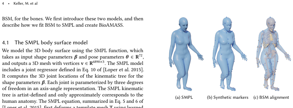
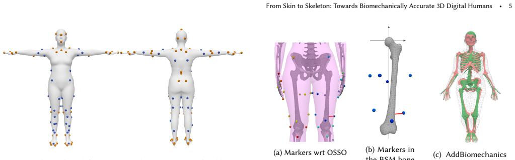
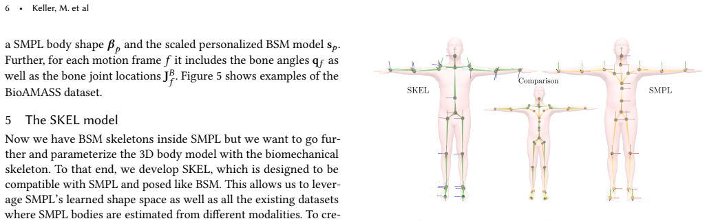
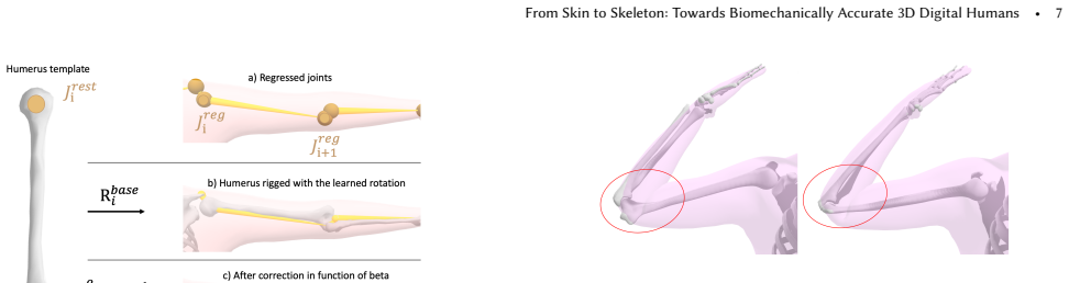
The load-bearing mechanism is a three-stage pipeline. First, a custom 24-bone biomechanical skeleton with 46 degrees of freedom—featuring a constant-curvature spine, an ellipsoid-sliding scapula, and a radius–ulna forearm—is fit inside posed SMPL meshes using virtual markers and a recent bilevel biomechanical optimization, producing the paired BioAMASS dataset. Second, a non-negative least-squares regressor maps SMPL mesh vertices to the 24 anatomical joint locations. Third, each bone's orientation is decomposed into a fixed base rotation learned from the dataset and a shape-dependent correction $R_{\boldsymbol{\beta}}^i(\boldsymbol{\beta})$ that aligns the bone with its parent–child joint segment; these pieces re-rig the SMPL mesh so the biomechanical pose parameters move skin and skeleton in synchrony. The decomposition is what lets SKEL keep SMPL's shape space while giving the bones anatomically coherent orientations inside every body shape.
What would settle it
Take a subject with both a CT or MRI scan and synchronized marker motion capture, fit SKEL to the motion, and compare its femur-head and humerus-head joint centers with the imaging-derived centers; a systematic offset, especially at the shoulder, would falsify the claimed biomechanical accuracy.
Extended reading notes
Core claim
SKEL is presented as the first model in which the body surface and the anatomical skeleton are directly controlled by the same shape and pose parameters. Trained on the BioAMASS dataset, SKEL places a properly scaled skeleton inside any SMPL body; its regressed joint locations are closer to functional anatomical joints than SMPL's artist-defined joints, and its bones fit inside the body surface better than earlier skeleton-from-skin methods. The model uses fewer pose parameters than SMPL (46 versus 72) but models the spine as a constant-curvature chain, the scapula as sliding on an ellipsoid around the thorax, and forearm pronation and supination as a radius–ulna motion, so its articulation is biomechanically constrained rather than a collection of ball joints.
Load-bearing premise
The whole result rests on the assumption that fitting the skeleton to markers painted on the skin of synthetic bodies yields essentially correct joint locations, even though those fits are only pseudo-ground-truth and are never checked against medical images or bone-pin measurements.
Editorial extensions
If this is right
- Existing SMPL-based vision pipelines can be post-processed by fitting SKEL, turning datasets such as 3DPW and BEDLAM into sources of biomechanical skeleton parameters.
- Regressing anatomical joints from skin (or fitting SKEL) yields joint locations closer to the functional joints than SMPL's own joints, with errors below one centimeter for most joints in the reported evaluation.
- Because SKEL has 46 biomechanical degrees of freedom instead of SMPL's 72 ball-joint degrees, posed bodies exhibit more realistic shoulder-blade sliding, spine bending, and forearm twist.
- Given a biomechanical skeleton, SKEL can generate a plausible skin surface while preserving the skeleton's bone lengths, allowing motion-capture data to be visualized with body shapes of different weights.
Reading between the lines
- The decisive test the paper does not run is against independent anatomy: registering SKEL's femur-head and humerus-head joints to CT or MRI-derived joint centers on the same subjects would show whether the pseudo-ground-truth fits are unbiased.
- If SKEL is used to train video-based biomechanics estimators, the natural downstream check is whether joint angles and moments computed from SKEL agree with marker-based motion capture to within clinical tolerances; that is the metric that would justify “biomechanics in the wild.”
- Since the learning and rigging pipeline is independent of the particular skeleton model, the same procedure could be rerun with a clinically validated skeleton the moment one exists, upgrading SKEL without architectural changes.
Signed reviews
Editorial analysis
A structured set of objections, weighed in public.
Referee Report
Summary. The paper introduces SKEL, a parametric body model that re-rigs the SMPL surface model with a biomechanical skeleton, BSM, so that skin and skeleton are controlled by the same pose vector q and shape vector β. To train SKEL, the authors construct BioAMASS, a dataset of about 9 hours of motion from 113 AMASS subjects, by placing virtual markers on SMPL meshes and fitting BSM with the AddBiomechanics optimization pipeline. They learn a non-negative joint regressor from SMPL vertices to BSM anatomical joint locations, estimate per-bone base rotations and shape-dependent corrective rotations, and define a new kinematic tree with 46 biomechanical degrees of freedom, including a constant-curvature spine, sliding scapulae, and radioulnar pronation/supination. The paper evaluates joint-location errors against the BSM fits used as pseudo-ground-truth, reports SKEL-to-SMPL surface fitting errors on DFAUST, and shows qualitative comparisons with OSSO on MOYO.
Significance. If the accuracy claims are taken at face value, SKEL is a valuable bridge between statistical body models and biomechanics: it is, to my knowledge, the first model in which skin and a biomechanical skeleton are driven by the same pose and shape parameters, and the BioAMASS dataset and the SMPL-to-BSM fitting pipeline are potentially useful resources. The re-rigging formulation is well motivated, and the paper includes several thoughtful design choices, such as using OSSO to personalize virtual marker offsets and modeling the shoulder blade with an ellipsoid constraint. The main limitation is that the central claim of biomechanical accuracy is evaluated against the same AddBiomechanics fitting pipeline that generated the training labels, and the paper's own Sec. 7 acknowledges this is pseudo-ground-truth. Consequently, the sub-centimeter reported errors demonstrate reproducibility of a fitting pipeline rather than independently established anatomical accuracy; the qualitative OSSO comparison also does not quantify the 'bones fit inside the body surface better' claim.
major comments (3)
- [Sec. 6.2, Sec. 4.3.2, Sec. 7] The evaluation of the joint regressor and the SKEL fits is circular. The joint locations J^B used as 'ground truth' in Fig. 10 and Fig. 11 are produced by the same AddBiomechanics optimization (Eq. (2)) that generated the BioAMASS training labels. The learned regressor and SKEL are therefore scored by their ability to reproduce the fitting pipeline, not by their anatomical correctness. The paper itself states in Sec. 7 that these fits 'should not be considered as actual ground truth, but rather a pseudo-ground truth.' To support the abstract's claim of 'more biomechanically accurate joint locations than SMPL,' the authors should add an external validation, for example comparing a subset of subjects against OSSO skeletons trained on medical images, functional joint-center estimates, or published anthropometric or imaging measurements; alternatively, the claims throughout the abstract, Sec. 1, and Sec. 6.2 should be softened to 'consistency with a marker-based biomechanical fitting pipeline.'
- [Sec. 6.4] The claim that the bones 'fit inside the body surface better than previous methods' is supported only by qualitative side-by-side images of OSSO and SKEL on MOYO. No quantitative metric is reported for bone containment, bone-skin penetration, or agreement of bone orientations with an independent reference. I recommend adding a scalar evaluation, such as the fraction of bone vertices inside the SMPL skin mesh, the signed distance of bone vertices to the skin surface, or a comparison of joint angles against the AddBiomechanics reference, on a held-out set of poses and subjects.
- [Sec. 4.3.2, Eq. (2)] The per-marker offsets δ and the marker weights λ_k are free parameters in the bi-level optimization that creates BioAMASS, and the paper does not state how δ is initialized or regularized. Unrestrained per-marker offsets can absorb systematic skin-to-bone offsets and shift the estimated joint centers, so the skeleton locations might be determined more by δ than by the marker trajectories. Please report the magnitude and variability of the learned offsets, and provide a sensitivity analysis showing that joint locations in BioAMASS are stable when δ is removed or differently regularized.
minor comments (5)
- [Sec. 4.2, Eq. (1)] The formula for t_spine is difficult to parse; the term '(r−cos(α)) ∗ −sin(q_z)' appears to have a missing operand or an unusual double sign, and the definitions of q_x, q_y, q_z are incomplete. Adding explicit parentheses and a short derivation would improve readability.
- [Sec. 5.2, Eq. (6)] The text refers to 'the green term' in the equation, but the printed equation is monochrome; please replace the color reference with an explicit description of the factors being discussed.
- [Sec. 5.2 and Sec. 6.3] Sec. 5.2 states that SKEL can match SMPL meshes 'with an average vertex-to-vertex error below 3 cm,' while Sec. 6.3 reports average mean differences of 1.1 cm for males and 0.9 cm for females on DFAUST. Please reconcile these numbers, and clarify whether the 3 cm figure refers to extreme poses outside DFAUST.
- [Sec. 4.3.2] The prior P(s,β) is said to use biological sex as in Werling et al., but the paper does not explain how biological sex is obtained from the SMPL shape parameters β. Please specify the sex estimation procedure or state that sex is assumed from the source dataset.
- [Fig. 12] The color bar is labeled 'Blue: 0 cm, Red: 2 cm,' yet the text reports a maximum female difference of 1.9 cm; please adjust the visualization range or the text so the figure and caption are consistent.
Circularity Check
The quantitative 'biomechanical accuracy' claim is benchmarked against the same AddBiomechanics pseudo-ground-truth that generated the training labels; no independent anatomical validation breaks the loop.
-
fitted input called prediction
[Sec. 4.3.2 (Eq. 2), Sec. 5.1, Sec. 6.2 and Figs. 10–11]
"We train these regressors from the posed vertices and joints of the BioAMASS dataset. ... For each frame of the DFAUST dataset, BioAMASS provides the anatomical joint locations J^B that we consider ground truth. Then, from the frame's SMPL mesh, we use our learned joint regressor to regress the anatomical joint location J^reg."
The training targets for the joint regressor are the BSM joint locations produced by solving Eq. (2) with AddBiomechanics. The evaluation in Sec. 6.2 uses the same kind of output ('J^B that we consider ground truth') as the reference for the regressed joints. The reported sub-centimeter errors therefore quantify how faithfully the learned regressor reproduces the AddBiomechanics/BSM fitting pipeline, not an independently established anatomical accuracy. The paper itself concedes in Sec.
full rationale
The construction of SKEL itself—learning anatomical joint regressors from BioAMASS, deriving bone orientations from Eq. (4), and re-rigging SMPL with BSM degrees of freedom—is a coherent, non-circular engineering pipeline. The regressor is trained on one subset of AMASS and evaluated on DFAUST, so the experiment does demonstrate generalization across subjects and poses relative to the chosen labels. The circularity is confined to the interpretation of those labels as 'ground truth' for biomechanical accuracy. The labels are outputs of AddBiomechanics fits to virtual markers placed on SMPL meshes; the same AddBiomechanics/BSM pipeline generated the training supervision. Thus the quantitative evaluation of 'more biomechanically accurate joint locations' (Figs. 10–11) measures consistency with the pseudo-ground-truth generator, not with an independent skeletal measurement such as medical imaging, bone-pin data, or functional joint-center tests. The paper explicitly acknowledges this limitation in Sec. 7, which is commendable but does not remove the self-referential nature of the benchmark. The qualitative OSSO comparison (Sec. 6.4) is not quantified and does not independently establish the 'bones fit inside the body surface better' claim. No uniqueness theorem, ansatz-smuggling via citation, or definitional equation identity was found in the rigging mathematics; the core issue is the closed evaluation loop around the pseudo-ground-truth.
Assumptions & free parameters
free parameters (7)
- BSM bone scales s =
Per subject, R^{24x3}, not reported in the paper
- Per-marker offsets δ =
Per marker per subject, R^{N_m x 3}
- Marker weights λ_k =
Low for soft markers, high for bony markers
- Joint regressor coefficients =
24 x 6890 x 3 matrix, learned
- Base bone rotations R_base^i =
Learned per bone from Eq. 4
- Spine arc length l =
Chosen by design, not reported
- Mapping from SKEL DOFs to SMPL pose correctives =
Hand-picked correspondence
assumptions (5)
- domain assumption AddBiomechanics optimization recovers sufficiently accurate skeletal joint locations from surface markers
- domain assumption The BSM kinematic structure (Rajagopal lower body, Seth scapula, custom constant-curvature spine) accurately represents human biomechanics
- domain assumption Skin markers, including soft markers, plus a scale prior, constrain the underlying skeleton pose and bone lengths
- ad hoc to paper Uniform body density can be assumed to estimate weight from SMPL shape
- ad hoc to paper SMPL pose-dependent deformations and skinning weights transfer to the SKEL rig with acceptable error
invented entities (2)
-
BSM (Biomechanical Skeleton Model)
-
Constant-curvature spine joint
Cite this review
Pith. "Pith review of From Skin to Skeleton: Towards Biomechanically Accurate 3D Digital Humans." pith.science (2026). https://pith.science/paper/6P6SPRFP
@misc{pith2026250906607,
  author       = {Pith},
  title        = {Pith review of: From Skin to Skeleton: Towards Biomechanically Accurate 3D Digital Humans},
  year         = {2026},
  howpublished = {\url{https://pith.science/paper/6P6SPRFP}},
  note         = {Machine review of arXiv:2509.06607}
}
read the original abstract
Great progress has been made in estimating 3D human pose and shape from images and video by training neural networks to directly regress the parameters of parametric human models like SMPL. However, existing body models have simplified kinematic structures that do not correspond to the true joint locations and articulations in the human skeletal system, limiting their potential use in biomechanics. On the other hand, methods for estimating biomechanically accurate skeletal motion typically rely on complex motion capture systems and expensive optimization methods. What is needed is a parametric 3D human model with a biomechanically accurate skeletal structure that can be easily posed. To that end, we develop SKEL, which re-rigs the SMPL body model with a biomechanics skeleton. To enable this, we need training data of skeletons inside SMPL meshes in diverse poses. We build such a dataset by optimizing biomechanically accurate skeletons inside SMPL meshes from AMASS sequences. We then learn a regressor from SMPL mesh vertices to the optimized joint locations and bone rotations. Finally, we re-parametrize the SMPL mesh with the new kinematic parameters. The resulting SKEL model is animatable like SMPL but with fewer, and biomechanically-realistic, degrees of freedom. We show that SKEL has more biomechanically accurate joint locations than SMPL, and the bones fit inside the body surface better than previous methods. By fitting SKEL to SMPL meshes we are able to "upgrade" existing human pose and shape datasets to include biomechanical parameters. SKEL provides a new tool to enable biomechanics in the wild, while also providing vision and graphics researchers with a better constrained and more realistic model of human articulation. The model, code, and data are available for research at https://skel.is.tue.mpg.de..
Figures

Figures from the paper (9 more)



Reference graph
Works this paper leans on
- [14]
-
[16]
DeepLabCut: markerless pose estimation of user-defined body parts with deep learning.Nature neuroscience 21, 9 (2018), 1281–1289. Laurie Needham, Murray Evans, Darren P Cosker, Logan Wade, Polly M McGuigan, James L Bilzon, and Steffi L Colyer
work page 2018
-
[18]
Scientific reports10, 1 (2020), 1–12
Efficient trajectory optimiza- tion for curved running using a 3D musculoskeletal model with implicit dynamics. Scientific reports10, 1 (2020), 1–12. Ahmed A. A. Osman, Timo Bolkart, and Michael J. Black
work page 2020
-
[19]
STAR: Sparse Trained Articulated Human Body Regressor. InEuropean Conf. on Computer Vision (ECCV), Vol. LNCS 12355. Springer, 598–613. https://doi.org/10.1007/978-3-030-58539-6_36 Ahmed A A Osman, Timo Bolkart, Dimitrios Tzionas, and Michael J. Black
-
[21]
Shunsuke Saito, Zi-Ye Zhou, and Ladislav Kavan
Full-body musculoskeletal model for muscle- driven simulation of human gait.IEEE Transactions on Biomedical Engineering63, 10 (2016), 2068–2079. Shunsuke Saito, Zi-Ye Zhou, and Ladislav Kavan
work page 2016
-
[22]
Computational bodybuilding: Anatomically-based modeling of human bodies.ACM Transactions on Graphics34, 4 (2015), 1–12. Robert Schleicher, Marlies Nitschke, Jana Martschinke, Marc Stamminger, Björn Es- kofier, Jochen Klucken, and Anne Koelewijn
work page 2015
-
[23]
BASH: Biomechanical Animated Skinned Human for Visualization of Kinematics and Muscle Activity. InProceedings of the 16th International Joint Conference on Computer Vision, Imaging and Computer Graphics Theory and Applications (VISIGRAPP)(2021-02-08/2021-02-10). Springer, 25–36. https://doi.org/10.5220/0010210600250036 Ajay Seth, Ricardo Matias, António P...
-
[24]
A Biomechanical Model of the Scapulothoracic Joint to Accurately Capture Scapular Kinematics during Shoulder Movements.PloS One11, 1 (01 2016), 1–18. https://doi.org/10.1371/ journal.pone.0141028 Karthik Shetty, Annette Birkhold, Srikrishna Jaganathan, Norbert Strobel, Bernhard Egger, Markus Kowarschik, and Andreas Maier
work page 2016
Show all 30 references
-
[25]
https: //doi.org/10.1016/j.compbiomed.2023.107383 Shashank Tripathi, Lea Müller, Chun-Hao P
BOSS: Bones, organs and skin shape model.Computers in Biology and Medicine165 (2023), 107383. https: //doi.org/10.1016/j.compbiomed.2023.107383 Shashank Tripathi, Lea Müller, Chun-Hao P. Huang, Omid Taheri, Michael Black, and Dimitrios Tzionas
2023
-
[26]
https: //doi.org/10.1101/2022.07.07.499061 Timo Von Marcard, Roberto Henschel, Michael J Black, Bodo Rosenhahn, and Gerard Pons-Moll
OpenCap: 3D human movement dynamics from smartphone videos.bioRxiv(2022). https: //doi.org/10.1101/2022.07.07.499061 Timo Von Marcard, Roberto Henschel, Michael J Black, Bodo Rosenhahn, and Gerard Pons-Moll
2022 doi
-
[28]
https://doi.org/10.1145/3306346.3322983 Haoyang Wang, Riza Alp Güler, Iasonas Kokkinos, George Papandreou, and Stefanos Zafeiriou
Hand Modeling and Simulation Using Stabilized Magnetic Resonance Imaging.ACM Transactions on Graphics38, 4, Article 115 (July 2019), 14 pages. https://doi.org/10.1145/3306346.3322983 Haoyang Wang, Riza Alp Güler, Iasonas Kokkinos, George Papandreou, and Stefanos Zafeiriou
2019
-
[29]
Rapid bilevel optimization to concurrently solve mus- culoskeletal scaling, marker registration, and inverse kinematic problems for human motion reconstruction.bioRxiv(2022). https://doi.org/10.1101/2022.08.22.504896 arXiv:https://www.biorxiv.org/content/early/2022/08/23/2022....
2022 doi
-
[30]
Adaptable Anatomical Models for Realistic Bone Motion Reconstruction.Comput. Graph. Forum34, 2 (2015), 459–471. https://doi.org/10.1111/cgf.12575
2015 doi
-
[1988]
Bohan Wang, George Matcuk, and Jernej Barbič
The effects of knee brace hinge design and placement on joint mechanics.Journal of Biomechanics21, 11 (1988), 965–974. Bohan Wang, George Matcuk, and Jernej Barbič
1988
-
[2000]
http://mocap
CMU Graphics Lab Motion Capture Database. http://mocap. cs.cmu.edu. Accessed: 2012-12-11. Scott L. Delp, Frank C. Anderson, Allison S. Arnold, Peter Loan, Ayman Habib, Chand T. John, Eran Guendelman, and Darryl G. Thelen
2012
-
[2003]
https://doi.org/10.1145/882262.882311 Dragomir Anguelov, Praveen Srinivasan, Daphne Koller, Sebastian Thrun, Jim Rodgers, and James Davis
The Space of Human Body Shapes: Reconstruction and Parameterization from Range Scans.ACM Transactions on Graphics22, 3 (July 2003), 587–594. https://doi.org/10.1145/882262.882311 Dragomir Anguelov, Praveen Srinivasan, Daphne Koller, Sebastian Thrun, Jim Rodgers, and James Davis
2003
-
[2005]
https://doi.org/10.1145/1073204
SCAPE: Shape Completion and Animation of People.ACM Transactions on Graphics24, 3 (July 2005), 408–416. https://doi.org/10.1145/1073204. 1073207 Marian Bittner, Wei-Tse Yang, Xucong Zhang, Ajay Seth, Jan van Gemert, and Frans CT van der Helm
2005 doi
-
[2007]
Hao-Shu Fang, Jiefeng Li, Hongyang Tang, Chao Xu, Haoyi Zhu, Yuliang Xiu, Yong- Lu Li, and Cewu Lu
OpenSim: Open-source soft- ware to create and analyze dynamic simulations of movement.IEEE Transactions on Biomedical Engineering54, 11 (2007), 1940–1950. Hao-Shu Fang, Jiefeng Li, Hongyang Tang, Chao Xu, Haoyi Zhu, Yuliang Xiu, Yong- Lu Li, and Cewu Lu
2007
-
[2010]
Creating and animating subject- specific anatomical models.Comput. Graph. Forum29, 8 (Dec. 2010), 2340–2351. https://doi.org/10.1111/j.1467-8659.2010.01718.x Petr Kadleček, Alexandru-Eugen Ichim, Tiantian Liu, Jaroslav Křivánek, and Ladislav Kavan
2010
-
[2012]
Marilyn Keller, Silvia Zuffi, Michael J
Zygote body: A new interactive 3-dimensional didactical tool for teaching anatomy.Webmedcentral Anatomy(2012). Marilyn Keller, Silvia Zuffi, Michael J. Black, and Sergi Pujades
2012
-
[2013]
2013), 8 pages
Anatomy transfer.ACM Transactions on Graphics32, 6, Article 188 (Nov. 2013), 8 pages. https://doi.org/10.1145/2508363. 2508415 Brett Allen, Brian Curless, and Zoran Popović
2013 doi
-
[2014]
2014), 220:1–220:13
MoSh: Motion and Shape Capture from Sparse Markers.ACM Transactions on Graphics33, 6 (Nov. 2014), 220:1–220:13. https://doi.org/10.1145/2661229.2661273 Naureen Mahmood, Nima Ghorbani, Nikolaus F. Troje, Gerard Pons-Moll, and Michael J. Black
2014
-
[2015]
InIEEE Conf
Pose-Conditioned Joint Angle Limits for 3D Human Pose Reconstruction. InIEEE Conf. on Computer Vision and Pattern Recognition (CVPR). IEEE, 1446–1455. https://doi.org/10.1109/CVPR.2015.7298751 Dicko Ali-Hamadi, Tiantian Liu, Benjamin Gilles, Ladislav Kavan, François Faure, Oli...
2015
-
[2016]
2016), 13 pages
Reconstructing Personalized Anatomical Models for Physics-Based Body Animation.ACM Transactions on Graphics35, 6, Article 213 (Nov. 2016), 13 pages. https://doi.org/10.1145/2980179.2982438 Angjoo Kanazawa, Michael J. Black, David W. Jacobs, and Jitendra Malik
2016
-
[2018]
InIEEE Conf
End- to-end Recovery of Human Shape and Pose. InIEEE Conf. on Computer Vision and Pattern Recognition (CVPR). IEEE, 7122–7131. Manuel Kaufmann, Velko Vechev, and Dario Mylonopoulos. 2022.aitviewer. https: //doi.org/10.5281/zenodo.1234 Robi Kelc
2022 doi
-
[2019]
The Virtual Caliper: Rapid Creation of Metrically Accurate Avatars from 3D Measurements.IEEE Trans. Vis. Comput. Graph.25, 5 (2019), 1887–1897. https://doi.org/10.1109/TVCG.2019.2898748 Apoorva Rajagopal, Christopher L Dembia, Matthew S DeMers, Denny D Delp, Jen- nifer L Hicks...
2019
-
[2020]
InIEEE Conf
VIBE: Video Inference for Human Body Pose and Shape Estimation. InIEEE Conf. on Computer Vision and Pattern Recognition (CVPR). IEEE, Piscataway, NJ, 5252–5262. https: //doi.org/10.1109/CVPR42600.2020.00530 Charles L Lawson and Richard J Hanson. 1995.Solving least squares prob...
2020
-
[2021]
Marlies Nitschke, Eva Dorschky, Dieter Heinrich, Heiko Schlarb, Bjoern M Eskofier, Anne D Koelewijn, and Antonie J van den Bogert
The accuracy of several pose estimation methods for 3D joint centre localisation.Scientific reports11, 1 (2021), 20673. Marlies Nitschke, Eva Dorschky, Dieter Heinrich, Heiko Schlarb, Bjoern M Eskofier, Anne D Koelewijn, and Antonie J van den Bogert
2021
-
[2022]
Towards Single Camera Human 3D-Kinematics.Sensors23, 1 (2022),
2022
-
[2023]
https://doi.org/10.1109/TPAMI.2022
AlphaPose: Whole-Body Regional Multi-Person Pose Estimation and Tracking in Real-Time.IEEE Transactions on Pattern Analysis and Machine Intelligence45, 6 (2023), 7157–7173. https://doi.org/10.1109/TPAMI.2022. 3222784 Benjamin Gilles, Lionel Reveret, and Dinesh Pai
2023 doi
Reviewed August 15, 2026 · model on record in the stance chip above.
Discussion (0). Continue with ORCID to comment.