REVIEW 3 major objections 5 minor 46 references
Deep Learning for Estimating Synaptic Health of Primary Neuronal Cell Culture
T0 review · 3 major / 5 minor · reviewed 2026-08-14 · deepseek-v4-flash
Pith's one-line read A fine-tuned residual CNN classifies raw cytoskeleton-channel images of rodent neurons as untreated or exposed to 30 µM Aβ(25–35) with 99.58% validation accuracy, then screens 36 compounds and finds none substantially protective.
desk verdict A useful screening application undermined by a train/test arithmetic inconsistency and an unvalidated proxy assumption; the 99.58% accuracy is not credible as reported. read the letter →
The pith
A machine-rendered reading of the paper's core claim, the machinery that carries it, and where it could break.
The reading
What carries the argument
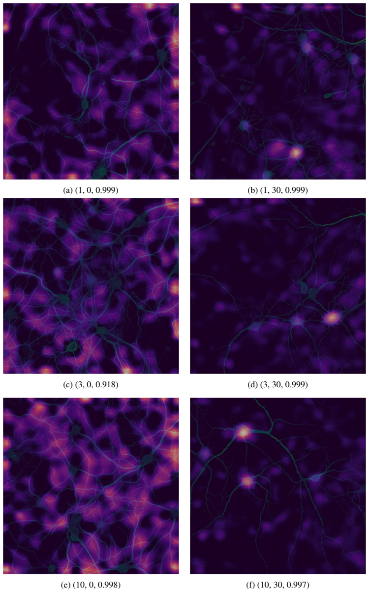
The carrying object is a fine-tuned ResNet18, a residual convolutional neural network with skip connections, pre-trained on a large natural-image corpus and re-trained on 2048×2048 Cy5 fluorescence images of neuronal cytoskeleton. Transfer learning, data augmentation, and dropout let the network learn features directly from pixels; Grad-CAM then maps which pixels drive each decision, showing attention on neurites. In screening, the network's per-image output is averaged over field views and wells to give a treatment-level score used to assign each condition to the untreated or Aβ-treated class.
What would settle it
Run the trained model on images from a plate containing a compound with independently confirmed synaptic rescue, for example by PSD-95 puncta counts; if those wells still receive Aβ-treated scores while the independent measure shows restored synapses, the screening criterion fails. Alternatively, compute validation accuracy separately for every held-out plate; if any plate's accuracy falls near chance, the 99.58% figure does not generalize across plates.
Extended reading notes
Core claim
On the paper's own terms, the central discovery is that the morphological signature of Aβ(25–35) toxicity in primary neuronal culture is learnable from raw Cy5-channel pixels: a ResNet18 initialized with pretrained weights and fine-tuned on 6,480 untreated and 6,480 treated images separates the two conditions with 99.58% validation accuracy. The same model, with predictions averaged over field views and wells, classifies cultures treated with compound plus Aβ as Aβ-treated unless the compound restores an untreated-like morphology. Using that criterion, none of the 36 screened compounds at 1, 3, or 10 µM showed substantial protection, and this negative result was corroborated by the standard feature-extraction and statistical-testing pipeline.
Load-bearing premise
The screen can only detect protection that makes an Aβ-exposed culture look like an untreated one; a compound that preserves synapses through a different visible morphology would be scored as ineffective.
Editorial extensions
If this is right
- A high-throughput screen can be run on raw images: about 60 seconds per plate in inference versus roughly 15 hours for the traditional feature-extraction pipeline on 48 CPUs, the paper reports.
- None of 36 candidate compounds at three doses substantially protected against Aβ(25–35)-induced synaptic loss under this assay's conditions.
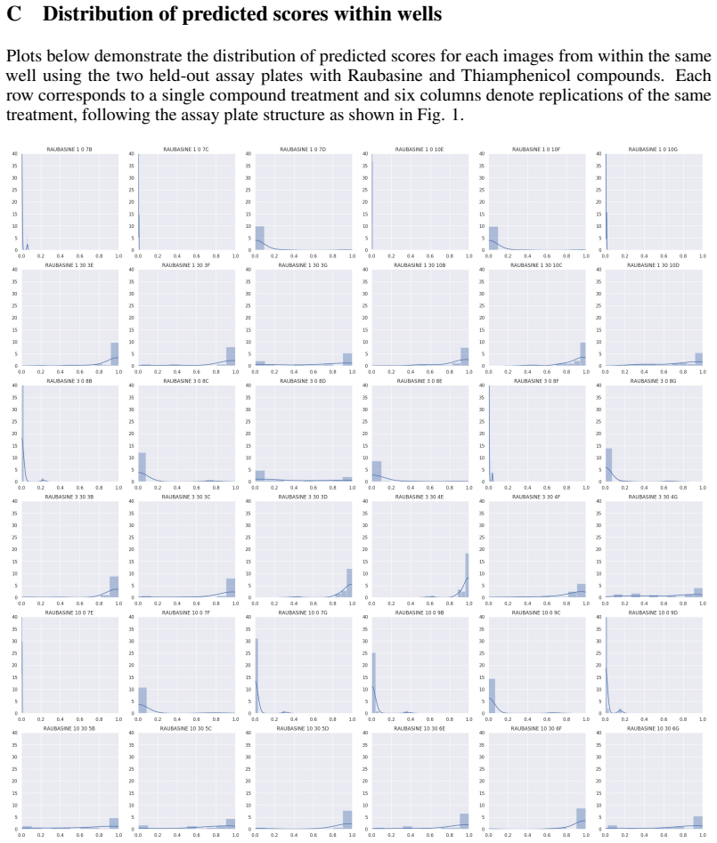
- The binary model's score can flag ambiguous wells for manual inspection, since most wells score near 0 or 1 while contaminated or blurry wells fall in between.
- The approach is designed to transfer to other assays and, ultimately, human cells, since only raw pixels are needed and pretrained weights are reused.
Reading between the lines
- Because the classifier is trained only on vehicle- and Aβ-treated endpoints, its screening utility depends on protection 'looking like' the untreated class; a compound that protects synapses through a different morphology would be scored as non-protective even if biologically effective.
- A quantitative extension the paper does not report would correlate the model's continuous prediction score with independent synapse-density measures, such as PSD-95 puncta counts, across doses, turning the screen from binary to graded.
- The single 34/2 plate split could be stress-tested by per-plate cross-validation; if accuracy varies sharply across plates, the 99.58% figure is a property of the chosen plates rather than the assay generally.
- Feeding all four stains, including nuclear, pre-synaptic, and post-synaptic channels, into a multi-channel model could reveal whether the cytoskeleton channel alone carries the full signal or whether the other stains add independent predictive information.
Editorial analysis
A structured set of objections, weighed in public.
Referee Report
Summary. The paper presents a binary image classifier, based on a fine-tuned ResNet18, that distinguishes vehicle-control primary rodent neuronal cultures from cultures treated with 30 µM of Aβ(25-35), using raw Cy5-channel microscopy images. The authors report a validation accuracy of 99.58% and then apply the classifier to screen 36 candidate compounds for protective effects against Aβ-induced synaptic loss, concluding that none shows a substantial protective effect. The paper also includes Grad-CAM visualizations as a sanity check that the model attends to neurite-like structures and reports a computational speed advantage over a CellProfiler-based pipeline.
Significance. If the accuracy estimate were reliable, the claimed result would be practically useful: a raw-pixel CNN classifier that is as accurate as claimed and runs in about 60 seconds per plate could substantially accelerate high-throughput screening of compounds for morphological neuroprotection. The paper also deserves credit for applying Grad-CAM to check that decisions are not driven by obvious artifacts, and for being explicit about the screening logic. However, the central quantitative claim is undermined by an arithmetic inconsistency in the training/test split, and the screening conclusion rests on a strong interpretive assumption that is not validated. As written, the 99.58% figure is not an established out-of-sample estimate, and the paper's main conclusions therefore require revision rather than being directly acceptable.
major comments (3)
- [§3.1, §3.2, and §4] The reported validation accuracy is not established as an out-of-sample estimate because the stated training-set size is inconsistent with the described plate split. Section 3.1 states that each well is imaged 30 times, and Table 1 gives six vehicle-control wells and six Aβ-only wells per plate; Section 4 states that 34 of 36 plates were used for training. That yields 34 × 6 × 30 = 6,120 images per class, not the 6,480 per class reported in Section 3.2. The stated 6,480 corresponds to using all 36 plates. Therefore either the two 'held-out' plates were included in fine-tuning, or the validation numbers in Table 2 were computed with training data leakage. In either case, the 99.58% validation accuracy cannot be interpreted as an independent test of generalization, and no code or data are provided to audit the split.
- [§3.2 and §5] The screening conclusion does not follow from the classification accuracy alone. Section 3.2 states that the trained model was used to determine whether cells treated with a compound plus Aβ(25-35) are 'more similar' to cells treated with Aβ(25-35) alone, and Section 5 concludes that none of the 36 compounds has a substantial protective effect. Because the classifier was trained only on the two extreme conditions, it cannot distinguish partial protection, protective mechanisms that do not restore the specific morphology of the untreated class, or off-target effects that change image features for unrelated reasons. The conclusion is a domain assumption about what protection should look like in the learned feature space, and the paper provides no independent validation of that assumption.
- [§5] The claim that the screening result was 'confirmed' by a CellProfiler-based statistical pipeline is not supported by any reported analysis. Section 5 states this confirmation, but the paper does not give the CellProfiler feature measurements, the statistical test results, or a comparison table for the 36 compounds. As written, this confirmation is unverifiable and cannot be used to strengthen the main screening conclusion.
minor comments (5)
- [§3.2] There is a typo: 'the the model was applied to screen candidate compounds' should read 'the model was applied to screen candidate compounds.'
- [Captions of Figures 2 and 3] The figure captions describe the second component of each triple as 'Aβ(25−30)', but the text and methods consistently use 'Aβ(25−35)'; the captions should be corrected for consistency.
- [§5] The word 're-suing' in the last paragraph should be 're-using.'
- [§4 and Appendix A] The reference to 'Table 5 in A' should refer to 'Appendix A,' and Table 5 should be placed in the appendix rather than left dangling in the results section.
- [§3.2] The model name is written inconsistently as 'Resnet18' in the text and 'ResNet18' in the abstract; please standardize the capitalization.
Circularity Check
No circular derivation: the 99.58% accuracy is an externally benchmarked classification result and the screening conclusion is an explicitly stated biological assumption, not a construction that renames the model's inputs.
full rationale
The paper's central accuracy claim (Table 2: validation accuracy 99.58%) is measured against independent treatment labels ('untreated' vehicle control vs. 30 µM Aβ(25-35)) on images that were not used for training (two plates held out, per Section 4). This is an external benchmark and not a fitted input masquerading as a prediction. The screening step is the only place where a conclusion could in principle be generated by the classifier's own labels: Section 3.2 states that once trained, the model was applied 'to determine whether images of cells treated with a compound + Aβ(25-35) were more similar to those treated with Aβ(25-35) alone.' That is a substantive biological assumption (protection implies untreated-like morphology), not a circular derivation; the conclusion is not guaranteed by the model's construction, and the paper separately claims confirmation via CellProfiler. Self-citations in the manuscript (e.g., refs. 34, 38, 39) concern the unrelated signature-method future-work discussion and do not support the classification or screening claims. The arithmetic discrepancy noted in the skeptical summary (34 plates × 6 wells × 30 field views = 6,120 images per class, not 6,480) is a data-integrity/validation concern outside the circularity rubric, since it does not show that the accuracy was defined into existence. Accordingly, no circular step meeting the evidentiary bar is identified.
Assumptions & free parameters
free parameters (4)
- Decision threshold =
0.5
- Fine-tuning learning rates =
1e-3 then 1e-4
- Number of training epochs =
10 + 10
- Dropout rate =
0.35
assumptions (5)
- domain assumption ImageNet-pretrained weights transfer to fluorescence microscopy images.
- domain assumption The Cy5 channel alone captures the morphological changes induced by Aβ(25-35).
- domain assumption The two held-out assay plates are representative of plate-to-plate variability.
- domain assumption A protective compound shifts cell morphology toward the untreated class.
- standard math Standard stochastic gradient descent training converges to a generalizing solution.
Cite this review
Pith. "Pith review of Deep Learning for Estimating Synaptic Health of Primary Neuronal Cell Culture." pith.science (2026). https://pith.science/paper/77M3WRQB
@misc{pith2026190811399,
  author       = {Pith},
  title        = {Pith review of: Deep Learning for Estimating Synaptic Health of Primary Neuronal Cell Culture},
  year         = {2026},
  howpublished = {\url{https://pith.science/paper/77M3WRQB}},
  note         = {Machine review of arXiv:1908.11399}
}
abstract
Understanding the morphological changes of primary neuronal cells induced by chemical compounds is essential for drug discovery. Using the data from a single high-throughput imaging assay, a classification model for predicting the biological activity of candidate compounds was introduced. The image recognition model which is based on deep convolutional neural network (CNN) architecture with residual connections achieved accuracy of 99.6$\%$ on a binary classification task of distinguishing untreated and treated rodent primary neuronal cells with Amyloid-$\beta_{(25-35)}$.
Figures


Figures from the paper (2 more)

Reference graph
Works this paper leans on
-
[1]
Contribution of alzheimer disease to mortality in the united states
Bryan D James, Sue E Leurgans, Liesi E Hebert, Paul A Scherr, Kristine Yaffe, and David A Bennett. Contribution of alzheimer disease to mortality in the united states. Neurology, 82(12):1045–1050, 2014
work page 2014
-
[2]
Uk research spend in 2008 and 2012: comparing stroke, cancer, coronary heart disease and dementia
Ramon Luengo-Fernandez, Jose Leal, and Alastair Gray. Uk research spend in 2008 and 2012: comparing stroke, cancer, coronary heart disease and dementia. BMJ open, 5(4):e006648, 2015
work page 2008
-
[3]
Guy M McKhann, David S Knopman, Howard Chertkow, Bradley T Hyman, Clifford R Jack Jr, Claudia H Kawas, William E Klunk, Walter J Koroshetz, Jennifer J Manly, Richard Mayeux, et al. The diagnosis of dementia due to alzheimer’s disease: Recommendations from the national institute on aging-alzheimer’s association workgroups on diagnostic guidelines for alzhe...
work page 2011
-
[4]
The amyloid beta peptide: a chemist’s perspective
Ian W Hamley. The amyloid beta peptide: a chemist’s perspective. role in alzheimer’s and fibrillization. Chemical reviews, 112(10):5147–5192, 2012
work page 2012
-
[5]
Sarah Westwood, Alison L Baird, Sneha N Anand, Liu Shi, Alejo J Nevado-Holgado, Andrey Kormilitzin, Abdul Hye, Nicholas J Ashton, Angharad Morgan, Samuel Touchard, et al. Discovery, replication and extension study of plasma proteomic biomarkers relating to brain amyloid burden and alzheimer’s disease progression. Alzheimer’s & Dementia: The Journal of the...
work page 2018
-
[6]
Overview of deep learning in medical imaging
Kenji Suzuki. Overview of deep learning in medical imaging. Radiological physics and technology, 10(3):257–273, 2017
work page 2017
-
[7]
Detecting cancer metastases on gigapixel pathology images
Yun Liu, Krishna Gadepalli, Mohammad Norouzi, George E Dahl, Timo Kohlberger, Aleksey Boyko, Subhashini Venugopalan, Aleksei Timofeev, Philip Q Nelson, Greg S Corrado, et al. Detecting cancer metastases on gigapixel pathology images. arXiv preprint arXiv:1703.02442, 2017
arXiv 2017
-
[8]
Yun Liu, Timo Kohlberger, Mohammad Norouzi, George E Dahl, Jenny L Smith, Arash Mohtashamian, Niels Olson, Lily H Peng, Jason D Hipp, and Martin C Stumpe. Artificial intelligence–based breast cancer nodal metastasis detection: Insights into the black box for pathologists. Archives of Pathology & Laboratory Medicine
Show all 46 references
-
[9]
Deep learning in image cytometry: a review
Anindya Gupta, Philip J Harrison, Håkan Wieslander, Nicolas Pielawski, Kimmo Kartasalo, Gabriele Partel, Leslie Solorzano, Amit Suveer, Anna H Klemm, Ola Spjuth, et al. Deep learning in image cytometry: a review. Cytometry Part A, 95(4):366–380, 2019
2019
-
[10]
Machine learning methods for histopathological image analysis
Daisuke Komura and Shumpei Ishikawa. Machine learning methods for histopathological image analysis. Computational and structural biotechnology journal , 16:34–42, 2018
2018
-
[11]
Deep learning-based virtual histology staining using auto-fluorescence of label-free tissue
Yair Rivenson, Hongda Wang, Zhensong Wei, Yibo Zhang, Harun Gunaydin, and Aydogan Ozcan. Deep learning-based virtual histology staining using auto-fluorescence of label-free tissue. arXiv preprint arXiv:1803.11293, 2018
2018 arXiv
-
[12]
Virtual histological staining of unlabelled tissue-autofluorescence images via deep learning
Yair Rivenson, Hongda Wang, Zhensong Wei, Kevin de Haan, Yibo Zhang, Yichen Wu, Harun Günaydın, Jonathan E Zuckerman, Thomas Chong, Anthony E Sisk, et al. Virtual histological staining of unlabelled tissue-autofluorescence images via deep learning. Nature biomedical engineering...
2019
-
[13]
Deep learning enables cross-modality super-resolution in fluorescence microscopy
Hongda Wang, Yair Rivenson, Yiyin Jin, Zhensong Wei, Ronald Gao, Harun Günaydın, Laurent A Bentolila, Comert Kural, and Aydogan Ozcan. Deep learning enables cross-modality super-resolution in fluorescence microscopy. Nat. Methods, 16:103–110, 2019
2019
-
[14]
Reconstructing cell cycle and disease progression using deep learning
Philipp Eulenberg, Niklas Köhler, Thomas Blasi, Andrew Filby, Anne E Carpenter, Paul Rees, Fabian J Theis, and F Alexander Wolf. Reconstructing cell cycle and disease progression using deep learning. Nature communications, 8(1):463, 2017
2017
-
[15]
Repurposing high-throughput image assays enables biological activity prediction for drug discovery
Jaak Simm, Günter Klambauer, Adam Arany, Marvin Steijaert, Jörg Kurt Wegner, Emmanuel Gustin, Vladimir Chupakhin, Yolanda T Chong, Jorge Vialard, Peter Buijnsters, et al. Repurposing high-throughput image assays enables biological activity prediction for drug discovery. Cell c...
2018
-
[16]
Cellprofiler: image analysis software for identifying and quantifying cell phenotypes
Anne E Carpenter, Thouis R Jones, Michael R Lamprecht, Colin Clarke, In Han Kang, Ola Friman, David A Guertin, Joo Han Chang, Robert A Lindquist, Jason Moffat, et al. Cellprofiler: image analysis software for identifying and quantifying cell phenotypes. Genome biology, 7(10):R100, 2006
2006
-
[17]
Deep learning
Yann LeCun, Yoshua Bengio, and Geoffrey Hinton. Deep learning. nature, 521(7553):436, 2015
2015
-
[18]
Deep residual learning for image recognition
Kaiming He, Xiangyu Zhang, Shaoqing Ren, and Jian Sun. Deep residual learning for image recognition. In Proceedings of the IEEE conference on computer vision and pattern recognition , pages 770–778, 2016
2016
-
[19]
J. Deng, W. Dong, R. Socher, L.-J. Li, K. Li, and L. Fei-Fei. ImageNet: A Large-Scale Hierarchical Image Database. In CVPR09, 2009
2009
-
[20]
Automatic differentiation in pytorch
Adam Paszke, Sam Gross, Soumith Chintala, Gregory Chanan, Edward Yang, Zachary DeVito, Zeming Lin, Alban Desmaison, Luca Antiga, and Adam Lerer. Automatic differentiation in pytorch. 2017
2017
-
[21]
Jeremy Howard et al. fastai. https://github.com/fastai/fastai, 2018
2018
-
[22]
Improving neural networks by preventing co-adaptation of feature detectors
Geoffrey E Hinton, Nitish Srivastava, Alex Krizhevsky, Ilya Sutskever, and Ruslan R Salakhutdinov. Improving neural networks by preventing co-adaptation of feature detectors. arXiv preprint arXiv:1207.0580, 2012
2012 arXiv
-
[23]
A disciplined approach to neural network hyper-parameters: Part 1–learning rate, batch size, momentum, and weight decay
Leslie N Smith. A disciplined approach to neural network hyper-parameters: Part 1–learning rate, batch size, momentum, and weight decay. arXiv preprint arXiv:1803.09820, 2018
2018 arXiv
-
[24]
Universal language model fine-tuning for text classification
Jeremy Howard and Sebastian Ruder. Universal language model fine-tuning for text classification. arXiv preprint arXiv:1801.06146, 2018
2018 arXiv
-
[25]
Visualizing the loss landscape of neural nets
Hao Li, Zheng Xu, Gavin Taylor, Christoph Studer, and Tom Goldstein. Visualizing the loss landscape of neural nets. In Advances in Neural Information Processing Systems , pages 6389–6399, 2018
2018
-
[26]
Searching for activation functions
Prajit Ramachandran, Barret Zoph, and Quoc V Le. Searching for activation functions. arXiv preprint arXiv:1710.05941, 2017
2017 arXiv
-
[27]
Grad-cam: Visual explanations from deep networks via gradient- based localization
Ramprasaath R Selvaraju, Michael Cogswell, Abhishek Das, Ramakrishna Vedantam, Devi Parikh, Dhruv Batra, et al. Grad-cam: Visual explanations from deep networks via gradient- based localization. In ICCV, pages 618–626, 2017
2017
-
[28]
Aβ influences cytoskeletal signaling cascades with consequences to alzheimer’s disease
Ana Gabriela Henriques, Joana Machado Oliveira, Liliana Patrícia Carvalho, and Odete AB da Cruz e Silva. Aβ influences cytoskeletal signaling cascades with consequences to alzheimer’s disease. Molecular neurobiology, 52(3):1391–1407, 2015
2015
-
[29]
Alexander B. Jung. imgaug. https://github.com/aleju/imgaug, 2018. [Online; accessed 25-Aug-2019]
2018
-
[30]
Segem: efficient image analysis for high-resolution connectomics
Manuel Berning, Kevin M Boergens, and Moritz Helmstaedter. Segem: efficient image analysis for high-resolution connectomics. Neuron, 87(6):1193–1206, 2015
2015
-
[31]
Analyzing image segmentation for connectomics
Stephen M Plaza and Jan Funke. Analyzing image segmentation for connectomics. Frontiers in Neural Circuits, 12, 2018
2018
-
[32]
Synapse-aware skeleton generation for neural circuits
Brian Matejek, Donglai Wei, Xueying Wang, Jinglin Zhao, Kálmán Palágyi, and Hanspeter Pfister. Synapse-aware skeleton generation for neural circuits
-
[33]
Rough paths, signatures and the modelling of functions on streams
Terry Lyons. Rough paths, signatures and the modelling of functions on streams. arXiv preprint arXiv:1405.4537, 2014. 6
2014 arXiv
-
[34]
A primer on the signature method in machine learning
Ilya Chevyrev and Andrey Kormilitzin. A primer on the signature method in machine learning. arXiv preprint arXiv:1603.03788, 2016
2016
-
[35]
Sparse arrays of signatures for online character recognition
Benjamin Graham. Sparse arrays of signatures for online character recognition. arXiv preprint arXiv:1308.0371, 2013
2013 arXiv
-
[36]
Learning spatial- semantic context with fully convolutional recurrent network for online handwritten chinese text recognition
Zecheng Xie, Zenghui Sun, Lianwen Jin, Hao Ni, and Terry Lyons. Learning spatial- semantic context with fully convolutional recurrent network for online handwritten chinese text recognition. IEEE transactions on pattern analysis and machine intelligence , 40(8):1903–1917, 2017
1903
-
[37]
Leveraging the path signature for skeleton-based human action recognition
Weixin Yang, Terry Lyons, Hao Ni, Cordelia Schmid, Lianwen Jin, and Jiawei Chang. Leveraging the path signature for skeleton-based human action recognition. arXiv preprint arXiv:1707.03993, 2017
2017 arXiv
-
[38]
Application of the signature method to pattern recognition in the cequel clinical trial
AB Kormilitzin, KEA Saunders, PJ Harrison, JR Geddes, and TJ Lyons. Application of the signature method to pattern recognition in the cequel clinical trial. arXiv preprint arXiv:1606.02074, 2016
2016 arXiv
-
[39]
Detecting early signs of depressive and manic episodes in patients with bipolar disorder using the signature-based model
Andrey Kormilitzin, Kate EA Saunders, Paul J Harrison, John R Geddes, and Terry Lyons. Detecting early signs of depressive and manic episodes in patients with bipolar disorder using the signature-based model. arXiv preprint arXiv:1708.01206, 2017
2017 arXiv
-
[40]
A signature-based machine learning model for bipolar disorder and borderline personality disorder
Imanol Perez Arribas, Kate Saunders, Guy Goodwin, and Terry Lyons. A signature-based machine learning model for bipolar disorder and borderline personality disorder. arXiv preprint arXiv:1707.07124, 2017
2017 arXiv
-
[41]
Using path signatures to predict a diagnosis of alzheimer’s disease
PJ Moore, J Gallacher, and TJ Lyons. Using path signatures to predict a diagnosis of alzheimer’s disease. arXiv preprint arXiv:1808.05865, 2018
2018 arXiv
-
[42]
Random forest prediction of alzheimer’s disease using pairwise selection from time series data
PJ Moore, TJ Lyons, John Gallacher, Alzheimer’s Disease Neuroimaging Initiative, et al. Random forest prediction of alzheimer’s disease using pairwise selection from time series data. PloS one, 14(2):e0211558, 2019
2019
-
[43]
Derivatives pricing using signature payoffs
Imanol Perez Arribas. Derivatives pricing using signature payoffs. arXiv preprint arXiv:1809.09466, 2018
2018 arXiv
-
[44]
Extracting information from the signature of a financial data stream
Lajos Gergely Gyurkó, Terry Lyons, Mark Kontkowski, and Jonathan Field. Extracting information from the signature of a financial data stream. arXiv preprint arXiv:1307.7244 , 2013
2013 arXiv
-
[45]
Learning from the past, predicting the statistics for the future, learning an evolving system
Daniel Levin, Terry Lyons, and Hao Ni. Learning from the past, predicting the statistics for the future, learning an evolving system. arXiv preprint arXiv:1309.0260, 2013
2013 arXiv
-
[46]
Deep signatures
Patric Bonnier, Patrick Kidger, Imanol Perez Arribas, Cristopher Salvi, and Terry Lyons. Deep signatures. arXiv preprint arXiv:1905.08494, 2019. A List of compounds used for training and screening Amprolium Bucladesine Camptothecin Carbimazol Cefapirin Chloropyrazine Chloroqui...
1905 arXiv
Reviewed August 14, 2026 · model on record in the stance chip above.
Discussion (0). Continue with ORCID to comment.