REVIEW 4 major objections 6 minor 42 references
Rethink Domain Generalization in Heterogeneous Sequence MRI Segmentation
T0 review · 4 major / 6 minor · reviewed 2026-08-06 · deepseek-v4-flash
Pith's one-line read Sequence differences, not hospitals, dominate MRI domain shift — and unlabeled pretraining bridges them.
desk verdict Valuable dataset and promising results, but the pretraining corpus provenance must be disclosed before the cross-sequence DG claim can be believed. read the letter →
The pith
A machine-rendered reading of the paper's core claim, the machinery that carries it, and where it could break.
The reading
What carries the argument
The load-bearing mechanism is semi-supervised pretraining of a promptable 3D segmentation network built on the nnU-Net encoder-decoder architecture, trained on 6,500 unlabeled MRI volumes before task-specific fine-tuning. The pretraining uses an Exponential Moving Average teacher-student setup: the teacher generates pseudo-labels from randomly sampled foreground points, the student learns from them, and the Felzenszwalb-Huttenlocher graph segmentation of the same volume is added as a regularizer to prevent trivial solutions. Fine-tuning only the last two blocks, with an optional bounding-box prompt, preserves cross-sequence robustness, while ablations show that fine-tuning more blocks improves same-sequence performance but degrades out-of-sequence performance.
What would settle it
Check the scan-level provenance of the 6,500 unlabeled pretraining volumes for any out-of-phase T1 MRI originating from the two test centers; finding even one such scan would undermine the claimed cross-sequence generalization. Ideally, rerun the full pipeline with those volumes excluded from pretraining and compare the Dice results.
Extended reading notes
Core claim
The central discovery is that the visual gap between MRI sequences — T1 venous phase versus T1 out-of-phase, for instance — behaves differently from the gap between hospitals. With more training epochs or more model parameters, same-sequence performance at every center keeps improving while cross-sequence performance degrades, so source-domain gains do not transfer across sequences. The proposed remedy is not a new domain-invariance loss but an initialization: a prompt-capable 3D segmentation network pretrained semi-supervisedly on 6,500 unlabeled MRI scans from diverse centers and sequences, using an EMA teacher-student loop with pseudo-labels and an unsupervised graph-segmentation regularizer to avoid collapse. After fine-tuning on venous-phase data, the model reaches 70.39% and 66.61% Dice on two unseen out-of-phase test sets, roughly 1.6 and 1.9 times the baseline Dice and far above every compared domain-generalization method.
Load-bearing premise
The 6,500 unlabeled MRI scans used for pretraining are described only as from diverse centers and sequences, and the paper never states whether this pool contains out-of-phase scans from the two test centers; if it does, the reported cross-sequence gains are partly in-domain rather than true out-of-distribution generalization.
Editorial extensions
If this is right
- Cross-sequence MRI generalization should be evaluated separately from cross-center generalization, since the paper finds the two respond oppositely to more training, more capacity, and more fine-tuning.
- Large unlabeled MRI corpora can substitute for labeled data in building sequence-invariant features: the semi-supervised pretrained model beats every supervised baseline tested, including the best standard domain-generalization method.
- General-purpose foundation segmentation models and CT-pretrained medical segmentation models do not transfer to cross-sequence MRI pancreas segmentation as well as a modest encoder-decoder pretrained on unlabeled MRI does.
- Fine-tuning strategy is a genuine hyperparameter for cross-sequence robustness: freezing more blocks trades same-sequence accuracy for out-of-sequence accuracy, so model selection needs target-like validation data.
- The dataset itself establishes a benchmark in which small test sets inflate apparent domain shifts, urging future cross-center claims to be checked against training size and case-level variance.
Reading between the lines
- The paper does not test the same recipe on other sequence pairs, but its mechanism is anatomical invariance rather than pancreas-specific detail, so a natural extension is to apply the pretrained initialization to T1-versus-T2, arterial-versus-delayed, or multi-organ abdominal tasks.
- Because the pretraining corpus is only described as diverse, a direct next step is to release scan-level provenance and rerun training with the target centers' out-of-phase volumes removed; that separates true out-of-distribution generalization from in-domain exposure.
- The authors' observation that a bounding-box prompt adds several Dice points suggests a practical clinical workflow where a radiologist supplies one box and the model segments the organ with much lower error.
- The finding that cross-center gaps shrink with more training data implies that some published cross-center domain-generalization claims may reflect small-sample variance rather than real shift; reanalyzing existing benchmarks at larger sample sizes would test this.
Editorial analysis
A structured set of objections, weighed in public.
Referee Report
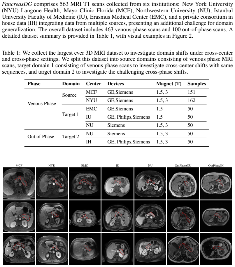
Summary. The paper introduces PancreasDG, a 563-case multi-institution 3D MRI pancreas segmentation dataset with 463 venous-phase and 100 out-of-phase T1 scans, and uses it to study domain generalization. It reports three insights: apparent cross-center shifts shrink with more training and test data; cross-center accuracy tracks source-domain accuracy; and cross-phase shifts behave differently and are not helped by longer training or larger models. To address cross-phase shift, the authors propose an EMA teacher-student semi-supervised pretraining scheme on 6.5K unlabeled MRI scans (along with 25 CT and 10 MRI annotated datasets), followed by nnU-Net fine-tuning on venous-phase data. The method is reported to achieve 70.39% Dice on NU out-of-phase and 66.61% on IH out-of-phase, versus 43.55% and 35.62% for the nnU-Net baseline.
Significance. If the headline result holds under controlled evaluation, this would be a meaningful advance: it would be the first demonstration that large-scale unlabeled MRI pretraining bridges cross-sequence shifts that conventional domain generalization methods cannot, and the dataset would be a valuable benchmark for the community. The paper also contains a useful negative result about cross-center 'domain shifts' being partly attributable to case-level sampling noise. Strengths include the double-blind two-pass annotation protocol, the multi-center and multi-sequence coverage, and the explicit separation of source, cross-center target, and cross-phase target domains. The current evidence is weakened by an uncontrolled comparison (prompted versus prompt-free models) and an undisclosed unlabeled-pretraining corpus, so the central claim is not yet fully verifiable.
major comments (4)
- [Section 4.3, Tables 2-4] The headline comparison is not controlled. The 'Ours' row in Table 2 (Dice 70.39 on NU) and in Table 3 (Dice 66.61 on IH) corresponds to the fine-tuned model with box prompts at inference, as shown in Table 4 ('+ Box'), while all domain generalization baselines and the nnU-Net baseline are prompt-free. Box prompts provide extra information about the target structure, so the comparison conflates prompt-free segmentation with interactive or guided segmentation. The prompt-free variant in Table 4 (65.70 NU, 60.95 IH) should be the primary comparison against the baselines; prompted variants should be reported as a separate setting, and ideally the same prompting protocol should be applied to the baselines.
- [Section 4.1] The unlabeled pretraining corpus is not specified: the paper states only that it uses '6.5K unlabeled 3D MRI scans from diverse centers and sequences' and does not list the centers, scanners, or sequence types. Since the two test domains in Section 3.2 are out-of-phase T1 scans from NU and IH, and since IH is described as in-house data, there is a live risk that out-of-phase scans from the test centers are present in the unlabeled pretraining pool. Without an explicit exclusion statement and a provenance breakdown, the reported cross-sequence gains cannot be interpreted as generalization to unseen domains. Please provide a data sheet for the 6.5K volumes, including institution and sequence composition, and state whether any NU or IH out-of-phase scans were included.
- [Abstract and Section 4.3] The abstract's '61.63% Dice score improvements and 87.00%' are relative improvements, not absolute Dice improvements: (70.39-43.55)/43.55 = 61.6% and (66.61-35.62)/35.62 = 87.0%. This presentation inflates the apparent effect and makes the numbers hard to compare with the domain generalization literature, which typically reports absolute Dice or percentage-point differences. Please report absolute percentage-point differences (26.84 and 30.99) alongside the relative changes, or clearly label the numbers as relative improvements.
- [Tables 2 and 3] The statistical evidence is incomplete. Table 2 reports standard deviations, but Table 3 reports only point estimates, and no significance tests are provided in either table. Given the large per-case variance in Table 2 (e.g., baseline 43.55 +/- 26.33), the difference between the proposed method and the best baseline should be assessed with paired tests (e.g., paired bootstrap or Wilcoxon signed-rank) on per-case Dice, and Table 3 should also report standard deviations or confidence intervals.
minor comments (6)
- [References] Several references are duplicated: [22] and [23] are both the SAM paper, [16] and [17] are both STU-Net, [8] and [9] are both SAM-Med2D, and [11] and [12] are both SegVol. Please consolidate them into single entries.
- [Section 4.1] The Felzenszwalb-Huttenlocher graph segmentation parameters (sigma, k, min_size) and the exact preprocessing used to create the regularization signal are not reported, so this component of the method is not reproducible.
- [Section 5, Figure 7] The fine-tuning-blocks experiment is presented only as a figure with no quantitative values; please include a table so that the claim that 'fine-tuning the last two blocks achieves the best balance' can be independently verified.
- [Section 3.2 and throughout] The terms 'cross-phase' and 'cross-sequence' are used interchangeably; please define them once and use a single consistent term throughout the paper.
- [Table 1] The claim that PancreasDG is 'the largest ever 3D MRI dataset' for cross-center shifts should be qualified with a comparison to existing public MRI segmentation datasets, since the basis for this claim is not stated.
- [Availability] During review, please provide an anonymized access mechanism for the dataset and code rather than only a public project URL, so that reviewers can verify the data splits and the pretraining corpus.
Circularity Check
No demonstrated circularity: the semi-supervised pretraining result is empirical and not equivalent to its inputs by construction; the main unresolved issue is the undisclosed composition of the 6.5K unlabeled pretraining pool, a data-contamination risk rather than a logical circularity.
full rationale
The claimed derivation is empirical: an EMA teacher-student model is pretrained on unlabeled volumes with graph-segmentation regularization (Section 4.1), then fine-tuned on venous-phase source data (Section 4.2), and evaluated on out-of-phase NU/IH test sets (Tables 2-3). None of the reported Dice gains follow algebraically from the loss or from the data definitions, so there is no self-definitional or fitted-input reduction. The dataset's source split is transparently built from the authors' prior work ('reorganized and expanded from [42]'), but this self-citation is provenance, not a load-bearing argument: it does not force the cross-sequence result. The pretraining pool does include the authors' own CirrMRI600 collection, but only as one of many datasets, so the conclusion is not carried by self-citation alone. The one serious caveat is in Section 4.1: 'we leverage 6.5K unlabeled 3D MRI scans from diverse centers and sequences' with no provenance breakdown, while Section 3.2 only guarantees that 'no data from target domains 1 or 2 will participate in any model's validation.' Because the unlabeled pool is not explicitly restricted to exclude out-of-phase NU/IH scans, the benchmark's 'unseen domain' claim could be weakened if such scans were present. This is a data-provenance and evaluation-validity risk, not a circularity that can be exhibited as an equation or a fitted parameter renamed as a prediction; therefore no circular step is scored.
Assumptions & free parameters
free parameters (2)
- Felzenszwalb-Huttenlocher graph segmentation parameters (sigma, k, min_size) =
not reported
- Pretraining hyperparameters (EMA decay, number of epochs, learning rate) =
10 epochs, lr=1e-5; EMA decay unspecified
assumptions (2)
- domain assumption Large-scale pretraining on diverse unlabeled MRI improves cross-sequence generalization without target-domain data.
- domain assumption nnU-Net is a sufficient baseline and backbone for fair comparison.
Cite this review
Pith. "Pith review of Rethink Domain Generalization in Heterogeneous Sequence MRI Segmentation." pith.science (2026). https://pith.science/paper/7DPDOTPL
@misc{pith2026250723110,
  author       = {Pith},
  title        = {Pith review of: Rethink Domain Generalization in Heterogeneous Sequence MRI Segmentation},
  year         = {2026},
  howpublished = {\url{https://pith.science/paper/7DPDOTPL}},
  note         = {Machine review of arXiv:2507.23110}
}
read the original abstract
Clinical magnetic-resonance (MR) protocols generate many T1 and T2 sequences whose appearance differs more than the acquisition sites that produce them. Existing domain-generalization benchmarks focus almost on cross-center shifts and overlook this dominant source of variability. Pancreas segmentation remains a major challenge in abdominal imaging: the gland is small, irregularly, surrounded by organs and fat, and often suffers from low T1 contrast. State-of-the-art deep networks that already achieve >90% Dice on the liver or kidneys still miss 20-30% of the pancreas. The organ is also systematically under-represented in public cross-domain benchmarks, despite its clinical importance in early cancer detection, surgery, and diabetes research. To close this gap, we present PancreasDG, a large-scale multi-center 3D MRI pancreas segmentation dataset for investigating domain generalization in medical imaging. The dataset comprises 563 MRI scans from six institutions, spanning both venous phase and out-of-phase sequences, enabling study of both cross-center and cross-sequence variations with pixel-accurate pancreas masks created by a double-blind, two-pass protocol. Through comprehensive analysis, we reveal three insights: (i) limited sampling introduces significant variance that may be mistaken for distribution shifts, (ii) cross-center performance correlates with source domain performance for identical sequences, and (iii) cross-sequence shifts require specialized solutions. We also propose a semi-supervised approach that leverages anatomical invariances, significantly outperforming state-of-the-art domain generalization techniques with 61.63% Dice score improvements and 87.00% on two test centers for cross-sequence segmentation. PancreasDG sets a new benchmark for domain generalization in medical imaging. Dataset, code, and models will be available at https://pancreasdg.netlify.app.
Figures


Figures from the paper (4 more)


Reference graph
Works this paper leans on
-
[1]
Invariance principle meets information bottleneck for out-of-distribution generalization
Kartik Ahuja, Ethan Caballero, Dinghuai Zhang, Jean-Christophe Gagnon-Audet, Yoshua Bengio, Ioannis Mitliagkas, and Irina Rish. Invariance principle meets information bottleneck for out-of-distribution generalization. Advances in Neural Information Processing Systems, 34:3438–3450, 2021
work page 2021
-
[2]
Pedro RAS Bassi, Wenxuan Li, Yucheng Tang, Fabian Isensee, Zifu Wang, Jieneng Chen, Yu-Cheng Chou, Yannick Kirchhoff, Maximilian Rokuss, Ziyan Huang, et al. Touchstone benchmark: Are we on the right way for evaluating ai algorithms for medical segmentation? arXiv preprint arXiv:2411.03670, 2024
arXiv 2024
-
[3]
Olivier Bernard, Alain Lalande, Clement Zotti, Frederick Cervenansky, Xin Yang, Pheng-Ann Heng, Irem Cetin, Karim Lekadir, Oscar Camara, Miguel Angel Gonzalez Ballester, et al. Deep learning techniques for automatic mri cardiac multi-structures segmentation and diagnosis: is the problem solved? IEEE transactions on medical imaging, 37(11):2514–2525, 2018
work page 2018
-
[4]
Multi-centre, multi-vendor and multi- disease cardiac segmentation: the m&ms challenge
Victor M Campello, Polyxeni Gkontra, Cristian Izquierdo, Carlos Martin-Isla, Alireza Sojoudi, Peter M Full, Klaus Maier-Hein, Yao Zhang, Zhiqiang He, Jun Ma, et al. Multi-centre, multi-vendor and multi- disease cardiac segmentation: the m&ms challenge. IEEE Transactions on Medical Imaging , 40(12): 3543–3554, 2021
work page 2021
-
[5]
Monai: An open-source framework for deep learning in healthcare
M Jorge Cardoso, Wenqi Li, Richard Brown, Nic Ma, Eric Kerfoot, Yiheng Wang, Benjamin Murrey, Andriy Myronenko, Can Zhao, Dong Yang, et al. Monai: An open-source framework for deep learning in healthcare. arXiv preprint arXiv:2211.02701, 2022
arXiv 2022
-
[6]
Realistic adversarial data augmentation for mr image segmentation
Chen Chen, Chen Qin, Huaqi Qiu, Cheng Ouyang, Shuo Wang, Liang Chen, Giacomo Tarroni, Wenjia Bai, and Daniel Rueckert. Realistic adversarial data augmentation for mr image segmentation. In Medical Image Computing and Computer Assisted Intervention–MICCAI 2020: 23rd International Conference, Lima, Peru, October 4–8, 2020, Proceedings, Part I 23, pages 667–...
work page 2020
-
[7]
Tianrun Chen, Lanyun Zhu, Chaotao Ding, Runlong Cao, Yan Wang, Zejian Li, Lingyun Sun, Papa Mao, and Ying Zang. Sam fails to segment anything?–sam-adapter: Adapting sam in underperformed scenes: Camouflage, shadow, medical image segmentation, and more. arXiv preprint arXiv:2304.09148, 2023
arXiv 2023
- [9]
Show all 42 references
-
[10]
Totalsegmentator mri: Sequence-independent segmentation of 59 anatomical structures in mr images
Tugba Akinci D’Antonoli, Lucas K Berger, Ashraya K Indrakanti, Nathan Vishwanathan, Jakob Weiß, Matthias Jung, Zeynep Berkarda, Alexander Rau, Marco Reisert, Thomas Küstner, et al. Totalsegmentator mri: Sequence-independent segmentation of 59 anatomical structures in mr images...
2024 arXiv
-
[11]
Segvol: Universal and interactive volumetric medical image segmentation
Yuxin Du, Fan Bai, Tiejun Huang, and Bo Zhao. Segvol: Universal and interactive volumetric medical image segmentation. arXiv preprint arXiv:2311.13385, 2023
2023 arXiv
-
[12]
Segvol: Universal and interactive volumetric medical image segmentation
Yuxin Du, Fan Bai, Tiejun Huang, and Bo Zhao. Segvol: Universal and interactive volumetric medical image segmentation. Advances in Neural Information Processing Systems, 37:110746–110783, 2025
2025
-
[13]
Probable domain generalization via quantile risk minimization.Advances in Neural Information Processing Systems, 35:17340–17358, 2022
Cian Eastwood, Alexander Robey, Shashank Singh, Julius V on Kügelgen, Hamed Hassani, George J Pappas, and Bernhard Schölkopf. Probable domain generalization via quantile risk minimization.Advances in Neural Information Processing Systems, 35:17340–17358, 2022
2022
-
[14]
In search of lost domain generalization
Ishaan Gulrajani and David Lopez-Paz. In search of lost domain generalization. arXiv preprint arXiv:2007.01434, 2020
2007 arXiv
-
[15]
Unetr: Transformers for 3d medical image segmentation
Ali Hatamizadeh, Yucheng Tang, Vishwesh Nath, Dong Yang, Andriy Myronenko, Bennett Landman, Holger R Roth, and Daguang Xu. Unetr: Transformers for 3d medical image segmentation. In Proceedings of the IEEE/CVF winter conference on applications of computer vision, pages 574–584, 2022
2022
-
[17]
Stu-net: Scalable and transferable medical image segmentation models empowered by large-scale supervised pre-training
Ziyan Huang, Haoyu Wang, Zhongying Deng, Jin Ye, Yanzhou Su, Hui Sun, Junjun He, Yun Gu, Lixu Gu, Shaoting Zhang, et al. Stu-net: Scalable and transferable medical image segmentation models empowered by large-scale supervised pre-training. arXiv preprint arXiv:2304.06716, 2023. 11
2023 arXiv
-
[18]
nnu-net: a self-configuring method for deep learning-based biomedical image segmentation
Fabian Isensee, Paul F Jaeger, Simon AA Kohl, Jens Petersen, and Klaus H Maier-Hein. nnu-net: a self-configuring method for deep learning-based biomedical image segmentation. Nature methods, 18(2): 203–211, 2021
2021
-
[19]
Cirrmri600+: Large scale mri collection and segmentation of cirrhotic liver
Debesh Jha, Onkar Kishor Susladkar, Vandan Gorade, Elif Keles, Matthew Antalek, Deniz Seyithanoglu, Timurhan Cebeci, Halil Ertugrul Aktas, Gulbiz Dagoglu Kartal, Sabahattin Kaymakoglu, et al. Cirrmri600+: Large scale mri collection and segmentation of cirrhotic liver. arXiv pr...
-
[20]
Amos: A large-scale abdominal multi-organ benchmark for versatile medical image segmentation
Yuanfeng Ji, Haotian Bai, Chongjian Ge, Jie Yang, Ye Zhu, Ruimao Zhang, Zhen Li, Lingyan Zhanng, Wanling Ma, Xiang Wan, et al. Amos: A large-scale abdominal multi-organ benchmark for versatile medical image segmentation. Advances in neural information processing systems, 35:36...
2022
-
[21]
Domain generalizer: A few-shot meta learning framework for domain generalization in medical imaging
Pulkit Khandelwal and Paul Yushkevich. Domain generalizer: A few-shot meta learning framework for domain generalization in medical imaging. In Domain Adaptation and Representation Transfer, and Distributed and Collaborative Learning: Second MICCAI Workshop, DART 2020, and Firs...
2020
-
[22]
Segment anything
Alexander Kirillov, Eric Mintun, Nikhila Ravi, Hanzi Mao, Chloe Rolland, Laura Gustafson, Tete Xiao, Spencer Whitehead, Alexander C Berg, Wan-Yen Lo, et al. Segment anything. In Proceedings of the IEEE/CVF international conference on computer vision, pages 4015–4026, 2023
2023
-
[23]
Segment anything
Alexander Kirillov, Eric Mintun, Nikhila Ravi, Hanzi Mao, Chloe Rolland, Laura Gustafson, Tete Xiao, Spencer Whitehead, Alexander C Berg, Wan-Yen Lo, et al. Segment anything. In Proceedings of the IEEE/CVF International Conference on Computer Vision, pages 4015–4026, 2023
2023
-
[24]
Out-of-distribution generalization via risk extrapolation (rex)
David Krueger, Ethan Caballero, Joern-Henrik Jacobsen, Amy Zhang, Jonathan Binas, Dinghuai Zhang, Remi Le Priol, and Aaron Courville. Out-of-distribution generalization via risk extrapolation (rex). In International conference on machine learning, pages 5815–5826. PMLR, 2021
2021
-
[25]
Shape-aware meta-learning for generalizing prostate mri segmentation to unseen domains
Quande Liu, Qi Dou, and Pheng-Ann Heng. Shape-aware meta-learning for generalizing prostate mri segmentation to unseen domains. In Medical Image Computing and Computer Assisted Intervention– MICCAI 2020: 23rd International Conference, Lima, Peru, October 4–8, 2020, Proceedings...
2020
-
[26]
Ms-net: Multi-site network for improving prostate segmentation with heterogeneous mri data
Quande Liu, Qi Dou, Lequan Yu, and Pheng Ann Heng. Ms-net: Multi-site network for improving prostate segmentation with heterogeneous mri data. IEEE Transactions on Medical Imaging, 2020
2020
-
[27]
Feddg: Federated domain generaliza- tion on medical image segmentation via episodic learning in continuous frequency space
Quande Liu, Cheng Chen, Jing Qin, Qi Dou, and Pheng-Ann Heng. Feddg: Federated domain generaliza- tion on medical image segmentation via episodic learning in continuous frequency space. In Proceedings of the IEEE/CVF conference on computer vision and pattern recognition, pages...
2021
-
[28]
Segment anything in medical images
Jun Ma, Yuting He, Feifei Li, Lin Han, Chenyu You, and Bo Wang. Segment anything in medical images. Nature Communications, 15(1):654, 2024
2024
-
[29]
Duke liver dataset: A publicly available liver mri dataset with liver segmentation masks and series labels
Jacob A Macdonald, Zhe Zhu, Brandon Konkel, Maciej A Mazurowski, Walter F Wiggins, and Mustafa R Bashir. Duke liver dataset: A publicly available liver mri dataset with liver segmentation masks and series labels. Radiology: Artificial Intelligence, 5(5):e220275, 2023
2023
-
[30]
The multimodal brain tumor image segmentation benchmark (brats)
Bjoern H Menze, Andras Jakab, Stefan Bauer, Jayashree Kalpathy-Cramer, Keyvan Farahani, Justin Kirby, Yuliya Burren, Nicole Porz, Johannes Slotboom, Roland Wiest, et al. The multimodal brain tumor image segmentation benchmark (brats). IEEE transactions on medical imaging, 34(1...
1993
-
[31]
Gradient starvation: A learning proclivity in neural networks
Mohammad Pezeshki, Oumar Kaba, Yoshua Bengio, Aaron C Courville, Doina Precup, and Guillaume Lajoie. Gradient starvation: A learning proclivity in neural networks. Advances in Neural Information Processing Systems, 34:1256–1272, 2021
2021
-
[32]
Learning transferable visual models from natural language supervision
Alec Radford, Jong Wook Kim, Chris Hallacy, Aditya Ramesh, Gabriel Goh, Sandhini Agarwal, Girish Sastry, Amanda Askell, Pamela Mishkin, Jack Clark, et al. Learning transferable visual models from natural language supervision. In International conference on machine learning, pa...
2021
-
[33]
U-net: Convolutional networks for biomedical image segmentation
Olaf Ronneberger, Philipp Fischer, and Thomas Brox. U-net: Convolutional networks for biomedical image segmentation. In Medical image computing and computer-assisted intervention–MICCAI 2015: 18th international conference, Munich, Germany, October 5-9, 2015, proceedings, part ...
2015
-
[34]
Distributionally robust neural networks for group shifts: On the importance of regularization for worst-case generalization.arXiv preprint arXiv:1911.08731, 2019
Shiori Sagawa, Pang Wei Koh, Tatsunori B Hashimoto, and Percy Liang. Distributionally robust neural networks for group shifts: On the importance of regularization for worst-case generalization.arXiv preprint arXiv:1911.08731, 2019
1911 arXiv
-
[35]
Sam-med3d: towards general- purpose segmentation models for volumetric medical images.Preprint at https://arxiv
Haoyu Wang, Sizheng Guo, Jin Ye, Zhongyi Deng, Junlong Cheng, et al. Sam-med3d: towards general- purpose segmentation models for volumetric medical images.Preprint at https://arxiv. org/abs/2310.15161, 2024
2024 arXiv
-
[36]
Generalizing to unseen domains: A survey on domain generalization
Jindong Wang, Cuiling Lan, Chang Liu, Yidong Ouyang, Tao Qin, Wang Lu, Yiqiang Chen, Wenjun Zeng, and Philip Yu. Generalizing to unseen domains: A survey on domain generalization. IEEE Transactions on Knowledge and Data Engineering, 2022
2022
-
[37]
Totalsegmentator: robust segmentation of 104 anatomic structures in ct images
Jakob Wasserthal, Hanns-Christian Breit, Manfred T Meyer, Maurice Pradella, Daniel Hinck, Alexander W Sauter, Tobias Heye, Daniel T Boll, Joshy Cyriac, Shan Yang, et al. Totalsegmentator: robust segmentation of 104 anatomic structures in ct images. Radiology: Artificial Intell...
2023
-
[38]
Robust and generalizable visual representation learning via random convolutions
Zhenlin Xu, Deyi Liu, Junlin Yang, Colin Raffel, and Marc Niethammer. Robust and generalizable visual representation learning via random convolutions. In International Conference on Learning Representations, 2021
2021
-
[39]
Generalizing deep learning for medical image segmentation to unseen domains via deep stacked transformation
Ling Zhang, Xiaosong Wang, Dong Yang, Thomas Sanford, Stephanie Harmon, Baris Turkbey, Bradford J Wood, Holger Roth, Andriy Myronenko, Daguang Xu, et al. Generalizing deep learning for medical image segmentation to unseen domains via deep stacked transformation. IEEE Transacti...
2020
-
[40]
Domain generalization with adversarial intensity attack for medical image segmentation
Zheyuan Zhang, Bin Wang, Lanhong Yao, Ugur Demir, Debesh Jha, Ismail Baris Turkbey, Boqing Gong, and Ulas Bagci. Domain generalization with adversarial intensity attack for medical image segmentation. arXiv preprint arXiv:2304.02720, 2023
2023 arXiv
-
[41]
Deep learning algorithms for pancreas segmentation from radiology scans: A review
Zheyuan Zhang, Lanhong Yao, Elif Keles, Yury Velichko, and Ulas Bagci. Deep learning algorithms for pancreas segmentation from radiology scans: A review. Advances in Clinical Radiology, 5(1):31–52, 2023
2023
-
[42]
Zheyuan Zhang, Elif Keles, Gorkem Durak, Yavuz Taktak, Onkar Susladkar, Vandan Gorade, Debesh Jha, Asli C. Ormeci, Alpay Medetalibeyoglu, Lanhong Yao, Bin Wang, Ilkin Sevgi Isler, Linkai Peng, Hongyi Pan, Camila Lopes Vendrami, Amir Bourhani, Yury Velichko, Boqing Gong, Concet...
2025
-
[43]
Domain generalization with mixstyle
Kaiyang Zhou, Yongxin Yang, Yu Qiao, and Tao Xiang. Domain generalization with mixstyle. In International Conference on Learning Representations, 2021
2021
-
[44]
Domain generalization: A survey
Kaiyang Zhou, Ziwei Liu, Yu Qiao, Tao Xiang, and Chen Change Loy. Domain generalization: A survey. IEEE Transactions on Pattern Analysis and Machine Intelligence, 2022. A Technical Appendices and Supplementary Material 13 Dice: 43.81Baseline Dice: 43.61Baseline Dice: 43.07Base...
2022
Reviewed August 6, 2026 · model on record in the stance chip above.
Discussion (0). Sign in to comment.