REVIEW 5 major objections 5 minor 62 references
Rethinking Decoder Design: Improving Biomarker Segmentation Using Depth-to-Space Restoration and Residual Linear Attention
T0 review · 5 major / 5 minor · reviewed 2026-08-15 · deepseek-v4-flash
Pith's one-line read A decoder redesign lifts biomarker segmentation by up to 4 percent IoU, without pre-trained encoders.
desk verdict Solid empirical decoder paper whose SOTA claims are undercut by missing error bars and a patch-size asymmetry; worth reviewing, but needs revision. read the letter →
The pith
A machine-rendered reading of the paper's core claim, the machinery that carries it, and where it could break.
The reading
What carries the argument
The load-bearing mechanism is depth-to-space upsampling combined with residual linear attention. DSUB computes the filter count needed for a depth-to-space (sub-pixel) rearrangement—filters equal output channels times the square of the downsampling factor—so that the feature map is reshaped from channel-heavy to spatially detailed without content loss. RLAB iteratively refines each skip connection with a residual block, concatenates it with the decoder feature map, projects to queries, keys, and values, applies scaled dot-product attention, and adds the attended output back through a residual connection. CASAB completes the pathway by combining channel attention (global average and max pooling) with spatial attention built from mean, max, min, and sum pooling.
What would settle it
Run MCADS and the strongest baseline (EMCAD) on MoNuSeg for ten independent seeds and compare the per-seed IoU distributions; if the mean difference is within one pooled standard deviation, the claimed state-of-the-art gain is not statistically separable from noise. Alternatively, an independent reproduction using the released code with the paper's exact preprocessing and patch protocol should reproduce the 2.76% margin.
Extended reading notes
Core claim
The central claim is that three decoder components—Depth-to-Space Upsampling Blocks (DSUB) applied in the first two stages, an Effective Upsampling Block (EUB) in later stages, a Channel and Spatial Attention Block (CASAB), and a Residual Linear Attention Block (RLAB) on skip connections—jointly produce more accurate biomarker segmentation than existing decoders. On the paper's own terms, the depth-to-space operation preserves encoder detail that nearest-neighbor upsampling loses, while RLAB lets the decoder integrate refined encoder features without discarding its own, and CASAB selects the channels and spatial regions that matter. The result is a decoder that is encoder-agnostic and, when paired with a modified U2-Net encoder, achieves the reported gains.
Load-bearing premise
The paper assumes the reproduced baseline scores, averaged over five runs, reflect each method's best achievable performance under the same tuning protocol used for MCADS, and that the reported 2-4 point IoU margins exceed run-to-run variability; no standard deviations or significance tests are reported (Section 4.2, Tables 1-4).
Editorial extensions
If this is right
- End-to-end training from scratch can match or beat pre-trained-encoder methods on four medical segmentation benchmarks, so pre-training is not a prerequisite for state-of-the-art accuracy.
- The decoder's gains transfer across CNN and Transformer encoders (ResNet-50, MaxViT-B, PVT v2-B2, modified U2-Net), so it can be dropped into existing architectures.
- The ablation studies indicate the first two decoder stages carry most of the upsampling benefit; later DSUB stages add under one IoU point while roughly tripling GFlops.
- Boundary metrics (HD95, ASD) improve by larger relative amounts than IoU, implying fewer fragmented or over-segmented regions.
Reading between the lines
- A direct test of the paper's encoder-limitation argument would be a scaling curve: holding the MCADS decoder fixed while scaling encoder width and depth, to see where accuracy plateaus.
- The same decoder recipe could be applied to other dense prediction tasks (instance masks, depth, surface normals) where high-frequency detail is lost during upsampling.
- Because no variance is reported, the central numerical claim is falsifiable by re-running with many seeds; the published margins, not the architecture, are the quantity to scrutinize.
Signed reviews
Editorial analysis
A structured set of objections, weighed in public.
Referee Report
Summary. The paper proposes MCADS, a new decoder for medical image segmentation that combines depth-to-space upsampling (DSUB), an effective upsampling block (EUB), channel and spatial attention (CASAB), and a residual linear attention block (RLAB), together with a modified U2-Net encoder that incorporates inner attention and CASAB. The method is evaluated on four datasets (MoNuSeg, DSB, EM, TNBC) against CNN-, transformer-, and SAM-based baselines, and the authors report absolute IoU gains of 2.76–4.03 points over prior state-of-the-art methods. Ablation studies on MoNuSeg examine the contribution of each architectural component and the placement of DSUB stages. The paper also reports inference-time and complexity comparisons and provides a public code link.
Significance. If the reported gains are statistically robust, the paper makes a useful empirical contribution: it shows that a carefully designed decoder can yield competitive or better segmentation accuracy than methods relying on pre-trained backbones, and it provides a systematic ablation of the decoder components. Strengths include the breadth of datasets, the reproduced baselines (for most entries), the ablation coverage, and the public code release. However, the central claim of outperforming state-of-the-art methods rests on five-run averages without variance estimates or significance tests, and the zero-shot comparison on TNBC is not cleanly specified. These issues currently weaken the evidentiary value of the headline margins and need to be addressed before the comparative claims can be fully assessed.
major comments (5)
- [Section 4.2, Tables 1–4] The central claim of outperforming SOTA by 2.76–4.03 IoU points rests on five-run averages reported as point estimates without standard deviations, confidence intervals, or significance tests. Since the reported margins are only a few points and the baseline spread is unknown, the claim is not yet supported. Please report variance across runs and perform paired significance tests (e.g., paired t-test or Wilcoxon signed-rank test over runs, or bootstrap over test images) against the closest baselines such as EMCAD and PVT-CASCADE.
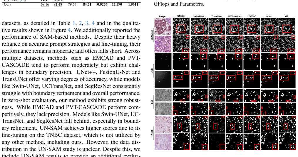
- [Section 4.2, Table 4] The TNBC 'zero-shot' comparison is under-specified and partly invalid: the paper does not state which training split the zero-shot models were trained on, and it includes UN-SAM, which the text acknowledges was fine-tuned on TNBC, in the same table without a distinguishing marker. Because UN-SAM (72.27 IoU) is not a zero-shot result, its inclusion makes the comparative table misleading. Please clarify the exact training set used for all zero-shot models, and either remove UN-SAM from the zero-shot table or clearly mark it as a fine-tuned reference.
- [Section 4.2, Tables 1–4] The statement 'we reproduced all results' in Section 4.2 and the same claim in the table captions is contradicted by the asterisk-marked entries (InstaSAM, All-in-SAM, UN-SAM, SAC 0-expert in Tables 1, 2, and 4) which are copied from original papers rather than reproduced. This discrepancy undermines the fairness of the comparisons because the copied entries may follow different evaluation protocols. Please state exactly which entries were reproduced and which were taken from the literature, and justify that the evaluation protocols are otherwise comparable.
- [Section 4.2, patch-size protocol] The experimental setup uses 256x256 training patches for the proposed method and end-to-end baselines, but 224x224 patches for methods with pre-trained backbones. Patch size can affect segmentation IoU independently of decoder quality, so this asymmetry introduces a confound in the comparisons against pre-trained-backbone methods (e.g., PVT-CASCADE, EMCAD, UCTransNet). Please either use a common patch size for all methods or provide an ablation showing that the reported margins are insensitive to patch size (for example, report the proposed method at 224x224 and at least one major baseline at 256x256).
- [Table 6 and Section 5.2] The ablation study chooses DSUB in the first two decoder stages as the final configuration, but the table shows that using DSUB in more stages continues to improve IoU (from 74.04 to 74.88 when DSUB is used in all five stages). The selection is justified by a performance/complexity tradeoff, which is a legitimate design choice, but the paper should state explicitly that the selected configuration is not the accuracy-optimal one and should report the sensitivity of the main SOTA comparison to this choice, since a different point on the tradeoff could affect the reported margins.
minor comments (5)
- [Section 3.1.2, Equation (2)] The text says the number of filters F is the product of output channels and the square of the downsampling factor, but writes 'F=C prev×2 d'; please disambiguate the notation (e.g., F = C_prev × 2^d) and clarify which 'output channels' are meant.
- [Section 3.1.5, Equation (8)] Equation (8) omits the iteration index i that appears in the surrounding text; please make the notation consistent so each residual iteration is clearly indexed.
- [Section 3.2, Figure 2] 'M repents the encoder stage' appears to be a typo for 'M represents the encoder stage'.
- [Acknowledgments] 'Coummunications' is misspelled; it should be 'Communications'.
- [Section 4.3, Computational Cost] The inference-time comparison reports only EMCAD and the proposed method; please clarify whether the comparison is on the same hardware and whether the times are averaged over the same number of runs, and consider reporting the variance of these timing measurements.
Circularity Check
No significant circularity: the decoder design and benchmark claims are self-contained; the only self-citation (Histoseg) is a non-load-bearing related-work mention.
full rationale
The paper is an empirical architecture paper, not a derivation from assumptions. The claimed SOTA gains (abstract; Tables 1-4) are measured IoU/Dice on held-out test sets after training on the corresponding train splits, so the reported numbers are not defined in terms of the method's own parameters or outputs. The MCADS decoder components (DSUB, EUB, CASAB, RLAB) are specified by independent equations (Eqs. 1-11) that do not reference the benchmark metrics or the final claims; ablations in Section 5 vary components and measure performance, rather than fitting a parameter and renaming it a prediction. The only self-citation is [51] (Wazir and Fraz, Histoseg) in the Related Work sentence "Studies [28, 44, 51, 59] have demonstrated that incorporating features from multiple deep layers can improve results." That citation is a related-work attribution, not a load-bearing justification of the decoder design or of any empirical result. Concerns about baseline tuning fairness, absent standard deviations, the patch-size asymmetry, and the TNBC zero-shot protocol (Tables 1-4, Section 4.2) are potential statistical or comparison-fairness issues, not instances of a result reducing by construction to its inputs. The footnote "Results marked with * were copied from the original papers" alongside the claim "we reproduced all results" is an internal reporting inconsistency, but it does not make any prediction equal to its input. No step in the paper's derivation chain is equivalent to its own input, so there is no circularity beyond a minor, non-load-bearing self-citation.
Assumptions & free parameters
free parameters (2)
- RLAB iteration schedule per stage =
[5, 4, 3, 2, 1]
- DSUB placement (first two decoder stages) =
Bridge and Stage 4 use DSUB; Stages 3-1 use EUB
assumptions (4)
- domain assumption Depth-to-space upsampling preserves information better than nearest-neighbor or bilinear upsampling in early decoder stages.
- domain assumption Softmax dot-product attention (Eq. 10) can effectively fuse encoder and decoder features across scales.
- domain assumption Binary cross-entropy summed over six deep-supervision maps is an appropriate training objective.
- domain assumption End-to-end training without pre-trained encoders can achieve state-of-the-art accuracy.
Cite this review
Pith. "Pith review of Rethinking Decoder Design: Improving Biomarker Segmentation Using Depth-to-Space Restoration and Residual Linear Attention." pith.science (2026). https://pith.science/paper/7IUXPJ2D
@misc{pith2026250618335,
  author       = {Pith},
  title        = {Pith review of: Rethinking Decoder Design: Improving Biomarker Segmentation Using Depth-to-Space Restoration and Residual Linear Attention},
  year         = {2026},
  howpublished = {\url{https://pith.science/paper/7IUXPJ2D}},
  note         = {Machine review of arXiv:2506.18335}
}
read the original abstract
Segmenting biomarkers in medical images is crucial for various biotech applications. Despite advances, Transformer and CNN based methods often struggle with variations in staining and morphology, limiting feature extraction. In medical image segmentation, where datasets often have limited sample availability, recent state-of-the-art (SOTA) methods achieve higher accuracy by leveraging pre-trained encoders, whereas end-to-end methods tend to underperform. This is due to challenges in effectively transferring rich multiscale features from encoders to decoders, as well as limitations in decoder efficiency. To address these issues, we propose an architecture that captures multi-scale local and global contextual information and a novel decoder design, which effectively integrates features from the encoder, emphasizes important channels and regions, and reconstructs spatial dimensions to enhance segmentation accuracy. Our method, compatible with various encoders, outperforms SOTA methods, as demonstrated by experiments on four datasets and ablation studies. Specifically, our method achieves absolute performance gains of 2.76% on MoNuSeg, 3.12% on DSB, 2.87% on Electron Microscopy, and 4.03% on TNBC datasets compared to existing SOTA methods. Code: https://github.com/saadwazir/MCADS-Decoder
Figures


Figures from the paper (1 more)
Reference graph
Works this paper leans on
-
[1]
Intuitive explanation of skip connections in deep learning.https://theaisummer.com/, 2020
Nikolas Adaloglou. Intuitive explanation of skip connections in deep learning.https://theaisummer.com/, 2020. 6
work page 2020
-
[2]
Ibtihaj Ahmad, Yong Xia, Hengfei Cui, and Zain Ul Islam. Dan-nucnet: A dual attention based framework for nuclei segmentation in cancer histology images under wild clinical conditions.Expert Systems with Applications, 213:118945,
-
[3]
Md Zahangir Alom, Mahmudul Hasan, Chris Yakopcic, Tarek M Taha, and Vijayan K Asari. Recurrent residual con- volutional neural network based on u-net (r2u-net) for med- ical image segmentation.arXiv preprint arXiv:1802.06955,
-
[4]
Yutong Cai and Yong Wang. Ma-unet: An improved ver- sion of unet based on multi-scale and attention mechanism for medical image segmentation. InThird international conference on electronics and communication; network and computer technology (ECNCT 2021), pages 205–211. SPIE,
work page 2021
-
[5]
Juan C Caicedo, Allen Goodman, Kyle W Karhohs, Beth A Cimini, Jeanelle Ackerman, Marzieh Haghighi, CherKeng Heng, Tim Becker, Minh Doan, Claire McQuin, et al. Nu- cleus segmentation across imaging experiments: the 2018 data science bowl.Nature methods, 16(12):1247–1253,
work page 2018
-
[6]
Swin-unet: Unet-like pure transformer for medical image segmentation
Hu Cao, Yueyue Wang, Joy Chen, Dongsheng Jiang, Xi- aopeng Zhang, Qi Tian, and Manning Wang. Swin-unet: Unet-like pure transformer for medical image segmentation. InEuropean conference on computer vision, pages 205–218. Springer, 2022. 2, 6, 7
work page 2022
-
[7]
Gongping Chen, Yuming Liu, Jiang Qian, Jianxun Zhang, Xiaotao Yin, Liang Cui, and Yu Dai. Dseu-net: A novel deep supervision seu-net for medical ultrasound image seg- mentation.Expert Systems with Applications, 223:119939,
-
[8]
Jieneng Chen, Yongyi Lu, Qihang Yu, Xiangde Luo, Ehsan Adeli, Yan Wang, Le Lu, Alan L Yuille, and Yuyin Zhou. Transunet: Transformers make strong encoders for medi- cal image segmentation.arXiv preprint arXiv:2102.04306,
Show all 62 references
-
[9]
Liang-Chieh Chen, George Papandreou, Iasonas Kokkinos, Kevin Murphy, and Alan L Yuille. Deeplab: Semantic image segmentation with deep convolutional nets, atrous convolu- tion, and fully connected crfs.IEEE transactions on pattern analysis and machine intelligence, 40(4):834–8...
2017
-
[10]
Un-sam: Universal prompt-free segmentation for generalized nuclei images.arXiv preprint arXiv:2402.16663, 2024
Zhen Chen, Qing Xu, Xinyu Liu, and Yixuan Yuan. Un-sam: Universal prompt-free segmentation for generalized nuclei images.arXiv preprint arXiv:2402.16663, 2024. 3, 6, 7
2024 arXiv
-
[11]
Conditional positional encodings for vision transformers.arXiv preprint arXiv:2102.10882, 2021
Xiangxiang Chu, Zhi Tian, Bo Zhang, Xinlong Wang, and Chunhua Shen. Conditional positional encodings for vision transformers.arXiv preprint arXiv:2102.10882, 2021. 1, 2
2021 arXiv
-
[12]
All-in-sam: from weak annota- tion to pixel-wise nuclei segmentation with prompt-based finetuning
Can Cui, Ruining Deng, Quan Liu, Tianyuan Yao, Shunx- ing Bao, Lucas W Remedios, Bennett A Landman, Yucheng Tang, and Yuankai Huo. All-in-sam: from weak annota- tion to pixel-wise nuclei segmentation with prompt-based finetuning. InJournal of Physics: Conference Series, page 0...
2024
-
[13]
Polyp-pvt: Polyp segmen- tation with pyramid vision transformers.arXiv preprint arXiv:2108.06932, 2021
Bo Dong, Wenhai Wang, Deng-Ping Fan, Jinpeng Li, Huazhu Fu, and Ling Shao. Polyp-pvt: Polyp segmen- tation with pyramid vision transformers.arXiv preprint arXiv:2108.06932, 2021. 2
2021 arXiv
-
[14]
Pamsnet: A medical image segmentation network based on spatial pyramid and attention mecha- nism.Biomedical Signal Processing and Control, 94: 106285, 2024
Yuncong Feng, Xiaoyan Zhu, Xiaoli Zhang, Yang Li, and Huimin Lu. Pamsnet: A medical image segmentation network based on spatial pyramid and attention mecha- nism.Biomedical Signal Processing and Control, 94: 106285, 2024. 1
2024
-
[15]
An enhanced u-network by combining ppm and cbam for med- ical image segmentation.IEEE Access, 12:107098–107112,
Zhongming Fu, Hejian Chen, Mengsi He, and Li Liu. An enhanced u-network by combining ppm and cbam for med- ical image segmentation.IEEE Access, 12:107098–107112,
-
[16]
Double encoder-decoder networks for gastroin- testinal polyp segmentation
Adrian Galdran, Gustavo Carneiro, and Miguel A Gonz ´alez Ballester. Double encoder-decoder networks for gastroin- testinal polyp segmentation. InPattern Recognition. ICPR International Workshops and Challenges: Virtual Event, January 10–15, 2021, Proceedings, Part I, pages 29...
2021
-
[17]
Hover-net: Simultaneous segmentation and classi- fication of nuclei in multi-tissue histology images.Medical image analysis, 58:101563, 2019
Simon Graham, Quoc Dang Vu, Shan E Ahmed Raza, Ayesha Azam, Yee Wah Tsang, Jin Tae Kwak, and Nasir Rajpoot. Hover-net: Simultaneous segmentation and classi- fication of nuclei in multi-tissue histology images.Medical image analysis, 58:101563, 2019. 2, 6, 7
2019
-
[18]
Squeeze-and-excitation net- works
Jie Hu, Li Shen, and Gang Sun. Squeeze-and-excitation net- works. InProceedings of the IEEE conference on computer vision and pattern recognition, pages 7132–7141, 2018. 2
2018
-
[19]
Unet 3+: A full-scale connected unet for medical image segmentation
Huimin Huang, Lanfen Lin, Ruofeng Tong, Hongjie Hu, Qiaowei Zhang, Yutaro Iwamoto, Xianhua Han, Yen-Wei Chen, and Jian Wu. Unet 3+: A full-scale connected unet for medical image segmentation. InICASSP 2020-2020 IEEE international conference on acoustics, speech and sig- nal pr...
2020
-
[20]
nnu-net: a self-configuring method for deep learning-based biomedical image segmen- tation.Nature methods, 18(2):203–211, 2021
Fabian Isensee, Paul F Jaeger, Simon AA Kohl, Jens Pe- tersen, and Klaus H Maier-Hein. nnu-net: a self-configuring method for deep learning-based biomedical image segmen- tation.Nature methods, 18(2):203–211, 2021. 2, 6, 7
2021
-
[21]
How much position information do convolutional neural networks en- code?arXiv preprint arXiv:2001.08248, 2020
Md Amirul Islam, Sen Jia, and Neil DB Bruce. How much position information do convolutional neural networks en- code?arXiv preprint arXiv:2001.08248, 2020. 1, 2
2001 arXiv
-
[22]
A survey of loss functions for semantic seg- mentation
Shruti Jadon. A survey of loss functions for semantic seg- mentation. In2020 IEEE Conference on Computational Intelligence in Bioinformatics and Computational Biology (CIBCB), pages 1–7, 2020. 6
2020
-
[23]
Attresdu- net: Medical image segmentation using attention-based residual double u-net.arXiv preprint arXiv:2306.14255,
Akib Mohammed Khan, Alif Ashrafee, Fahim Shahriar Khan, Md Bakhtiar Hasan, and Md Hasanul Kabir. Attresdu- net: Medical image segmentation using attention-based residual double u-net.arXiv preprint arXiv:2306.14255,
-
[24]
Segment any- thing
Alexander Kirillov, Eric Mintun, Nikhila Ravi, Hanzi Mao, Chloe Rolland, Laura Gustafson, Tete Xiao, Spencer White- head, Alexander C Berg, Wan-Yen Lo, et al. Segment any- thing. InProceedings of the IEEE/CVF International Con- ference on Computer Vision, pages 4015–4026, 2023. 3
2023
-
[25]
A dataset and a technique for generalized nuclear segmentation for computational pathology.IEEE transactions on medical imaging, 36(7):1550–1560, 2017
Neeraj Kumar, Ruchika Verma, Sanuj Sharma, Surabhi Bhargava, Abhishek Vahadane, and Amit Sethi. A dataset and a technique for generalized nuclear segmentation for computational pathology.IEEE transactions on medical imaging, 36(7):1550–1560, 2017. 6
2017
-
[26]
FusionU-Net: U- Net with enhanced skip connection for pathology image seg- mentation
Zongyi Li, Hongbing Lyu, and Jun Wang. FusionU-Net: U- Net with enhanced skip connection for pathology image seg- mentation. InProceedings of the 15th Asian Conference on Machine Learning, pages 694–706. PMLR, 2024. 6, 7
2024
-
[27]
Rethinking skip connection with layer normalization in transformers and resnets.arXiv preprint arXiv:2105.07205, 2021
Fenglin Liu, Xuancheng Ren, Zhiyuan Zhang, Xu Sun, and Yuexian Zou. Rethinking skip connection with layer normalization in transformers and resnets.arXiv preprint arXiv:2105.07205, 2021. 5
2021 arXiv
-
[28]
Fully convolutional networks for semantic segmentation
Jonathan Long, Evan Shelhamer, and Trevor Darrell. Fully convolutional networks for semantic segmentation. InPro- ceedings of the IEEE conference on computer vision and pat- tern recognition, pages 3431–3440, 2015. 1, 2
2015
-
[29]
Learning for structured prediction using approximate subgradient descent with working sets
Aurelien Lucchi, Yunpeng Li, and Pascal Fua. Learning for structured prediction using approximate subgradient descent with working sets. InProceedings of the IEEE Conference on Computer Vision and Pattern Recognition (CVPR), 2013. 6
2013
-
[30]
Segment anything in medical images.Nature Communications, 15(1):654, 2024
Jun Ma, Yuting He, Feifei Li, Lin Han, Chenyu You, and Bo Wang. Segment anything in medical images.Nature Communications, 15(1):654, 2024. 3
2024
-
[31]
Alrelu: A different approach on leaky relu activation function to improve neural networks performance.arXiv preprint arXiv:2012.07564, 2020
Stamatis Mastromichalakis. Alrelu: A different approach on leaky relu activation function to improve neural networks performance.arXiv preprint arXiv:2012.07564, 2020. 3
2012 arXiv
-
[32]
3d mri brain tumor segmentation using autoencoder regularization
Andriy Myronenko. 3d mri brain tumor segmentation using autoencoder regularization. InBrainlesion: Glioma, Multi- ple Sclerosis, Stroke and Traumatic Brain Injuries: 4th In- ternational Workshop, BrainLes 2018, Held in Conjunction with MICCAI 2018, Granada, Spain, September 16...
2018
-
[33]
Segment any cell: A sam-based auto- prompting fine-tuning framework for nuclei segmentation
Saiyang Na, Yuzhi Guo, Feng Jiang, Hehuan Ma, and Junzhou Huang. Segment any cell: A sam-based auto- prompting fine-tuning framework for nuclei segmentation. arXiv preprint arXiv:2401.13220, 2024. 3, 6
2024 arXiv
-
[34]
In- stasam: Instance-aware segment any nuclei model with point annotations
Siwoo Nam, Hyun Namgung, Jaehoon Jeong, Miguel Luna, Soopil Kim, Philip Chikontwe, and Sang Hyun Park. In- stasam: Instance-aware segment any nuclei model with point annotations. InInternational Conference on Medical Image Computing and Computer-Assisted Intervention, pages 232–
-
[35]
Segmentation of nuclei in histopathology images by deep re- gression of the distance map.IEEE transactions on medical imaging, 38(2):448–459, 2018
Peter Naylor, Marick La ´e, Fabien Reyal, and Thomas Walter. Segmentation of nuclei in histopathology images by deep re- gression of the distance map.IEEE transactions on medical imaging, 38(2):448–459, 2018. 6
2018
-
[36]
Raunet: Residual attention u-net for semantic segmentation of cataract surgical instruments
Zhen-Liang Ni, Gui-Bin Bian, Xiao-Hu Zhou, Zeng-Guang Hou, Xiao-Liang Xie, Chen Wang, Yan-Jie Zhou, Rui-Qi Li, and Zhen Li. Raunet: Residual attention u-net for semantic segmentation of cataract surgical instruments. InInterna- tional Conference on Neural Information Processin...
2019
-
[37]
Atten- tion u-net: Learning where to look for the pancreas.arXiv preprint arXiv:1804.03999, 2018
Ozan Oktay, Jo Schlemper, Loic Le Folgoc, Matthew Lee, Mattias Heinrich, Kazunari Misawa, Kensaku Mori, Steven McDonagh, Nils Y Hammerla, Bernhard Kainz, et al. Atten- tion u-net: Learning where to look for the pancreas.arXiv preprint arXiv:1804.03999, 2018. 1, 2, 5
2018 arXiv
-
[38]
U2-net: Go- ing deeper with nested u-structure for salient object detec- tion.Pattern recognition, 106:107404, 2020
Xuebin Qin, Zichen Zhang, Chenyang Huang, Masood De- hghan, Osmar R Zaiane, and Martin Jagersand. U2-net: Go- ing deeper with nested u-structure for salient object detec- tion.Pattern recognition, 106:107404, 2020. 1, 2, 5, 6, 7
2020
-
[39]
Rahman, S
M. Rahman, S. Shokouhmand, S. Bhatt, and M. Faezipour. Mist: Medical image segmentation transformer with convo- lutional attention mixing (cam) decoder. In2024 IEEE/CVF Winter Conference on Applications of Computer Vision (WACV), pages 403–412, Los Alamitos, CA, USA, 2024. IEE...
2024
-
[40]
Medical im- age segmentation via cascaded attention decoding
Md Mostafijur Rahman and Radu Marculescu. Medical im- age segmentation via cascaded attention decoding. InPro- ceedings of the IEEE/CVF Winter Conference on Applica- tions of Computer Vision (WACV), pages 6222–6231, 2023. 2, 6, 7
2023
-
[41]
Emcad: Efficient multi-scale convolutional atten- tion decoding for medical image segmentation
Md Mostafijur Rahman, Mustafa Munir, and Radu Mar- culescu. Emcad: Efficient multi-scale convolutional atten- tion decoding for medical image segmentation. InProceed- ings of the IEEE/CVF Conference on Computer Vision and Pattern Recognition, pages 11769–11779, 2024. 2, 6, 7
2024
-
[42]
Mist: Medical image segmentation trans- former with convolutional attention mixing (cam) decoder
Md Motiur Rahman, Shiva Shokouhmand, Smriti Bhatt, and Miad Faezipour. Mist: Medical image segmentation trans- former with convolutional attention mixing (cam) decoder. InProceedings of the IEEE/CVF Winter Conference on Ap- plications of Computer Vision, pages 404–413, 2024. 2
2024
-
[43]
Searching for activation functions.arXiv preprint arXiv:1710.05941, 2017
Prajit Ramachandran, Barret Zoph, and Quoc V Le. Searching for activation functions.arXiv preprint arXiv:1710.05941, 2017. 4
2017 arXiv
-
[44]
U- net: Convolutional networks for biomedical image segmen- tation
Olaf Ronneberger, Philipp Fischer, and Thomas Brox. U- net: Convolutional networks for biomedical image segmen- tation. InMedical image computing and computer-assisted intervention–MICCAI 2015: 18th international conference, Munich, Germany, October 5-9, 2015, proceedings, par...
2015
-
[45]
Real-time single image and video super-resolution using an efficient sub-pixel convolutional neural network
Wenzhe Shi, Jose Caballero, Ferenc Husz ´ar, Johannes Totz, Andrew P Aitken, Rob Bishop, Daniel Rueckert, and Zehan Wang. Real-time single image and video super-resolution using an efficient sub-pixel convolutional neural network. In Proceedings of the IEEE conference on compu...
2016
-
[46]
Unleashing the power of prompt-driven nu- cleus instance segmentation
Zhongyi Shui, Yunlong Zhang, Kai Yao, Chenglu Zhu, Sunyi Zheng, Jingxiong Li, Honglin Li, Yuxuan Sun, Ruizhe Guo, and Lin Yang. Unleashing the power of prompt-driven nu- cleus instance segmentation. InEuropean Conference on Computer Vision, pages 288–304. Springer, 2025. 3
2025
-
[47]
Ddanet: Dual decoder attention network for auto- matic polyp segmentation
Nikhil Kumar Tomar, Debesh Jha, Sharib Ali, H ˚avard D Johansen, Dag Johansen, Michael A Riegler, and P ˚al Halvorsen. Ddanet: Dual decoder attention network for auto- matic polyp segmentation. InPattern Recognition. ICPR In- ternational Workshops and Challenges: Virtual Event...
2021
-
[48]
Uctransnet: rethinking the skip connections in u-net from a channel-wise perspective with transformer
Haonan Wang, Peng Cao, Jiaqi Wang, and Osmar R Zaiane. Uctransnet: rethinking the skip connections in u-net from a channel-wise perspective with transformer. InProceed- ings of the AAAI conference on artificial intelligence, pages 2441–2449, 2022. 2, 6, 7
2022
-
[49]
U-net using stacked dilated convolutions for medical image segmenta- tion.arXiv preprint arXiv:2004.03466, 2020
Shuhang Wang, Szu-Yeu Hu, Eugene Cheah, Xiaohong Wang, Jingchao Wang, Lei Chen, Masoud Baikpour, Ar- inc Ozturk, Qian Li, Shinn-Huey Chou, et al. U-net using stacked dilated convolutions for medical image segmenta- tion.arXiv preprint arXiv:2004.03466, 2020. 1
2004 arXiv
-
[50]
Uformer: A general u-shaped transformer for image restoration
Zhendong Wang, Xiaodong Cun, Jianmin Bao, Wengang Zhou, Jianzhuang Liu, and Houqiang Li. Uformer: A general u-shaped transformer for image restoration. InProceedings of the IEEE/CVF conference on computer vision and pattern recognition, pages 17683–17693, 2022. 1
2022
-
[51]
Histoseg: Quick attention with multi-loss function for multi-structure seg- mentation in digital histology images
Saad Wazir and Muhammad Moazam Fraz. Histoseg: Quick attention with multi-loss function for multi-structure seg- mentation in digital histology images. In2022 12th Interna- tional Conference on Pattern Recognition Systems (ICPRS), pages 1–7. IEEE, 2022. 2
2022
-
[52]
Cbam: Convolutional block attention module
Sanghyun Woo, Jongchan Park, Joon-Young Lee, and In So Kweon. Cbam: Convolutional block attention module. In Proceedings of the European conference on computer vision (ECCV), pages 3–19, 2018. 2
2018
-
[53]
Segformer: Simple and efficient design for semantic segmentation with transform- ers.Advances in neural information processing systems, 34: 12077–12090, 2021
Enze Xie, Wenhai Wang, Zhiding Yu, Anima Anandkumar, Jose M Alvarez, and Ping Luo. Segformer: Simple and efficient design for semantic segmentation with transform- ers.Advances in neural information processing systems, 34: 12077–12090, 2021. 1, 2
2021
-
[54]
Sea-net: medical image segmentation network based on spi- ral squeeze-and-excitation and attention modules.BMC Medical Imaging, 24(1):17, 2024
Liangli Xiong, Chen Yi, Qiliang Xiong, and Shaofeng Jiang. Sea-net: medical image segmentation network based on spi- ral squeeze-and-excitation and attention modules.BMC Medical Imaging, 24(1):17, 2024. 1
2024
-
[55]
A medical image segmentation method based on improved unet 3+ network.Diagnostics, 13(3):576, 2023
Yang Xu, Shike Hou, Xiangyu Wang, Duo Li, and Lu Lu. A medical image segmentation method based on improved unet 3+ network.Diagnostics, 13(3):576, 2023. 2
2023
-
[56]
Deep layer aggregation
Fisher Yu, Dequan Wang, Evan Shelhamer, and Trevor Dar- rell. Deep layer aggregation. InProceedings of the IEEE Conference on Computer Vision and Pattern Recognition (CVPR), 2018. 2
2018
-
[57]
mu-net: Medical image segmentation using efficient and effective deep supervision
Di Yuan, Zhenghua Xu, Biao Tian, Hening Wang, Yuefu Zhan, and Thomas Lukasiewicz. mu-net: Medical image segmentation using efficient and effective deep supervision. Computers in Biology and Medicine, 160:106963, 2023. 2
2023
-
[58]
Fsa-net: Re- thinking the attention mechanisms in medical image segmen- tation from releasing global suppressed information.Com- puters in Biology and Medicine, 161:106932, 2023
Bangcheng Zhan, Enmin Song, and Hong Liu. Fsa-net: Re- thinking the attention mechanisms in medical image segmen- tation from releasing global suppressed information.Com- puters in Biology and Medicine, 161:106932, 2023. 1
2023
-
[59]
Amulet: Aggregating multi-level convolu- tional features for salient object detection
Pingping Zhang, Dong Wang, Huchuan Lu, Hongyu Wang, and Xiang Ruan. Amulet: Aggregating multi-level convolu- tional features for salient object detection. InProceedings of the IEEE international conference on computer vision, pages 202–211, 2017. 2
2017
-
[60]
Scau-net: spatial-channel attention u-net for gland segmen- tation.Frontiers in Bioengineering and Biotechnology, 8: 670, 2020
Peng Zhao, Jindi Zhang, Weijia Fang, and Shuiguang Deng. Scau-net: spatial-channel attention u-net for gland segmen- tation.Frontiers in Bioengineering and Biotechnology, 8: 670, 2020. 2
2020
-
[61]
Unet++: A nested u-net ar- chitecture for medical image segmentation
Zongwei Zhou, Md Mahfuzur Rahman Siddiquee, Nima Tajbakhsh, and Jianming Liang. Unet++: A nested u-net ar- chitecture for medical image segmentation. InDeep Learn- ing in Medical Image Analysis and Multimodal Learning for Clinical Decision Support: 4th International Workshop, ...
2018
-
[242]
Springer, 2024. 3, 6
2024
Reviewed August 15, 2026 · model on record in the stance chip above.
Discussion (0). Continue with ORCID to comment.