REVIEW 4 major objections 6 minor 43 references
VEMamba: Efficient Isotropic Reconstruction of Volume Electron Microscopy with Axial-Lateral Consistent Mamba
T0 review · 4 major / 6 minor · reviewed 2026-08-02 · deepseek-v4-flash
Pith's one-line read VEMamba claims that a Mamba-based state-space backbone, with deliberately reordered axial-lateral scans, can reconstruct isotropic 3D electron microscopy volumes from anisotropic stacks more accurately and far more cheaply than current tran
desk verdict Plausible and efficient Mamba-based architecture for VEM isotropic reconstruction, but the CREMI evaluation is ambiguous and the real-world anisotropic claim needs axial-plane validation before it holds. read the letter →
The pith
A machine-rendered reading of the paper's core claim, the machinery that carries it, and where it could break.
The reading
What carries the argument
The load-bearing piece is the Axial-Lateral Chunking Selective Scan Module (ALCSSM), a channel-chunked, eight-directional 3D-to-1D scan that feeds both axial and lateral dependencies to the selective SSM. By chunking channels and scanning along eight trajectories (axial-to-lateral, lateral-to-axial, and their reverses), it converts 3D context modeling into a set of 1D sequence problems Mamba handles in linear time. The Dynamic Weights Aggregation Module (DWAM) adaptively recombines those scans, and the Volume Degradation Injection Module (VDIM) injects the learned degradation representation via channel-wise affine transformations.
What would settle it
Reconstruct axial slices of a real anisotropic volume for which true isotropic ground truth exists (e.g., a FIB-SEM volume synthetically decimated along z, or serial-section TEM co-registered with FIB-SEM), and compare VEMamba against the transformer and diffusion baselines on axial-plane PSNR/SSIM. If VEMamba does not outperform on that true axial test, the core claim of axial-lateral consistency on real data collapses.
Extended reading notes
Core claim
VEMamba claims isotropic reconstruction of volume electron microscopy can be reformulated as a 3D dependency-reordering problem solvable by a linear-complexity state-space model. Its Axial-Lateral Chunking Selective Scan Module (ALCSSM) splits the feature map along channels and scans each chunk along eight continuous trajectories linking axial and lateral directions, so the SSM reads intra-slice and inter-slice information together. The Dynamic Weights Aggregation Module then learns adaptive fusion weights for the reordered sequences, and a momentum-contrast branch injects a learned degradation representation via channel-wise affine transforms. On EPFL and CREMI data at ×4–×10 upscaling, the
Load-bearing premise
The evaluation on the real CREMI dataset trains and evaluates on lateral planes without specifying how ground-truth axial slices are obtained, so the claim of superiority on genuinely anisotropic data rests on an unverified protocol.
Editorial extensions
If this is right
- If the reported numbers hold, Mamba-based backbones offer a viable substitute for attention-heavy 3D transformers in volumetric restoration, with roughly 2x fewer FLOPs than the transformer baseline and 80x fewer than the diffusion baseline.
- Since inter-slice and intra-slice dependencies are processed in the same scan, the model should reduce slice-to-slice artifacts that plague 2D slice-wise methods.
- The realistic blur-downsample-noise degradation simulation plus MoCo-driven degradation injection should transfer better to real anisotropic data than training with simple downsampling.
- Higher reconstruction quality carries through to downstream analysis: mitochondria segmentation IoU on VEMamba output is within about 0.003 of segmentation on isotropic ground truth.
- With 0.94 M parameters, the architecture is light enough to deploy on high-resolution volumes where transformer and diffusion baselines become intractable.
Reading between the lines
- The same ALCSSM reordering could transfer to other 3D restoration tasks, such as MRI/CT super-resolution or volume rendering, wherever axial sampling is coarser than lateral, because the mechanism is agnostic to image modality.
- The MoCo-trained degradation descriptor is a standalone object: once learned, it could condition any restoration backbone, not just Mamba, as a plug-in prior.
- A direct test would be to ablate the number of scan directions (e.g., four vs eight) at ×8 and ×10; if the margin holds, the eight-path design is the operative factor rather than overall model capacity.
- The 0.28 TFLOPs footprint makes VEMamba-style models plausible for on-the-fly reconstruction during large connectomics acquisition, where transformer- or diffusion-based approaches would be too slow.
Editorial analysis
A structured set of objections, weighed in public.
Referee Report
Summary. The paper proposes VEMamba, a Mamba-based framework for isotropic reconstruction of volume electron microscopy (VEM) data. The architecture introduces an Axial-Lateral Chunking Selective Scan Module (ALCSSM) that reorders 3D spatial dependencies into 1D sequences via eight directional axial/lateral scans, and a Dynamic Weights Aggregation Module (DWAM) that fuses multi-directional outputs. A degradation-simulation pipeline with MoCo-based representation learning is used to improve robustness. Experiments on EPFL (synthetic anisotropic degradation) and CREMI (real anisotropic data) compare against interpolation, IsoVEM, and EMDiffuse, with PSNR/SSIM/LPIPS, computational cost, downstream mitochondria segmentation, and ablations. The paper claims highly competitive performance with a lower computational footprint.
Significance. If fully substantiated, the work would provide a useful efficient 3D-native architecture for isotropic VEM reconstruction, addressing a real limitation of 2D slice-wise methods and the heavy cost of 3D transformers/diffusion models. The linear-complexity Mamba design and the explicit axial-lateral scanning are sensible and potentially valuable for the microscopy community. Strengths include a released code repository, clear architectural diagrams, a downstream segmentation evaluation, and explicit reporting of parameter count and FLOPs. However, the empirical support is not yet at the level needed for the paper's claims: the real-data CREMI evaluation is ambiguously defined, statistical robustness is not demonstrated, and the ablation attribution is internally inconsistent. The core architecture is defensible, but the external-validity claims require additional evidence.
major comments (4)
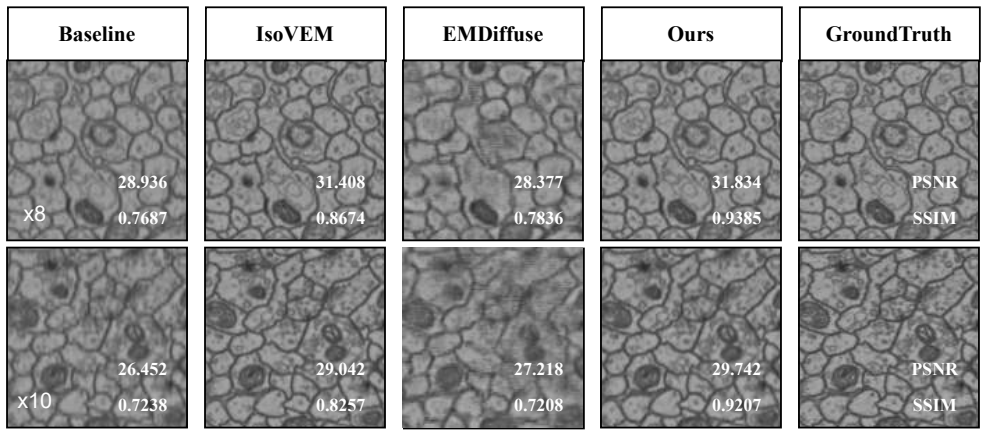
- [Sec. 4.1 (Datasets), Table 1, Fig. 5] The CREMI protocol is load-bearing for the 'real-world anisotropic VEM' claim, but it is not defined precisely enough. The paper states, 'Due to the sparse axial sampling, we conducted training and evaluation on the lateral planes for this dataset.' It does not explain how axial ground truth is obtained or whether any axial interpolation/super-resolution is evaluated. Since CREMI is anisotropic at 4x4x40 nm/voxel, an isotropic reconstruction method should be assessed on reconstructed axial slices (xz/yz), yet Fig. 5 shows only lateral (xy) sections. If 'lateral planes' means restoring synthetically degraded xy sections, then the CREMI rows of Table 1 measure 2D in-plane restoration, not axial isotropic reconstruction, and the claim of superiority on real anisotropic data is unsupported. The authors should either provide an explicit axial-plane evaluation with a clearly described ground-t
- [Sec. 4.2, Table 1] The quantitative comparison has no error bars, no multiple seeds, and no statistical tests. The reported PSNR advantages are often around 0.2–0.3 dB, and for some metrics/settings VEMamba is not best (e.g., CREMI LPIPS at x4–x10 is substantially worse than EMDiffuse: 0.2021 vs. 0.1362 at x4). With only two deep-learning baselines and no variance estimates, 'state-of-the-art' and 'highly competitive' are not robustly supported. At minimum, the authors should add run-to-run variability (e.g., 3 seeds) and paired significance tests, and ideally include additional recent 3D reconstruction baselines.
- [Sec. 4.4, Table 3] The ablation table and its textual description do not agree. Starting from the full model PSNR of 29.442, the three ablated rows have drops of 0.061, 0.070, and 0.046 dB. The text attributes these drops to replacing ALCSSM (0.07), substituting DWAM (0.061), and removing MoCo (0.046). Even if the table rows are ordered to match that reading, the drops attributed to DWAM and MoCo are swapped relative to the table as typeset: the row without MoCo appears to give 0.061, while the row without DWAM appears to give 0.046. The checkmark layout is also ambiguous. Since the paper credits each component with a specific contribution, the table must be corrected and clearly labeled so the reader can verify the claims.
- [Sec. 3.4, Sec. 4.1] The MoCo degradation representation is learned from the same synthetic degradation distribution (blur/downsampling/noise, parameters from DiffuseEM) that is used to create the training data for the main reconstruction network. Consequently, the MoCo branch does not provide independent evidence of robustness to real-world acquisition shifts; it can only align the feature representation with the training-time degradation prior. To support the claim of realistic degradation modeling, the authors should include at least one cross-domain test, e.g., training on one degradation model and evaluating on another, or a dataset with a genuinely different anisotropic acquisition geometry.
minor comments (6)
- [Sec. 4.1] The baseline 'interpolation' is never defined. Please state the exact method (e.g., bicubic, cubic spline, or linear interpolation) used to generate the numbers in Table 1.
- [Fig. 3 / Sec. 3.3] The eight scanning trajectories are described only verbally and illustrated with small arrows. A precise algorithmic definition (or a pseudocode listing) of the chunking and scan ordering would improve reproducibility.
- [Eq. (5)] The InfoNCE loss uses qi and ki, but the encoder architecture, projection head, and temperature hyper-parameter value are not reported in Section 3.4 or the implementation details. Please specify these.
- [Sec. 4.3, Table 2] The mitochondria segmentation IoU is reported only on the EPFL dataset. Since the downstream-task claim is part of the paper's significance, consider reporting the same analysis on CREMI (if the protocol ambiguity is resolved) or at least discussing why it is omitted.
- [Sec. 4.4, Fig. 6] The 'axial pixel differences' plot is illustrative but lacks units/error bars and does not indicate which volume/slice ranges are used. It would be stronger to report per-slice quantitative errors in a table or as a box plot.
- [Throughout] There are several typos and layout artifacts, e.g., 'efficientyeteffective' in the Introduction, 'V olume' spacing artifacts in figure captions, and truncated equations/labels in the ablation table. A careful proofread is recommended.
Circularity Check
No circularity found: VEMamba's claims rest on architectural design and empirical evaluation, not on self-referential derivations.
full rationale
The paper does not derive any prediction from an input that already contains that prediction. Its equations are standard training objectives and module formulations: Eq. (1) is a conventional L1+SSIM loss, Eq. (5) is the InfoNCE contrastive loss, and Eq. (6) defines channel-wise affine degradation injection. The ALCSSM and DWAM are architectural components; their descriptions do not reference the reconstruction targets as fitting parameters. The MoCo degradation representation is learned from the same synthetic degradation distribution used to generate training pairs, which is a domain-gap or representation-learning concern, not circular reasoning: the final reconstruction quality is still evaluated against external ground-truth volumes never used to define the degradation prior. There is no self-citation chain bearing the central claim; related work is cited for context and baselines, and the comparisons are retrained on the same data. The CREMI protocol ('training and evaluation on the lateral planes', Sec. 4.1) is an external-validity ambiguity regarding whether axial interpolation is actually tested, but it does not make any stated result true by construction. Therefore the appropriate circularity score is 0.
Assumptions & free parameters
free parameters (5)
- Number of RVMG and RVMB blocks =
4 groups, 4 blocks each (Sec. 4.1)
- Input subvolume sizes =
(32,128,128) for ×4; (16,128,128) for ×8; (16,160,160) for ×10 (Sec. 4.1)
- Loss weights =
1.0 for L1 and SSIM (Eq. 1)
- Degradation model parameters =
Following DiffuseEM (Sec. 4.1)
- Temperature tau in InfoNCE loss =
Not specified (Eq. 5)
assumptions (5)
- standard math InfoNCE loss provides useful degradation representations (Eq. 5)
- domain assumption Synthetic degradation model (blur+downsample+noise) approximates real VEM anisotropy
- domain assumption Training on high-resolution lateral planes transfers to axial reconstruction
- domain assumption MoCo-learned degradation representation improves generalization
- ad hoc to paper Chunked 8-direction scanning strategy is an optimal reordering of 3D dependencies
Cite this review
Pith. "Pith review of VEMamba: Efficient Isotropic Reconstruction of Volume Electron Microscopy with Axial-Lateral Consistent Mamba." pith.science (2026). https://pith.science/paper/7PO4TRYF
@misc{pith2026260300887,
  author       = {Pith},
  title        = {Pith review of: VEMamba: Efficient Isotropic Reconstruction of Volume Electron Microscopy with Axial-Lateral Consistent Mamba},
  year         = {2026},
  howpublished = {\url{https://pith.science/paper/7PO4TRYF}},
  note         = {Machine review of arXiv:2603.00887}
}
read the original abstract
Volume Electron Microscopy (VEM) is crucial for 3D tissue imaging but often produces anisotropic data with poor axial resolution, hindering visualization and downstream analysis. Existing methods for isotropic reconstruction often suffer from neglecting abundant axial information and employing simple downsampling to simulate anisotropic data. To address these limitations, we propose VEMamba, an efficient framework for isotropic reconstruction. The core of VEMamba is a novel 3D Dependency Reordering paradigm, implemented via two key components: an Axial-Lateral Chunking Selective Scan Module (ALCSSM), which intelligently re-maps complex 3D spatial dependencies (both axial and lateral) into optimized 1D sequences for efficient Mamba-based modeling, explicitly enforcing axial-lateral consistency; and a Dynamic Weights Aggregation Module (DWAM) to adaptively aggregate these reordered sequence outputs for enhanced representational power. Furthermore, we introduce a realistic degradation simulation and then leverage Momentum Contrast (MoCo) to integrate this degradation-aware knowledge into the network for superior reconstruction. Extensive experiments on both simulated and real-world anisotropic VEM datasets demonstrate that VEMamba achieves highly competitive performance across various metrics while maintaining a lower computational footprint. The source code is available on GitHub: https://github.com/I2-Multimedia-Lab/VEMamba
Figures


Figures from the paper (4 more)



Reference graph
Works this paper leans on
-
[1]
Three-dimensional or- ganization of transzonal projections and other cytoplasmic extensions in the mouse ovarian follicle.Scientific Reports, 9:1262, 2019
Veronica Baena and Mark Terasaki. Three-dimensional or- ganization of transzonal projections and other cytoplasmic extensions in the mouse ovarian follicle.Scientific Reports, 9:1262, 2019. 1
2019
-
[2]
Electron microscopy dataset, 2021
EPFL CVLab. Electron microscopy dataset, 2021. 5
2021
-
[3]
B. D. de Senneville, F. Z. Khoubai, M. Bevilacqua, and et al. Deciphering tumour tissue organization by 3d electron mi- croscopy and machine learning.Communications Biology, 4:1390, 2021. 1
2021
-
[4]
Isotropic Re- construction of 3D EM Images with Unsupervised Degra- dation Learning
Shiyu Deng, Xueyang Fu, Zhiwei Xiong, Chang Chen, Dong Liu, Xuejin Chen, Qing Ling, and Feng Wu. Isotropic Re- construction of 3D EM Images with Unsupervised Degra- dation Learning. InMedical Image Computing and Com- puter Assisted Intervention – MICCAI 2020, pages 163–173. Springer International Publishing, Cham, 2020. Series Title: Lecture Notes in Comp...
2020
-
[5]
Notes on noise contrastive estimation and nega- tive sampling
Chris Dyer. Notes on noise contrastive estimation and nega- tive sampling. InarXiv preprint arXiv:1410.8251, 2014. 5
arXiv 2014
-
[6]
Ferede, Ali Khalighifar, Jaison John, Krishnan Venkataraman, and Khaled Khairy
Fisseha A. Ferede, Ali Khalighifar, Jaison John, Krishnan Venkataraman, and Khaled Khairy. Z-upscaling: Optical Flow Guided Frame Interpolation for Isotropic Reconstruc- tion of 3D EM V olumes, 2024. arXiv:2410.07043 [eess]. 2
arXiv 2024
-
[7]
Funke and S
J. Funke and S. Saalfeld. Cremi dataset, 2016. 5
2016
-
[8]
Mamba: Linear-time sequence modeling with selective state spaces
Albert Gu and Tri Dao. Mamba: Linear-time sequence modeling with selective state spaces. InarXiv preprint arXiv:2312.00752, 2024. 2
arXiv 2024
Show all 43 references
-
[9]
Combining recurrent, con- volutional, and continuous-time models with linear state- space layers
Albert Gu, Isys Johnson, Karan Goel, Khaled Saab, Tri Dao, Atri Rudra, and Christopher Re. Combining recurrent, con- volutional, and continuous-time models with linear state- space layers. InarXiv preprint arXiv:2110.13985, 2021. 2
2021 arXiv
-
[10]
Efficiently mod- eling long sequences with structured state spaces
Albert Gu, Karan Goel, and Christopher Re. Efficiently mod- eling long sequences with structured state spaces. InarXiv preprint arXiv:2111.00396, 2022. 2
2022 arXiv
-
[11]
MambaIR: A Simple Base- line for Image Restoration with State-Space Model, 2024
Hang Guo, Jinmin Li, Tao Dai, Zhihao Ouyang, Xudong Ren, and Shu-Tao Xia. MambaIR: A Simple Base- line for Image Restoration with State-Space Model, 2024. arXiv:2402.15648 [cs]. 2
2024 arXiv
-
[12]
Super- resolution for asymmetric resolution of fib-sem 3d imaging using ai with deep learning.Scientific Reports, 8(1):5877,
Ken Hagita, Takashi Higuchi, and Hirofumi Jinnai. Super- resolution for asymmetric resolution of fib-sem 3d imaging using ai with deep learning.Scientific Reports, 8(1):5877,
-
[13]
Hayworth, C
Kenneth J. Hayworth, C. Shan Xu, Zhiyuan Lu, et al. Ultra- structurally smooth thick partitioning and volume stitching for large-scale connectomics.Nature Methods, 12(4):319– 322, 2015. 2
2015
-
[14]
MobileMamba: Lightweight Multi-Receptive Visual Mamba Network, 2024
Haoyang He, Jiangning Zhang, Yuxuan Cai, Hongxu Chen, Xiaobin Hu, Zhenye Gan, Yabiao Wang, Chengjie Wang, Yunsheng Wu, and Lei Xie. MobileMamba: Lightweight Multi-Receptive Visual Mamba Network, 2024. arXiv:2411.15941 [cs]. 4
2024 arXiv
-
[15]
Isovem: Isotropic reconstruction for volume electron mi- croscopy based on transformer.bioRxiv, 2023
Jia He, Yan Zhang, Wenhao Sun, Ge Yang, and Fei Sun. Isovem: Isotropic reconstruction for volume electron mi- croscopy based on transformer.bioRxiv, 2023. 2
2023
-
[16]
Momentum contrast for unsupervised visual repre- sentation learning
Kaiming He, Haoqi Fan, Yuxin Wu, Saining Xie, and Ross Girshick. Momentum contrast for unsupervised visual repre- sentation learning. InProceedings of the IEEE/CVF Confer- ence on Computer Vision and Pattern Recognition (CVPR), pages 9729–9738, 2020. 2
2020
-
[17]
Bogovic, and Stephan Saalfeld
Larissa Heinrich, John A. Bogovic, and Stephan Saalfeld. Deep learning for isotropic super-resolution from non- isotropic 3d electron microscopy. InMedical Image Com- puting and Computer-Assisted Intervention - MICCAI 2017, pages 135–143, Cham, 2017. Springer International Pub...
2017
-
[18]
Johnson and et al
Brett E. Johnson and et al. An omic and multidimensional spatial atlas from serial biopsies of an evolving metastatic breast cancer.Cell Reports Medicine, 3(2):100525, 2022. 1
2022
-
[19]
Self-Supervised Super-Resolution Approach for Isotropic Reconstruction of 3D Electron Microscopy Images from Anisotropic Acquisition, 2023
Mohammad Khateri, Morteza Ghahremani, Alejandra Sierra, and Jussi Tohka. Self-Supervised Super-Resolution Approach for Isotropic Reconstruction of 3D Electron Microscopy Images from Anisotropic Acquisition, 2023. arXiv:2309.10646 [eess]. 2
2023 arXiv
-
[20]
Kingma and Jimmy Ba
Diederik P. Kingma and Jimmy Ba. Adam: A method for stochastic optimization. InarXiv preprint arXiv:1412.6980,
-
[21]
Reference-Free Isotropic 3D EM Reconstruction using Diffusion Models,
Kyungryun Lee and Won-Ki Jeong. Reference-Free Isotropic 3D EM Reconstruction using Diffusion Models,
-
[22]
EAMamba: Efficient All-Around Vision State Space Model for Image Restoration, 2025
Yu-Cheng Lin, Yu-Syuan Xu, Hao-Wei Chen, Hsien-Kai Kuo, and Chun-Yi Lee. EAMamba: Efficient All-Around Vision State Space Model for Image Restoration, 2025. arXiv:2506.22246 [cs]. 2
2025 arXiv
-
[23]
Diffusion-based deep learn- ing method for augmenting ultrastructural imaging and vol- ume electron microscopy.Nature Communications, 15(1): 4677, 2024
Chixiang Lu, Kai Chen, Heng Qiu, Xiaojun Chen, Gu Chen, Xiaojuan Qi, and Haibo Jiang. Diffusion-based deep learn- ing method for augmenting ultrastructural imaging and vol- ume electron microscopy.Nature Communications, 15(1): 4677, 2024. 2
2024
-
[24]
Progress towards mammalian whole-brain cellular connectomics.Frontiers in Neuroanatomy, 10:62,
Shawn Mikula. Progress towards mammalian whole-brain cellular connectomics.Frontiers in Neuroanatomy, 10:62,
-
[25]
Morgan and Jeff W
Josh L. Morgan and Jeff W. Lichtman. An individual in- terneuron participates in many kinds of inhibition and inner- vates much of the mouse visual thalamus.Neuron, 106(3): 468–481.e2, 2020. 1
2020
-
[26]
Masaaki Nagai and et al. Glomerular cellular interac- tions following disruption of the glomerular basement mem- brane in iga nephropathy: Ultrastructural analyses by 3- dimensional serial block-face scanning electron microscopy. Kidney Medicine, 2(2):222–225, 2020. 1
2020
-
[27]
Diffu- seIR:Diffusion Models For Isotropic Reconstruction of 3D Microscopic Images, 2023
Mingjie Pan, Yulu Gan, Fangxu Zhou, Jiaming Liu, Aimin Wang, Shanghang Zhang, and Dawei Li. Diffu- seIR:Diffusion Models For Isotropic Reconstruction of 3D Microscopic Images, 2023. arXiv:2306.12109 [eess]. 2
2023 arXiv
-
[28]
U-net: Convolutional networks for biomedical image segmentation
Olaf Ronneberger, Philipp Fischer, and Thomas Brox. U-net: Convolutional networks for biomedical image segmentation. InarXiv preprint arXiv:1505.04597, 2015. 7
2015 arXiv
-
[29]
The cns connectome of a tad- pole larva ofCiona intestinalis(l.) highlights sidedness in the brain of a chordate sibling.eLife, 5:e16962, 2016
Kerrianne Ryan and et al. The cns connectome of a tad- pole larva ofCiona intestinalis(l.) highlights sidedness in the brain of a chordate sibling.eLife, 5:e16962, 2016. 1
2016
-
[30]
Self-supervised ControlNet with Spatio-Temporal Mamba for Real-world Video Super-resolution, 2025
Shijun Shi, Jing Xu, Lijing Lu, Zhihang Li, and Kai Hu. Self-supervised ControlNet with Spatio-Temporal Mamba for Real-world Video Super-resolution, 2025. arXiv:2506.01037 [cs]. 4
2025 arXiv
-
[31]
Primary ciliary dyskinesia with normal ultrastructure: three-dimensional tomography detects absence of dnah11.The European Respiratory Journal, 51 (2):1701809, 2018
Amelia Shoemark and et al. Primary ciliary dyskinesia with normal ultrastructure: three-dimensional tomography detects absence of dnah11.The European Respiratory Journal, 51 (2):1701809, 2018. 1
2018
-
[32]
Amelia Shoemark and et al. International consensus guide- line for reporting transmission electron microscopy results in the diagnosis of primary ciliary dyskinesia (beat pcd tem cri- teria).The European Respiratory Journal, 55(4):1900725,
-
[33]
Linderman
Jimmy Smith, Andrew Warrington, and Scott W. Linderman. Simplified state space layers for sequence modeling.ArXiv, abs/2208.04933, 2022. 2
2022 arXiv
-
[34]
Takashi Takaki and et al. Podocyte penetration of the glomerular basement membrane to contact on the mesan- gial cell at the lesion of mesangial interposition in lupus nephritis: a three-dimensional analysis by serial block-face scanning electron microscopy.Clinical and Experi...
2019
-
[35]
Terasaki
Mark et al. Terasaki. Stacked endoplasmic reticulum sheets are connected by helicoidal membrane motifs.Cell, 154(2): 285–296, 2013. 1
2013
-
[36]
Attention is all you need.arXiv, 2017
Ashish Vaswani, Noam Shazeer, Niki Parmar, Jakob Uszko- reit, Llion Jones, Aidan N Gomez, Lukasz Kaiser, and Illia Polosukhin. Attention is all you need.arXiv, 2017. 2
2017
-
[37]
Pvt v2: Improved baselines with pyramid vision transformer
Wenhai Wang, Enze Xie, Xiang Li, Deng-Ping Fan, Kaitao Song, Ding Liang, Tong Lu, Ping Luo, and Ling Shao. Pvt v2: Improved baselines with pyramid vision transformer. Computational Visual Media, 8(3):415–424, 2022. 3
2022
-
[38]
Temporal Spatial-Adaptive Interpolation with Deformable Refinement for Electron Microscopic Images,
Zejin Wang, Guodong Sun, Lina Zhang, Guoqing Li, and Hua Han. Temporal Spatial-Adaptive Interpolation with Deformable Refinement for Electron Microscopic Images,
-
[39]
Wilson and et al
Courtney E. Wilson and et al. Taste bud connectome: Im- plications for taste information processing.The Journal of Neuroscience, 42(5):804–816, 2022. 1
2022
-
[40]
Shtengel G
Pang S. Shtengel G. et al. Xu, C.S. An open-access volume electron microscopy atlas of whole cells and tissues.Nature, 599(7883):147–151, 2021. 1
2021
-
[41]
Shtengel G
Pang S. Shtengel G. et al. Xu, C.S. Publisher correction: An open-access volume electron microscopy atlas of whole cells and tissues.Nature, 599(7885):E5, 2021. 1
2021
-
[2021]
arXiv:2101.06771 [cs]. 2
-
[2023]
arXiv:2308.01594 [cs]. 2, 5
Reviewed August 2, 2026 · model on record in the stance chip above.
Discussion (0). Sign in to comment.