REVIEW 3 major objections 5 minor 53 references
Non-invasive measurement of local stress inside soft materials with programmed shear waves
T0 review · 3 major / 5 minor · reviewed 2026-08-07 · deepseek-v4-flash
Pith's one-line read This paper derives and demonstrates a constitutive-parameter-free formula that turns two perpendicular shear wave speeds into the principal stress difference in an incompressible soft material.
desk verdict A clean constitutive-independent stress formula with real experimental validation in aligned cases, but the coaxiality assumption leaves a genuine untested gap for realistic tissue architectures. read the letter →
The pith
A machine-rendered reading of the paper's core claim, the machinery that carries it, and where it could break.
The reading
What carries the argument
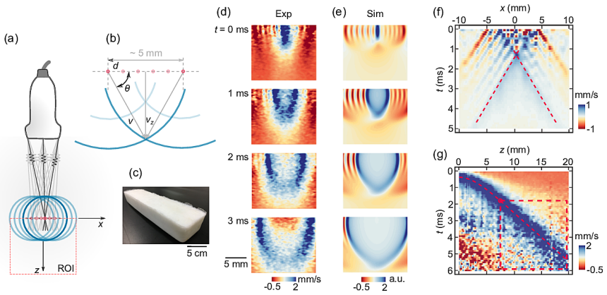
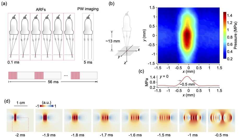
The machinery is equation (2) plus a way to excite and time two perpendicular shear waves. A focused ultrasound beam applies acoustic radiation force; by stepping the focus through six points at $d=1$ mm spacing, the authors create a supershear moving load (Mach number about 10) whose Huygens-Fresnel interference amplifies the otherwise weak vertical shear wave. Speeds $v_x$ and $v_z$ are extracted from spatiotemporal maps by Radon transformation, and because phase and group speeds coincide along the principal axes, the measured group speeds feed directly into equation (2). The derivation itself rests on the stress identity $\alpha-\gamma=\sigma_1-\sigma_3$ for incompressible solids and on the coaxiality condition that structure tensors, initial stress, and deformation commute.
What would settle it
Take a transversely isotropic phantom whose fibres are deliberately inclined at a known angle to an applied uniaxial stress, measure $v_x$ and $v_z$ along the transducer axes, and compare the value $\rho(v_x^2-v_z^2)/\cos 2\theta_0$ with the known applied principal stress difference. If the two agree within measurement error even when fibres and stress are misaligned, the coaxiality premise is unnecessary; if they diverge systematically with misalignment, the premise is load-bearing and the method's range is limited to aligned tissues.
Extended reading notes
Core claim
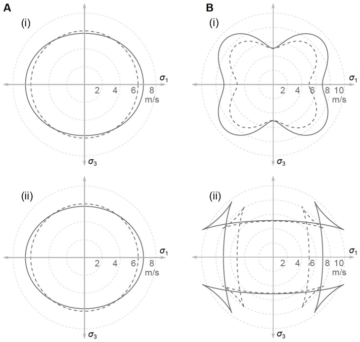
The central discovery is that for an incompressible soft solid, the principal stress difference is encoded in two shear wave speeds exactly through $\sigma_1-\sigma_3=\rho(v_x^2-v_z^2)/\cos 2\theta_0$, with no dependence on the strain-energy function or on third-order elastic constants. The identity comes from $\alpha-\gamma=\sigma_1-\sigma_3$, where $\alpha=A^0_{1313}$ and $\gamma=A^0_{3131}$ are Eulerian elastic moduli, combined with the wave-speed relations $\rho v_x^2=\alpha$ and $\rho v_z^2=\gamma$ for waves along the principal axes. It holds for arbitrary anisotropy, including fibre reinforcement and initial stress, as long as all sources of anisotropy are coaxial with the stress and deformation. The paper presents this as a parameter-free route to stress, validated by imaging uniaxial stress, bending stress, and passive muscle tension.
Load-bearing premise
The whole method collapses if the material's structural anisotropy, its initial stress, and its deformation are not aligned with one another, because then the two measured waves no longer sit exactly on the principal stress axes and equation (2) no longer follows.
Editorial extensions
If this is right
- In hydrogels and other weakly viscoelastic soft solids, uniaxial and bending stress fields can be imaged remotely with errors around 5%, without calibrating the constitutive law.
- The method works in anisotropic, fibre-reinforced tissues when fibres are aligned with the stress, as demonstrated by the passive muscle experiment, opening a route to in vivo stress estimation.
- Because only the principal stress difference is obtained, quantitative use requires either a known zero-stress direction, such as a free surface, or a second independent condition; the paper uses $\sigma_3=0$ for uniaxial cases.
- Viscoelasticity biases the inferred stress downward, but the paper's analysis with a quasi-linear model indicates errors below about 10% for soft materials whose stress relaxation is less than about 50%.
- The measured quantity is the group speed along the principal axes, where phase and group speeds coincide, so the method transfers directly to standard shear-wave-elastography systems.
Reading between the lines
- The identity gives only the difference of principal stresses; reconstructing the full in-plane stress tensor would require one more condition, such as a known stress-free boundary or an independent measurement of one principal stress, which the paper does not address.
- Because the acoustic-radiation-force excitation is only one way to launch shear waves, the same formula should work with mechanical shakers or other sources, making the method portable to non-ultrasound settings.
- A direct test of the coaxiality premise would be a phantom with fibres deliberately misaligned from the applied stress; the paper's muscle experiment only covers the aligned case.
- For living tissues, passive tension along fibres is the favourable case; active contraction or residual stresses that are not fibre-aligned may violate the condition, so extending to those settings would need a multi-angle version of the measurement.
Signed reviews
Editorial analysis
A structured set of objections, weighed in public.
Referee Report
Summary. The manuscript proposes and tests an acoustoelastic shear-wave method to measure the difference of principal stresses in soft materials without knowing their constitutive parameters. The theoretical basis is Eq. (2), derived from a constitutive-independent identity (SM Eq. 4) together with the assumption that all anisotropy is coaxial with the stress, so that waves along the two principal directions propagate as pure shear modes. The authors implement the method with a medical ultrasound transducer programmed to apply six successive acoustic radiation forces, producing both lateral and vertical shear waves; vertical speeds are extracted from z > 7 mm data with an interference correction. They validate the method on a PVA hydrogel under uniaxial and bending stress (errors ~5%) and on ex vivo porcine muscle under passive uniaxial stretch along the fiber direction (errors ~15%), and they analyze viscoelastic effects using a QLV model.
Significance. The stress identity and the basic experimental demonstration are valuable: if the coaxiality condition holds, the method offers a parameter-free route to local stress imaging, and the hydrogel and muscle experiments compare against externally applied loads rather than fitted stress values. The programmed-ARF excitation is a useful technical advance. However, the paper's broad claim of applicability to soft tissues is not yet supported for non-coaxial fiber/stress architectures, and the vertical-wave measurement relies on a correction/cutoff whose influence is not quantified. With those gaps addressed or the claims appropriately restricted, the work would be a solid contribution.
major comments (3)
- [SM Note 2 and main text Eq. (2)] SM Note 2 assumes the coaxiality conditions Cτ = τC, CMMᵀ = MMᵀC, and τMMᵀ = MMᵀτ, and Eq. (12) restricts the moduli; without these assumptions the measured ρv_x² and ρv_z² cannot be identified with A⁰₁₃₁₃ and A⁰₃₁₃₁. The muscle experiment stretches along the fiber, so it only validates the coaxial case. The abstract states that the method will find broad applications for diagnosing diseases that alter stresses in soft tissues, but in general tissue fibers need not be coaxial with the in vivo stress; in that case the x–z plane is not a symmetry plane and the tracked SV mode is not a pure mode, so Eq. (2) fails. The authors should either extend the theory to non-coaxial situations, provide a validation with fibers not aligned with the principal stress, or clearly restrict the claimed domain of applicability.
- [Generating shear waves / Fig. 2G] The vertical speed is not measured as a direct plane-wave speed: the source interference makes the apparent speed approach vz only for z > 7 mm, and the paper relies on this cutoff without a sensitivity analysis. Since Eq. (2) uses v_x² − v_z², a systematic bias in vz maps directly into a bias in the inferred stress. The zero-stress isotropy check (vx ≈ vz) and the hydrogel ground-truth comparisons provide indirect support, but a quantitative study of how the z cutoff and the interference correction affect the inferred stress would be needed to support the claimed accuracy in less controlled, anisotropic samples.
- [Scope of Eq. (2)] The central result provides only the difference of two in-plane principal stresses, σ₁ − σ₃; for a general biaxial stress state, individual stress components require additional assumptions or measurements, and only for uniaxial stress (σ₃ = 0) does Eq. (2) directly give σ₁. The paper sometimes refers broadly to measuring 'mechanical stresses', and this limitation should be stated more prominently in the abstract and discussion to avoid overstating what the method delivers in realistic tissue loading conditions.
minor comments (5)
- [Eq. (1) vs SM Eq. (13)] The powers in Eq. (1) of the main text appear different from those in SM Eq. (13): the main text writes ρv² = α cos²θ + 2β cos²θ sin²θ + γ sin²θ while the SM has cos⁴θ and sin⁴θ. Please align the notation and clarify whether 'cos2θ' means cos²θ or cos(2θ).
- [SM Note 2, Eq. (14)] Equation (14) in SM Note 2 writes σ₁ − σ₂ on the left-hand side, but the main text and the preceding derivation use σ₁ − σ₃; this appears to be a typo.
- [Results, 'Generating shear waves...' paragraph] The phrase 'the so-call longitudinal shear waves' should be 'the so-called longitudinal shear waves'.
- [Fig. 4C caption] The caption uses 'AFRs' where 'ARFs' (acoustic radiation forces) is meant.
- [Fig. 4E] The identified stresses in the muscle experiment are shown without error bars; reporting the standard deviation of the five measurements would help readers judge the statistical significance of the ~15% maximum error.
Circularity Check
No circularity found: Eq. (2) is derived from a constitutive-parameter-free stress identity and validated against externally applied loads.
full rationale
The central formula σ1−σ3 = ρ(vx²−vz²)/cos2θ0 is derived in the paper from the exact identity A0_1313−A0_3131=σ11−σ33 (SM Note 1), which follows algebraically from the universal expression for the Eulerian moduli and does not require a fitted constitutive law. The step connecting measured speeds to those moduli (SM Note 2, Eq. (11)) does invoke a coaxiality assumption, but this is a stated modeling condition, not a parameter fitted from the stress data; the muscle experiment enforces it by stretching along the fiber axis. The muscle-model parameters (c2, c4, Prony terms) are fitted to explain the measured vx and vz curves and to estimate viscoelastic corrections, but the reported stresses are computed directly from the measured wave speeds via Eq. (2), not from the fitted model. Validation is against externally applied loads and finite-element bending fields, so the predictions are not statistically forced by the inputs. The prior self-citations to Refs. [25], [50], and [51] support the acoustoelastic wave-speed expression, but the SM rederives the needed relations, so no load-bearing circular step exists.
Assumptions & free parameters
free parameters (3)
- Muscle strain-hardening parameters c2, c4 =
c2 ≈ 3.5, c4 ≈ 8
- Muscle QLV Prony parameters g1, tau1 =
g1 ≈ 0.79, tau1 ≈ 0.49 ms
- Hydrogel Prony series g1, tau1, g2, tau2 =
g1 = 0.07, tau1 = 0.08 s, g2 = 0.05, tau2 = 2.05 s
assumptions (5)
- domain assumption The soft material is incompressible.
- domain assumption Structural anisotropy, initial stress, and deformation are all coaxial with the principal stress directions, and the measurement axes are aligned with them.
- domain assumption The measured vertical shear wave is a plane SV wave propagating along z with polarization along x at sufficiently large z.
- standard math Group speed equals phase speed along the principal directions.
- domain assumption Mass density ρ is known and approximately 1 g/cm3.
Cite this review
Pith. "Pith review of Non-invasive measurement of local stress inside soft materials with programmed shear waves." pith.science (2026). https://pith.science/paper/7TEHIFI4
@misc{pith2026250603816,
  author       = {Pith},
  title        = {Pith review of: Non-invasive measurement of local stress inside soft materials with programmed shear waves},
  year         = {2026},
  howpublished = {\url{https://pith.science/paper/7TEHIFI4}},
  note         = {Machine review of arXiv:2506.03816}
}
read the original abstract
Mechanical stresses in soft materials across different length scales play a fundamental role in understanding the function of biological systems and in the use of artificial materials for engineering soft machines and biomedical devices. Yet it remains a great challenge to probe local mechanical stresses in situ in a non-invasive, non-destructive manner, in particular when the mechanical properties are unknown. To address this challenge, we propose an acoustoelastic imaging-based method to infer the local mechanical stresses in soft materials by measuring the speed of shear waves induced by custom-programmed acoustic radiation force. Using a medical ultrasound transducer to excite and track the shear waves remotely, we demonstrate the application of the method by imaging uniaxial stress and bending stress in an isotropic hydrogel, and the passive uniaxial stress in a skeletal muscle. These measurements were all done without the knowledge of the constitutive parameters of the materials. These examples indicate that our method will find broad applications, ranging from health monitoring of soft structures and machines, to the diagnosis of diseases that alter stresses in soft tissues.
Figures


Figures from the paper (9 more)



Reference graph
Works this paper leans on
-
[1]
N. L. Nerurkar, C. Lee, L. Mahadevan, C. J. Tabin, Nature 565, 480 (2019)
work page 2019
-
[2]
M. G´ omez-Gonz´ alez, E. Latorre, M. Arroyo, X. Trepat, Nat. Rev. Phys. 2, 300 (2020)
work page 2020
-
[3]
K. H. Vining, D. J. Mooney, Nat. Rev. Mol. Cell Biol. 18, 728 (2017)
work page 2017
- [4]
- [5]
-
[6]
J.-H. Lee, H. S. Park, D. P. Holmes, Phys. Rev. Lett. 127, 138102 (2021)
work page 2021
-
[7]
B. Li, F. Jia, Y.-P. Cao, X.-Q. Feng, H. Gao, Phys. Rev. Lett. 106, 234301 (2011)
work page 2011
-
[8]
S. K. Powers, M. J. Jackson, Physiol. Rev. 88, 1243 (2008)
work page 2008
Show all 53 references
-
[9]
J. A. Martin, et al., Nat. Commun. 9, 1 (2018)
2018
-
[10]
Lee, et al., Nat
J. Lee, et al., Nat. Electron. 4, 291 (2021)
2021
-
[11]
J. Y. Sun, et al., Nature 489, 133 (2012)
2012
-
[12]
J. Kim, G. Zhang, M. Shi, Z. Suo, Science 374, 212 (2021)
2021
-
[13]
Matsuhisa, et al., Nature 600, 246 (2021)
N. Matsuhisa, et al., Nature 600, 246 (2021)
2021
-
[14]
G. H. Lee, et al., Nat. Rev. Mater. 5, 149 (2020)
2020
-
[15]
R. Bai, J. Yang, Z. Suo, Eur. J. Mech. A/Solids 74, 337 (2019)
2019
-
[16]
J. A. Rogers, T. Someya, Y. Huang, Science 327, 1603 (2010)
2010
-
[17]
Wang, et al., Sci
Y. Wang, et al., Sci. Adv. 6, 7043 (2020)
2020
-
[18]
G. S. Schajer, Exp. Mech. 50, 245 (2009)
2009
-
[19]
N. S. Rossini, M. Dassisti, K. Y. Benyounis, A. G. Olabi, Mater. Des. 35, 572 (2012)
2012
-
[20]
C. O. Ruud, NDT E Int. 15, 15 (1982)
1982
-
[21]
D. S. Hughes, J. L. Kelly, Phys. Rev. 92, 1145 (1953)
1953
-
[22]
A. N. Guz, F. G. Makhort, Int. Appl. Mech. 36, 1119 (2000)
2000
-
[23]
F. Shi, J. E. Michaels, S. J. Lee, J. Acoust. Soc. Am. 133, 677 (2013)
2013
-
[24]
Gennisson, et al., J
J.-L. Gennisson, et al., J. Acoust. Soc. Am. 122, 3211 (2008)
2008
-
[25]
G.-Y. Li, A. Gower, M. Destrade, J. Acoust. Soc. Am. 148, 3963 (2020)
2020
-
[26]
Jiang, et al., Med
Y. Jiang, et al., Med. Image Anal. 20, 97 (2015)
2015
-
[27]
Creton, C
C. Creton, C. Matteo, Rep. Prog. Phys. 79, 046601 (2016). 27
2016
-
[28]
Shams, M
M. Shams, M. Destrade, R. W. Ogden, Wave Motion 48, 552 (2011)
2011
-
[29]
A. L. Gower, T. Shearer, P. Ciarletta, The Quarterly Journal of Mechanics and Applied Mathematics 70, 455 (2017)
2017
-
[30]
Destrade, M
M. Destrade, M. D. Gilchrist, R. W. Ogden, The Journal of the Acoustical Society of America 127, 2103 (2010)
2010
-
[31]
Deroy, M
C. Deroy, M. Destrade, A. Mc Alinden, A. N ´ ı Annaidh, Skin Research and Technol- ogy 23, 326 (2017)
2017
-
[32]
Hariton, G
I. Hariton, G. Debotton, T. C. Gasser, G. A. Holzapfel, Biomechanics and modeling in mechanobiology 6, 163 (2007)
2007
-
[33]
G. W. Jones, S. J. Chapman, Siam review 54, 52 (2012)
2012
-
[34]
N. C. Rouze, A. Caenen, K. R. Nightingale, Physics in Medicine & Biology 67, 095015 (2022)
2022
-
[35]
Bercoff, M
J. Bercoff, M. Tanter, M. Fink, IEEE Trans. Ultrason. Ferroelectr. Freq. Control 51, 396 (2004)
2004
-
[36]
A. P. Sarvazyan, O. V. Rudenko, S. D. Swanson, J. B. Fowlkes, S. Y. Emelianov, Ultrasound Med. Biol. 24, 1419 (1998)
1998
-
[37]
Catheline, N
S. Catheline, N. Benech, J. Acoust. Soc. Am. 137, EL200 (2015)
2015
-
[38]
Sandrin, M
L. Sandrin, M. Tanter, J.-L. Gennisson, S. Catheline, M. Fink, IEEE transactions on ultrasonics, ferroelectrics, and frequency control 49, 436 (2002)
2002
-
[39]
Sandrin, D
L. Sandrin, D. Cassereau, M. Fink, The Journal of the Acoustical Society of America 115, 73 (2004)
2004
-
[40]
Tanter, M
M. Tanter, M. Fink, IEEE Trans. Ultrason. Ferroelectr. Freq. Control61, 102 (2014)
2014
-
[41]
Rouze, M
N. Rouze, M. Wang, M. Palmeri, K. Nightingale, IEEE Trans. Ultrason. Ferroelectr. Freq. Control 57, 2662 (2010)
2010
-
[42]
J. P. Kerris, A. C. Betik, J. Li, G. K. McConell, J. Appl. Physiol. 126, 239 (2019)
2019
-
[43]
Calvo, et al., J
B. Calvo, et al., J. Biomech. 43, 318 (2010)
2010
-
[44]
J. P. Remeni´ eras, et al., Phys. Med. Biol.66, 145009 (2021)
2021
-
[45]
Berjamin, R
H. Berjamin, R. De Pascalis, International Journal of Solids and Structures 241, 111529 (2022)
2022
-
[46]
Montaldo, M
G. Montaldo, M. Tanter, J. Bercoff, N. Benech, M. Fink, IEEE transactions on ultrasonics, ferroelectrics, and frequency control 56, 489 (2009)
2009
-
[47]
Loupas, J
T. Loupas, J. Powers, R. W. Gill, IEEE transactions on ultrasonics, ferroelectrics, and frequency control 42, 672 (1995)
1995
-
[48]
T. J. Hall, M. Bilgen, M. F. Insana, T. A. Krouskop, IEEE Trans. Ultrason. Ferro- electr. Freq. Control 44, 1355 (1997). 28
1997
-
[49]
R. W. Ogden, Waves in Nonlinear Pre-Stressed Materials, M. Destrade, G. Sacco- mandi, eds. (Springer, Vienna, 2007), vol. 495, pp. 1–26
2007
-
[50]
G.-Y. Li, Y. Zheng, Y. Liu, M. Destrade, Y. Cao, J. Mech. Phys. Solids 96, 388 (2016)
2016
-
[51]
Destrade, M
M. Destrade, M. D. Gilchrist, G. Saccomandi, J. Acoust. Soc. Am. 127, 2759 (2010)
2010
-
[52]
G. Y. Li, et al., Philos. Trans. Royal Soc. A 377, 20180075 (2019)
2019
-
[53]
J. G. Murphy, Eur. J. Mech. A/Solids 42, 90 (2013). 29
2013
Reviewed August 7, 2026 · model on record in the stance chip above.
Discussion (0). Continue with ORCID to comment.