REVIEW 5 major objections 6 minor 43 references
A Physics-Inspired Deep Learning Framework with Polar Coordinate Attention for Ptychographic Imaging
T0 review · 5 major / 6 minor · reviewed 2026-08-12 · deepseek-v4-flash
Pith's one-line read A polar-coordinate attention network beats CNN and transformer baselines and accelerates ptychographic phase retrieval by over 1000x.
desk verdict A new attention mechanism for ptychographic imaging with modest real gains, but the speed and overlap claims are overstated and the physics rationale needs empirical support. read the letter →
The pith
A machine-rendered reading of the paper's core claim, the machinery that carries it, and where it could break.
The reading
What carries the argument
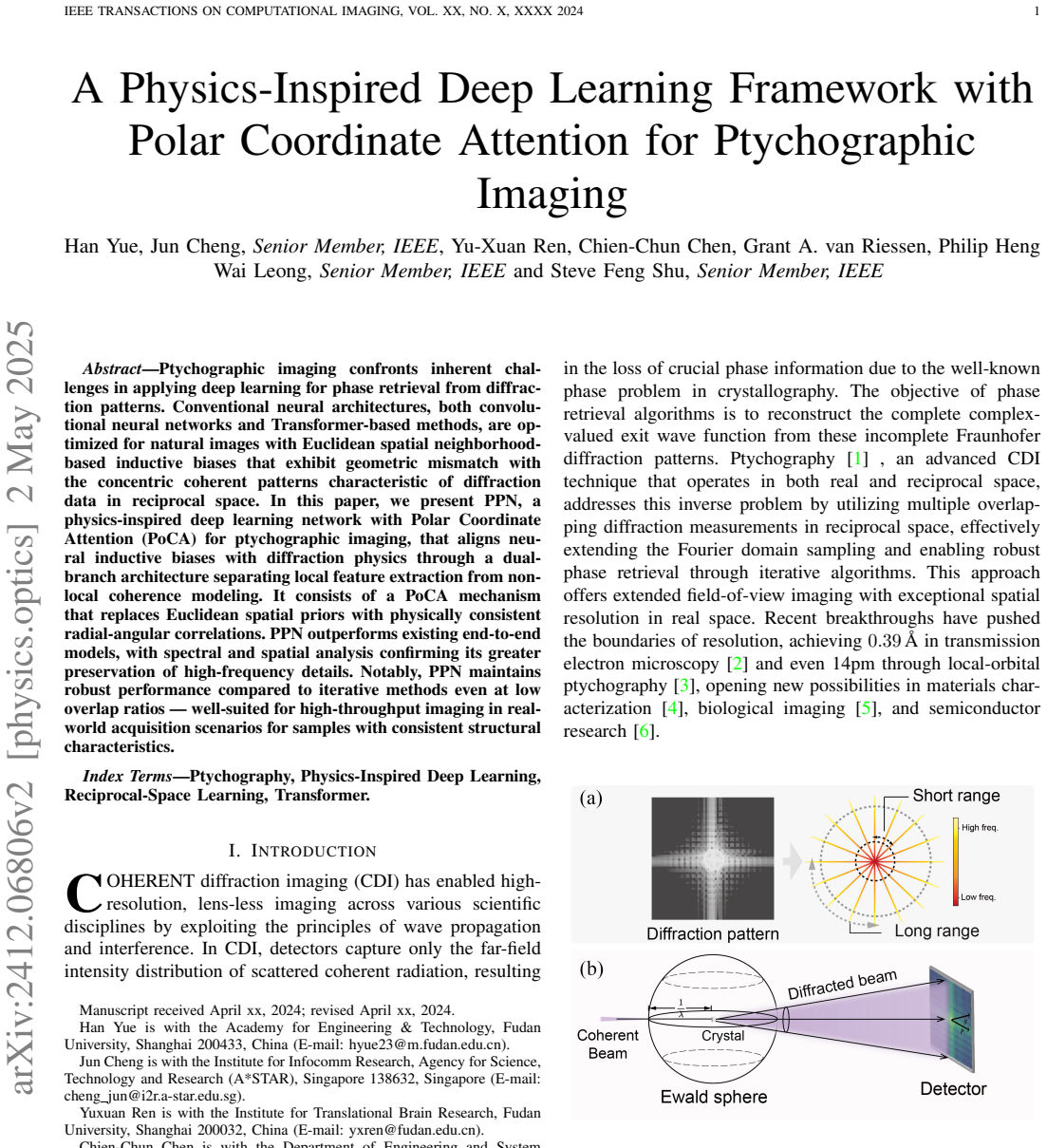
PoCA is the mechanism that carries the argument: a polar-coordinate attention layer that replaces the Euclidean position-based attention of standard transformers. Each pixel is mapped to a log-scaled radius $r_m$ and an angle $\theta_m$ around a learnable center $c=(W/2+\alpha_x W/2,\, H/2+\alpha_y H/2)$, and the base attention logits are multiplied by radial decay $\Phi^r_{mn}=1/(1+|r_m-r_n|)$ and angular continuity $\Phi^\theta_{mn}=\cos(\theta_m-\theta_n)$ before the softmax. This encodes the radial intensity falloff and angular symmetry of diffraction, which the paper connects to the Ewald-sphere construction. PoCA lives in the Non-Local Coherence Branch, a pixel-level transformer branch run in parallel with a patch-based Local Dependencies Branch; the branches are fused and decoded into separate amplitude and phase maps. The ablation study shows that removing PoCA, swapping it for standard multi-head self-attention, or removing the decoder each degrades the full model.
What would settle it
Train PPN and a matched-parameter Euclidean attention model on a diffraction dataset with strong non-radial structure (for example, off-axis illumination, split diffraction peaks, or a curved detector with known distortion), and check whether PPN's PSNR/SSIM advantage survives; if the polar model loses on that data, the claim that polar alignment is what causes the improvement is falsified.
Extended reading notes
Core claim
The central discovery is that the inductive bias of the attention mechanism is the main bottleneck for end-to-end ptychographic phase retrieval, and that replacing Euclidean attention with polar-coordinate attention aligned to reciprocal-space physics fixes it. PoCA parameterizes each pixel by $r_m = \log(1+\lVert p_m - c \rVert)/\log(1+r_{\max})$ and $\theta_m = \mathrm{arctan2}(y_m-c_y, x_m-c_x)$ around a learnable center $c$, then modulates the dot-product attention logits by $\Phi^r_{mn}=1/(1+|r_m-r_n|)$ and $\Phi^\theta_{mn}=\cos(\theta_m-\theta_n)$ before softmax. In the paper's experiments this yields the best metrics among end-to-end models, a 58.6% improvement in mid-frequency energy preservation over CNN baselines, and stable SSIM (0.86-0.92) across overlap ratios from 30% to 90%, while ePIE falls to 0.12 at 30% overlap. The paper interprets this as evidence that a physically consistent polar geometry, rather than additional parameters, is what lets the network retain high-frequency structure.
Load-bearing premise
The load-bearing premise is that the correlations a reconstruction network needs from a diffraction pattern are organized around a single physical center in polar coordinates, so the hand-designed radial and angular weighting functions match the true structure; if a real dataset has multiple centers, detector distortion, or substantial non-radial signal, this premise fails and the claimed high-frequency advantage could disappear.
Editorial extensions
If this is right
- Low-overlap scans, which reduce acquisition time and radiation dose, no longer force a large reconstruction quality penalty, so end-to-end ptychography becomes viable for high-throughput experiments.
- Attention mechanisms for reciprocal-space data should be designed with polar coordinate geometry rather than Euclidean neighborhoods, changing how transformer blocks are built for diffraction problems.
- The dual-branch separation suggests that local dependencies and global non-local coherence in diffraction patterns should be extracted separately and fused late rather than mixed in a single hierarchy.
- If the central claim holds, deep-learning phase retrieval can serve as a practical complement to iterative algorithms for time-sensitive measurements while retaining high-frequency detail.
Reading between the lines
- Extension: PoCA's single learned center and hand-designed radial/angular weights assume one well-defined diffraction center; datasets with multiple centers, detector distortion, or strong non-radial structure would be a natural stress test, and a multi-center or deformable polar parameterization would be the next step if it fails.
- Extension: the reported speed and low-overlap advantages are demonstrated on samples within the training feature distribution; a useful extension is measuring how the advantage decays as the test sample geometry moves farther from the training set.
- Extension: the same polar-geometry argument could be transferred to other Fourier-domain inverse problems with concentric structure, such as cryo-EM or astronomical speckle imaging, to test whether the benefit is specific to ptychography or general to diffraction physics.
Editorial analysis
A structured set of objections, weighed in public.
Referee Report
Summary. The paper proposes PPN, a dual-branch deep learning architecture for ptychographic phase retrieval. A local ViT branch extracts local features, while a non-local branch uses a new Polar Coordinate Attention (PoCA) mechanism that replaces Euclidean attention with hand-designed radial and angular weighting functions. The model is trained end-to-end on diffraction intensities to output amplitude and phase, and is evaluated on simulated USAF-like data and on experimental synchrotron data from Argonne. The authors claim superior reconstruction quality over CNN/Transformer baselines, better high-frequency preservation, low-overlap robustness compared with ePIE, and large speedups at inference time. Ablation studies and loss-function analyses are included, along with parameter/FLOP comparisons against existing models.
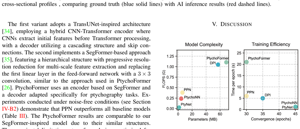
Significance. If the empirical claims are reproducible, PPN would be a practically useful contribution: it offers fast inference, competitive reconstruction quality, and a claimed robustness to low overlap that is relevant for high-throughput synchrotron imaging. The evaluation is more careful than typical for this area: multiple baselines, statistical testing with ten independent runs, ablations, and validation on both simulated and real experimental data. The parameter/FLOP efficiency gains over transformer-based baselines are also notable. However, the central claims contain numerical inconsistencies, the physical justification for the PoCA weighting functions is not supported by data, and the headline loss used in the main experiments is inconsistent with the loss analysis presented later. These issues are load-bearing and need to be resolved before the contribution can be accepted as stated.
major comments (5)
- [Abstract and Section I vs. Fig. 8] The claims in the abstract and in the contributions bullet (Section I) that PPN maintains '<5% performance degradation at 30% overlap ratio' and '>1000x faster inference' are not supported by the numbers reported in Section IV-E3 and Fig. 8. Fig. 8(a) states that PPN SSIM stays between 0.86 and 0.92 across overlap ratios; if the degradation is computed from the 90% value (0.92) to the 30% value (0.86), that is about 6.5%, not below 5%. The same figure reports an 852x speedup at 30% overlap (0.15s vs. 125s), not >1000x; the 1767x figure corresponds to 60% overlap. The authors should either correct the headline numbers or state precisely which quantities are used for the degradation and speedup claims.
- [Section IV-E3 and Fig. 8] The ePIE baseline at 30% overlap is reported to have SSIM=0.12, which is unusually low for a ptychographic reconstruction even at reduced overlap, and the comparison is therefore hard to assess. The manuscript does not give enough detail about the ePIE implementation: number of iterations, initialization, use of position correction, probe update strategy, or any regularization. Since the central claim that PPN is 'robust compared to iterative methods at low overlap' depends entirely on this baseline, the authors should specify the implementation and, ideally, repeat the comparison with a standard, publicly available ePIE code on the same diffraction data and report the resulting overlap curve.
- [Section III-B4 vs. Section V-B and Table IV] The main experiments in Tables I-III use the MSE loss defined in Eq. (12), but Section V-B later presents a combined loss L_combined(alpha) = alpha*L_MSE + (1-alpha)*L_NSSIM and reports in Table IV that alpha=0.9 substantially improves all metrics (e.g., amplitude PSNR 15.10 vs. 14.42 dB, amplitude SSIM 88.50% vs. 87.00%). If the combined loss is part of the proposed method, the headline results understate PPN; if it is not part of the proposed method, then the comparison against CNN baselines that use standard losses is not on equal footing. The authors should clarify which loss is used in the main comparisons and, if it is MSE, explain why the better combined loss is not adopted in the main results.
- [Section I and Section III-B2, Eqs. (6)-(7)] The physical justification for PoCA depends on an Ewald-sphere and Bragg-diffraction picture, but ptychographic diffraction patterns from general non-periodic objects are speckle patterns whose pixel-pixel correlations are not necessarily radial or angular in the sense of Phi_r = 1/(1+|rm-rn|) and Phi_theta = cos(theta_m-theta_n). The paper posits these weightings without an empirical analysis of the actual correlation structure of the diffraction data. The authors should either supply such an analysis (e.g., measured radial and angular correlation statistics for the datasets used) or reframe the contribution as an empirically motivated inductive bias rather than a physics-derived prior. This also bears on the generalization claims in Section IV-D, because object classes with strong non-radial structure may not benefit from this prior.
- [Section V-B and Table IV] The loss-function analysis introduces L_combined(alpha) and reports that alpha=0.9 is optimal, but the main results in Table IV are only given for eight loss configurations; the figure for 'Weighted Loss (alpha=0.9)' is not integrated into the earlier experiments. If the combined loss is intended as a component of the proposed method, the authors should include it in the main comparisons, or explicitly state that all baseline comparisons use MSE only. Otherwise, the claim that PPN 'outperforms existing end-to-end models' is made under a loss that the paper itself identifies as suboptimal.
minor comments (6)
- [Section IV-A1] The text says 'We set the initial learning rate to 1.0'; for Adam with MSE loss this is likely a typo for 0.001 or 0.0001. Please state the actual learning rate used.
- [Table II] The variant labeled 'Ours w/ MinN' is presumably 'Ours w/ MHSA' (multi-head self-attention); please rename the variant for clarity.
- [Figure 8 caption] The caption contains the typo 'ietrative method'; it should read 'iterative method'.
- [Section V-A] The units 'FLOPS' and 'FLOPs' are used inconsistently; please standardize the terminology.
- [Eq. (2)] The partial-coherence term (1-mu)P(r-rj-delta_rj)*E_r[O(r)] is not clearly defined; the meaning of the expectation E_r and the physical range of mu should be specified.
- [Section IV-E3] The claim that reducing overlap from 90% to 30% increases data acquisition efficiency by 'approximately 49 times' is stated without derivation; please spell out the calculation (probe size, step size, and the inverse-square relation) to make it verifiable.
Circularity Check
No significant circularity: PoCA is a hand-designed inductive bias validated on held-out data, not a fitted input renamed as a prediction.
full rationale
PPN is a supervised encoder–decoder that maps a measured (or simulated) diffraction pattern to amplitude and phase maps. The polar-coordinate attention in Eqs. (5)–(8) is an architectural inductive bias: Φr and Φθ are fixed functions of pixel coordinates, plus two learnable center offsets αx, αy and the ordinary network weights. None of the paper's claimed outputs (reconstructed amplitude/phase, PSD fidelity, overlap-ratio robustness, inference speed) is a parameter fitted to the test quantity and then reported as a prediction; the reported metrics are computed on a held-out 20% split against external ground truth. The Ewald-sphere discussion is an analogy used to motivate the coordinate system, not a 'uniqueness theorem' imported from the authors' prior work, and the paper contains no load-bearing chain of author self-citations. The ablation study (Table II) separates the PoCA contribution from the decoder contribution, and while one could dispute the attribution of the high-frequency advantage to the polar prior, that is a correctness/generalization risk rather than a circular reduction. No equation reduces to its own input by construction, and the loss function compares network outputs to ground-truth images rather than to functions of the fitted parameters. Therefore the derivation is self-contained with respect to circularity.
Assumptions & free parameters
free parameters (2)
- Learned diffraction center offsets alpha_x, alpha_y =
alpha_x, alpha_y in [-0.5, 0.5], initialized at 0
- Combined loss weight alpha =
0.9 (optimal)
assumptions (5)
- standard math Far-field Fraunhofer approximation: the recorded intensity is the squared modulus of the Fourier transform of the exit wave.
- domain assumption Poisson-plus-Gaussian noise model for diffraction intensity.
- ad hoc to paper A single learned center with log-scaled radial coordinate and cosine angular similarity captures the physically relevant correlations in diffraction patterns.
- domain assumption End-to-end supervised mapping from diffraction patterns to amplitude and phase is learnable and generalizes to samples with consistent structural characteristics.
- domain assumption Ground truth for experimental data produced by the PIE algorithm is treated as correct.
Cite this review
Pith. "Pith review of A Physics-Inspired Deep Learning Framework with Polar Coordinate Attention for Ptychographic Imaging." pith.science (2026). https://pith.science/paper/7XZSMV4A
@misc{pith2026241206806,
  author       = {Pith},
  title        = {Pith review of: A Physics-Inspired Deep Learning Framework with Polar Coordinate Attention for Ptychographic Imaging},
  year         = {2026},
  howpublished = {\url{https://pith.science/paper/7XZSMV4A}},
  note         = {Machine review of arXiv:2412.06806}
}
read the original abstract
Ptychographic imaging confronts inherent challenges in applying deep learning for phase retrieval from diffraction patterns. Conventional neural architectures, both convolutional neural networks and Transformer-based methods, are optimized for natural images with Euclidean spatial neighborhood-based inductive biases that exhibit geometric mismatch with the concentric coherent patterns characteristic of diffraction data in reciprocal space. In this paper, we present PPN, a physics-inspired deep learning network with Polar Coordinate Attention (PoCA) for ptychographic imaging, that aligns neural inductive biases with diffraction physics through a dual-branch architecture separating local feature extraction from non-local coherence modeling. It consists of a PoCA mechanism that replaces Euclidean spatial priors with physically consistent radial-angular correlations. PPN outperforms existing end-to-end models, with spectral and spatial analysis confirming its greater preservation of high-frequency details. Notably, PPN maintains robust performance compared to iterative methods even at low overlap ratios, making it well suited for high-throughput imaging in real-world acquisition scenarios for samples with consistent structural characteristics.
Figures


Figures from the paper (8 more)



Reference graph
Works this paper leans on
-
[1]
R. Hegerl and W. Hoppe, “Dynamische Theorie der Kristallstrukturanal- yse durch Elektronenbeugung im inhomogenen Prim ¨arstrahlwellenfeld,” Berichte der Bunsengesellschaft f ¨ur physikalische Chemie , vol. 74, no. 11, pp. 1148–1154, Nov. 1970
work page 1970
-
[2]
Electron ptychography of 2D materials to deep sub-˚angstr¨om resolution,
Y . Jiang, Z. Chen, Y . Han, P. Deb, H. Gao, S. Xie, P. Purohit, M. W. Tate, J. Park, and S. M. Gruner, “Electron ptychography of 2D materials to deep sub-˚angstr¨om resolution,” Nature, vol. 559, no. 7714, pp. 343–349, 2018
work page 2018
-
[3]
Local-orbital ptychography for ultrahigh-resolution imaging,
W. Yang, H. Sha, J. Cui, L. Mao, and R. Yu, “Local-orbital ptychography for ultrahigh-resolution imaging,” Nature Nanotechnology , pp. 1–6, 2024
work page 2024
-
[4]
E. B. L. Pedersen, D. Angmo, H. F. Dam, K. T. S. Thyd ´en, T. R. Andersen, E. T. B. Skjønsfjell, F. C. Krebs, M. Holler, A. Diaz, and M. Guizar-Sicairos, “Improving organic tandem solar cells based on water-processed nanoparticles by quantitative 3D nanoimaging,” Nanoscale, vol. 7, no. 32, pp. 13 765–13 774, 2015
work page 2015
-
[5]
R. Kasprowicz, R. Suman, and P. O’Toole, “Characterising live cell behaviour: Traditional label-free and quantitative phase imaging ap- proaches,” The international journal of biochemistry & cell biology , vol. 84, pp. 89–95, 2017. IEEE TRANSACTIONS ON COMPUTATIONAL IMAGING, VOL. XX, NO. X, XXXX 2024 13
work page 2017
-
[6]
High-resolution non-destructive three-dimensional imaging of integrated circuits,
M. Holler, M. Guizar-Sicairos, E. H. Tsai, R. Dinapoli, E. M ¨uller, O. Bunk, J. Raabe, and G. Aeppli, “High-resolution non-destructive three-dimensional imaging of integrated circuits,” Nature, vol. 543, no. 7645, pp. 402–406, 2017
work page 2017
-
[7]
Deep learning at the edge enables real-time streaming ptychographic imaging,
A. V . Babu, T. Zhou, S. Kandel, T. Bicer, Z. Liu, W. Judge, D. J. Ching, Y . Jiang, S. Veseli, and S. Henke, “Deep learning at the edge enables real-time streaming ptychographic imaging,” Nature Communications , vol. 14, no. 1, p. 7059, 2023
work page 2023
-
[8]
Sampling in x-ray ptychography,
T. B. Edo, D. J. Batey, A. M. Maiden, C. Rau, U. Wagner, Z. D. Pe ˇsi´c, T. A. Waigh, and J. M. Rodenburg, “Sampling in x-ray ptychography,” Physical Review A , vol. 87, no. 5, p. 053850, May 2013
work page 2013
Show all 43 references
-
[9]
AI-enabled high-resolution scanning coherent diffraction imaging,
M. J. Cherukara, T. Zhou, Y . Nashed, P. Enfedaque, A. Hexemer, R. J. Harder, and M. V . Holt, “AI-enabled high-resolution scanning coherent diffraction imaging,” Applied Physics Letters , vol. 117, no. 4, 2020
2020
-
[10]
Deep-Learning Electron Diffractive Imaging,
D. J. Chang, C. M. O’Leary, C. Su, D. A. Jacobs, S. Kahn, A. Zettl, J. Ciston, P. Ercius, and J. Miao, “Deep-Learning Electron Diffractive Imaging,” Physical Review Letters, vol. 130, no. 1, p. 016101, Jan. 2023
2023
-
[11]
An efficient ptychography reconstruction strategy through fine-tuning of large pre-trained deep learning model,
X. Pan, S. Wang, Z. Zhou, L. Zhou, P. Liu, C. Li, W. Wang, C. Zhang, Y . Dong, and Y . Zhang, “An efficient ptychography reconstruction strategy through fine-tuning of large pre-trained deep learning model,” Iscience, vol. 26, no. 12, 2023
2023
-
[12]
Learning to synthesize: Robust phase retrieval at low photon counts,
M. Deng, S. Li, A. Goy, I. Kang, and G. Barbastathis, “Learning to synthesize: Robust phase retrieval at low photon counts,” Light: Science & Applications, vol. 9, no. 1, p. 36, 2020
2020
-
[13]
Phase retrieval based on deep learning with bandpass filtering in holographic data storage,
R. Fan, J. Hao, R. Chen, J. Wang, Y . Lin, J. Jin, R. Yang, X. Zheng, K. Wang, and D. Lin, “Phase retrieval based on deep learning with bandpass filtering in holographic data storage,” Optics Express, vol. 32, no. 3, pp. 4498–4510, 2024
2024
-
[14]
On-the-fly scans for X-ray ptychography,
P. M. Pelz, M. Guizar-Sicairos, P. Thibault, I. Johnson, M. Holler, and A. Menzel, “On-the-fly scans for X-ray ptychography,” Applied Physics Letters, vol. 105, no. 25, 2014
2014
-
[15]
Noise-robust latent vector re- construction in ptychography using deep generative models,
J. Seifert, Y . Shao, and A. P. Mosk, “Noise-robust latent vector re- construction in ptychography using deep generative models,” Optics Express, vol. 32, no. 1, pp. 1020–1033, 2024
2024
-
[16]
Using a modified double deep image prior for crosstalk mitigation in multislice ptychography,
M. Du, X. Huang, and C. Jacobsen, “Using a modified double deep image prior for crosstalk mitigation in multislice ptychography,” Journal of synchrotron radiation, vol. 28, no. 4, pp. 1137–1145, 2021
2021
-
[17]
PtychoDV: Vision Transformer-Based Deep Unrolling Network for Ptychographic Image Reconstruction,
W. Gan, Q. Zhai, M. T. McCann, C. G. Cardona, U. S. Kamilov, and B. Wohlberg, “PtychoDV: Vision Transformer-Based Deep Unrolling Network for Ptychographic Image Reconstruction,” IEEE Open Journal of Signal Processing , 2024
2024
-
[18]
Very Deep Convolutional Networks for Large-Scale Image Recognition,
K. Simonyan and A. Zisserman, “Very Deep Convolutional Networks for Large-Scale Image Recognition,” Apr. 2015
2015
-
[19]
Imagenet classification with deep convolutional neural networks,
A. Krizhevsky, I. Sutskever, and G. E. Hinton, “Imagenet classification with deep convolutional neural networks,” Advances in neural informa- tion processing systems , vol. 25, 2012
2012
-
[20]
An Image is Worth 16x16 Words: Transformers for Image Recognition at Scale,
A. Dosovitskiy, L. Beyer, A. Kolesnikov, D. Weissenborn, X. Zhai, T. Unterthiner, M. Dehghani, M. Minderer, G. Heigold, S. Gelly, J. Uszkoreit, and N. Houlsby, “An Image is Worth 16x16 Words: Transformers for Image Recognition at Scale,” Jun. 2021
2021
-
[21]
Mvitv2: Improved multiscale vision transformers for classification and detection,
Y . Li, C.-Y . Wu, H. Fan, K. Mangalam, B. Xiong, J. Malik, and C. Feichtenhofer, “Mvitv2: Improved multiscale vision transformers for classification and detection,” in Proceedings of the IEEE/CVF Conference on Computer Vision and Pattern Recognition , 2022, pp. 4804–4814
2022
-
[22]
A survey on vision transformer,
K. Han, Y . Wang, H. Chen, X. Chen, J. Guo, Z. Liu, Y . Tang, A. Xiao, C. Xu, and Y . Xu, “A survey on vision transformer,” IEEE transactions on pattern analysis and machine intelligence, vol. 45, no. 1, pp. 87–110, 2022
2022
-
[23]
Medsegdiff-v2: Diffusion- based medical image segmentation with transformer,
J. Wu, W. Ji, H. Fu, M. Xu, Y . Jin, and Y . Xu, “Medsegdiff-v2: Diffusion- based medical image segmentation with transformer,” in Proceedings of the AAAI Conference on Artificial Intelligence , vol. 38, 2024, pp. 6030– 6038
2024
-
[24]
Climatelearn: Benchmarking machine learning for weather and climate modeling,
T. Nguyen, J. Jewik, H. Bansal, P. Sharma, and A. Grover, “Climatelearn: Benchmarking machine learning for weather and climate modeling,” Advances in Neural Information Processing Systems , vol. 36, 2024
2024
-
[25]
Bidirectional generation of structure and properties through a single molecular foundation model,
J. Chang and J. C. Ye, “Bidirectional generation of structure and properties through a single molecular foundation model,” Nature Com- munications, vol. 15, no. 1, p. 2323, 2024
2024
-
[26]
PtychoFormer: A Transformer-based Model for Ptychographic Phase Retrieval,
R. Nakahata, S. Zaman, M. Zhang, F. Lu, and K. Chiu, “PtychoFormer: A Transformer-based Model for Ptychographic Phase Retrieval,” Oct. 2024
2024
-
[27]
On the Fraunhofer (Far Field) Diffraction Patterns of Opaque and Transparent Objects with Coherent Background,
G. B. Parrent and B. J. Thompson, “On the Fraunhofer (Far Field) Diffraction Patterns of Opaque and Transparent Objects with Coherent Background,” Optica Acta: International Journal of Optics , vol. 11, no. 3, pp. 183–193, Jul. 1964
1964
-
[28]
Kirchhoff’s theory for optical diffrac- tion, its predecessor and subsequent development: The resilience of an inconsistent theory,
J. Z. Buchwald and C.-P. Yeang, “Kirchhoff’s theory for optical diffrac- tion, its predecessor and subsequent development: The resilience of an inconsistent theory,”Archive for History of Exact Sciences, vol. 70, no. 5, pp. 463–511, Sep. 2016
2016
-
[29]
An improved ptychographical phase retrieval algorithm for diffractive imaging,
A. M. Maiden and J. M. Rodenburg, “An improved ptychographical phase retrieval algorithm for diffractive imaging,” Ultramicroscopy, vol. 109, no. 10, pp. 1256–1262, 2009
2009
-
[30]
Differential programming enabled functional imaging with Lorentz transmission electron mi- croscopy,
T. Zhou, M. Cherukara, and C. Phatak, “Differential programming enabled functional imaging with Lorentz transmission electron mi- croscopy,” npj Computational Materials , vol. 7, no. 1, p. 141, 2021
2021
-
[31]
X-ray Ptychography Imaging of Hu- man Chromosomes After Low-dose Irradiation,
A. Bhartiya, D. Batey, S. Cipiccia, X. Shi, C. Rau, S. Botchway, M. Yusuf, and I. K. Robinson, “X-ray Ptychography Imaging of Hu- man Chromosomes After Low-dose Irradiation,” Chromosome Research, vol. 29, no. 1, pp. 107–126, Mar. 2021
2021
-
[32]
In situ X-ray-based imaging of nano materials,
J. N. Weker, X. Huang, and M. F. Toney, “In situ X-ray-based imaging of nano materials,” Current opinion in chemical engineering , vol. 12, pp. 14–21, 2016
2016
-
[33]
Robust uncertainty principles: Exact signal reconstruction from highly incomplete frequency informa- tion,
E. J. Cand `es, J. Romberg, and T. Tao, “Robust uncertainty principles: Exact signal reconstruction from highly incomplete frequency informa- tion,” IEEE Transactions on information theory, vol. 52, no. 2, pp. 489– 509, 2006
2006
-
[34]
TransUNet: Transformers Make Strong Encoders for Medical Image Segmentation,
J. Chen, Y . Lu, Q. Yu, X. Luo, E. Adeli, Y . Wang, L. Lu, A. L. Yuille, and Y . Zhou, “TransUNet: Transformers Make Strong Encoders for Medical Image Segmentation,” Feb. 2021
2021
-
[35]
SegFormer: Simple and efficient design for semantic segmentation with transformers,
E. Xie, W. Wang, Z. Yu, A. Anandkumar, J. M. Alvarez, and P. Luo, “SegFormer: Simple and efficient design for semantic segmentation with transformers,” Advances in neural information processing systems , vol. 34, pp. 12 077–12 090, 2021
2021
-
[36]
Cellpose3: One-click image restoration for improved cellular segmentation,
C. Stringer and M. Pachitariu, “Cellpose3: One-click image restoration for improved cellular segmentation,” Nature Methods, pp. 1–8, 2025
2025
-
[37]
PIXART-$$ \Sigma $$: Weak-to-Strong Training of Diffusion Transformer for 4K Text-to-Image Generation,
J. Chen, C. Ge, E. Xie, Y . Wu, L. Yao, X. Ren, Z. Wang, P. Luo, H. Lu, and Z. Li, “PIXART-$$ \Sigma $$: Weak-to-Strong Training of Diffusion Transformer for 4K Text-to-Image Generation,” in Computer Vision – ECCV 2024 , A. Leonardis, E. Ricci, S. Roth, O. Russakovsky, T. Satt...
2024
-
[38]
Medical image segmentation review: The success of u-net,
R. Azad, E. K. Aghdam, A. Rauland, Y . Jia, A. H. Avval, A. Bozorgpour, S. Karimijafarbigloo, J. P. Cohen, E. Adeli, and D. Merhof, “Medical image segmentation review: The success of u-net,” IEEE Transactions on Pattern Analysis and Machine Intelligence , 2024
2024
-
[39]
A foundation model for joint segmentation, detection and recognition of biomedical objects across nine modalities,
T. Zhao, Y . Gu, J. Yang, N. Usuyama, H. H. Lee, S. Kiblawi, T. Nau- mann, J. Gao, A. Crabtree, and J. Abel, “A foundation model for joint segmentation, detection and recognition of biomedical objects across nine modalities,” Nature methods, pp. 1–11, 2024
2024
-
[40]
Robust Estimation of a Location Parameter,
P. J. Huber, “Robust Estimation of a Location Parameter,” in Break- throughs in Statistics, S. Kotz and N. L. Johnson, Eds. New York, NY: Springer New York, 1992, pp. 492–518
1992
-
[41]
VII. Note on regression and inheritance in the case of two parents,
“VII. Note on regression and inheritance in the case of two parents,” Proceedings of the Royal Society of London , vol. 58, no. 347-352, pp. 240–242, Dec. 1895
-
[42]
Loss functions for image restoration with neural networks,
H. Zhao, O. Gallo, I. Frosio, and J. Kautz, “Loss functions for image restoration with neural networks,” IEEE Transactions on computational imaging, vol. 3, no. 1, pp. 47–57, 2016
2016
-
[43]
Image quality assessment: From error visibility to structural similarity,
Z. Wang, A. C. Bovik, H. R. Sheikh, and E. P. Simoncelli, “Image quality assessment: From error visibility to structural similarity,” IEEE transactions on image processing , vol. 13, no. 4, pp. 600–612, 2004
2004
Reviewed August 12, 2026 · model on record in the stance chip above.
Discussion (0). Continue with ORCID to comment.