REVIEW 5 major objections 6 minor 39 references
The-Bodega: A Matlab Toolbox for Biologically Dynamic Microbubble Simulations on Realistic Hemodynamic Microvascular Graphs
T0 review · 5 major / 6 minor · reviewed 2026-08-04 · deepseek-v4-flash
Pith's one-line read The paper presents The-Bodega, an open-source toolbox that simulates thousands of microbubbles flowing through realistic brain and heart vascular networks, and converts their trajectories into ground-truth ultrasound localization microscopy
desk verdict A well-engineered ULM simulation toolbox with a couple of self-consistent demonstrations that are described as validations; deserves review once code and data ship. read the letter →
The pith
A machine-rendered reading of the paper's core claim, the machinery that carries it, and where it could break.
The reading
What carries the argument
The carrying mechanism is a directed vascular graph coupled to a sequential Monte Carlo microbubble simulator. The graph supplies all possible trajectories; each microbubble is propagated along a randomly weighted path under a Poiseuille flow profile modulated by pulse-decomposition-analysis (PDA) waveforms that are precomputed per edge and shifted in time. The resulting microbubble positions, velocities, and radii are passed to a linear acoustic simulator (SIMUS) that renders RF and IQ data, with tissue and skull clutter added as separate scatterer blocks. This design preserves exact ground truth at every stage while allowing arbitrary vascular architectures and transducer configurations.
What would settle it
Record microbubble tracks in a living mouse brain with ULM and compare individual capillary transit times, velocity pulsatility phase lags, and branch choices to simulations run on the same vascular graph; a systematic mismatch would falsify the claim that the simulated ground truth is biologically realistic.
Extended reading notes
Core claim
The central claim is that The-Bodega provides an end-to-end, modular simulation framework that produces ULM ground truth of sufficient hemodynamic and anatomical realism to stand in for in vivo acquisitions. The authors show that by starting from directed vascular graphs annotated with vessel radii, flow, and pulse pressure, they can simulate stochastic microbubble trajectories, encode them in HDF5 with full ground truth, and then generate RF/IQ ultrasound data with tissue clutter and motion. They demonstrate that this lets them quantify how long it takes to populate the capillary mesh, how SVD clutter filtering ablates slow capillary signals and degrades resolution, how cardiac tissue motio
Load-bearing premise
The realism rests on treating blood as a simple pipe flow and microbubbles as passive spheres that only slow down or vanish in narrow vessels; if real microbubbles stick, deform, or interact with sound, the simulated ground truth will not match in vivo.
Editorial extensions
If this is right
- ULM algorithms can be benchmarked under controlled, known-truth conditions that separate the effects of microbubble concentration, tissue clutter, and tissue motion.
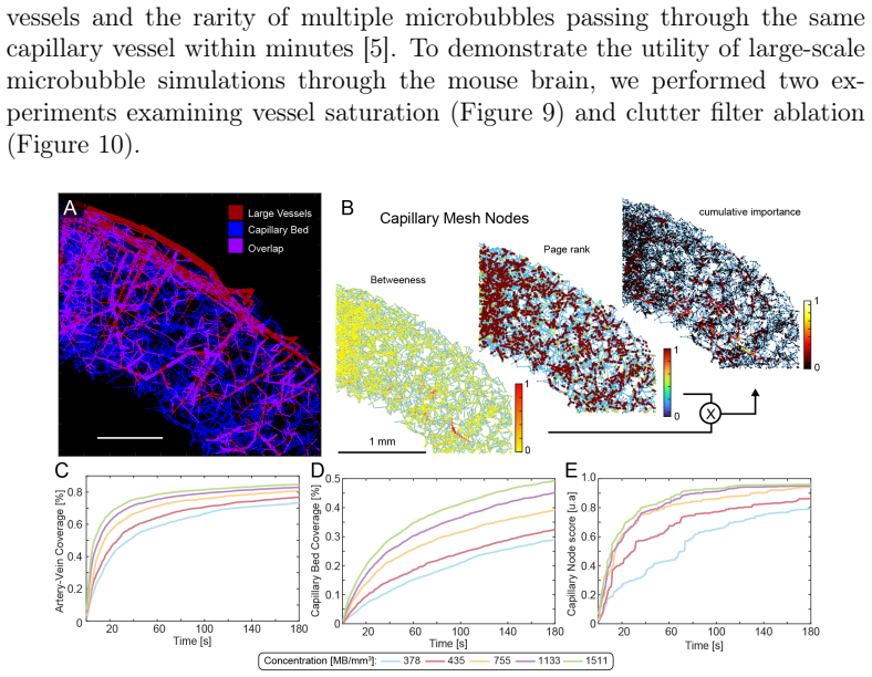
- The capillary saturation analysis suggests that roughly three to five minutes of acquisition may suffice to sample capillary function, even though full capillary mesh reconstruction takes much longer.
- SVD clutter filtering is shown to preferentially ablate slow capillary signals, and higher eigenvalue cutoffs degrade ULM resolution; this motivates alternative clutter-removal strategies.
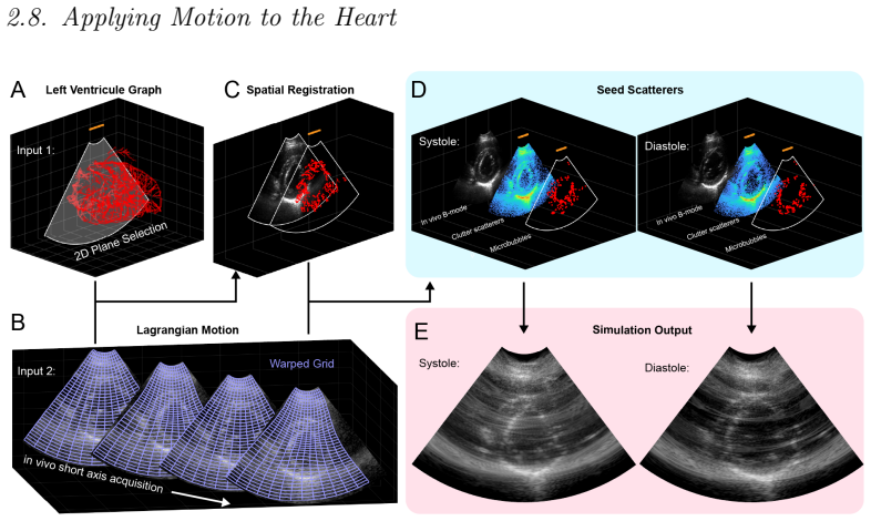
- In the beating heart, the simulations indicate that tissue clutter, not motion itself, is the dominant obstacle, because motion alone with correction preserves most vessels while motion plus clutter destroys them.
- Simulated neurovascular responses could serve as test data for functional ULM analysis pipelines and for studying the link between blood-volume changes and ULM-derived activation maps.
Reading between the lines
- The same pipeline could be used to stress-test adaptive or spatially-varying SVD filters, nonlinear contrast imaging, and motion-compensation algorithms before deploying them in vivo.
- Because the simulator accepts arbitrary directed graphs and user-defined pulse waveforms, it could generate pathological hemodynamic patterns to identify ULM-visible biomarkers of vascular disease.
- A direct experimental comparison between simulated microbubble trajectories and in vivo microbubble tracks in the same vascular network would be a natural next step; the paper currently validates pulsatility against its own imposed waveforms.
- The ground-truth labels and raw data could be used to train deep learning models for localization, tracking, and denoising without manual annotation, an application the authors note but leave largely undeveloped.
Editorial analysis
A structured set of objections, weighed in public.
Referee Report
Summary. The paper introduces The-Bodega, a Matlab toolbox for simulating microbubble trajectories on anatomically derived vascular graphs and generating synthetic Ultrasound Localization Microscopy (ULM) datasets. The pipeline combines sequential Monte Carlo particle simulation with Poiseuille flow profiles and PDA-derived pulsatile waveforms (Algorithm 1), then feeds resulting trajectories into a SIMUS-based linear ultrasound simulator with optional tissue clutter and motion. Demonstrations cover computational benchmarking, 3D pulsatility via dynamic ULM, capillary saturation and SVD clutter-filter ablation, cardiac motion artifacts, and simulated neurovascular responses for functional ULM. The authors position the toolbox as an open-source resource for generating realistic ground-truth ULM data for algorithm benchmarking and deep learning training.
Significance. If released with working code and data, The-Bodega would be a valuable community resource, complementing existing simulators such as PALA, BUFF, and PROTEUS. Its modular design, support for arbitrary vascular graphs, HDF5 output, and CPU/GPU parallelization are practical strengths. The capillary saturation times reported in Fig. 9D appear consistent with independent estimates [5,35], and the controlled SVD ablation and cardiac-motion experiments are plausible demonstrations of the kind of studies such a simulator enables. However, the central claim of hemodynamic realism is not yet externally validated; the current evidence supports a self-consistent digital phantom rather than a substitute for in vivo acquisitions. The computational scaling results (Fig. 7, Table 1) are useful and well presented.
major comments (5)
- [Sec. 3.2 with Algorithm 1 and Sec. 2.5] The pulsatility validation is a round-trip rather than an independent test. The PDA waveforms are an input (Algorithm 1), and dULM realignment uses the same simulated cardiac-phase information from the simulation (§2.5). The recovered pulsatility index is then described as validating "a model of pulsatility that mimics in vivo pulsatile flow." This only checks internal consistency. Please add external validation against in vivo microbubble trajectory or Doppler velocity waveforms (e.g., in the same mouse-brain or human-heart context), or explicitly reframe the demonstration as self-consistency, not hemodynamic validation.
- [Sec. 2.2] The microbubble-vessel interaction rule is an ad hoc heuristic: if a bubble's diameter exceeds vessel diameter, it decelerates until it shrinks or disappears. No experimental reference or validation is provided, and there is no sensitivity analysis to the parameters of this rule. Since this rule directly controls capillary occupancy and disappearance, it is load-bearing for the capillary saturation and SVD ablation conclusions. Please validate against measured microbubble trajectories or, at minimum, provide a sensitivity analysis and describe the rule as a heuristic whose impact on downstream metrics is quantified.
- [Sec. 2.6 and Fig. 9] The node-importance weighting is under-specified. The text states the importance is "a linear combination of the betweenness centrality and PageRank estimator for node connectivity," but the weights, normalization, and directed/undirected treatment are not given. The definition of the track "score" used in Fig. 9E is also missing. Without these details, the claim that 95% of highly influential capillary nodes are populated within 2-3 minutes cannot be reproduced or evaluated. Please provide the exact formula and parameter values.
- [Sec. 3 (Figs. 8-12)] Quantitative results are reported without repeats, error bars, or confidence intervals. Because the simulator is stochastic (random sampling of trajectories, bubble distributions, and subsets in Secs. 2.2-2.3), all reported curves and metrics depend on random seeds. This applies to Fig. 9 saturation curves, Fig. 10F Dice/Jaccard/sensitivity/specificity, Fig. 8G pulsatility index distributions, and Fig. 12 fULM correlations. The absence of variability estimates undermines quantitative conclusions such as "3-5 minute scan time may be sufficient" and the FRC resolution values. Please include multiple independent runs with corresponding statistics.
- [Intro, Sec. 2.1, Sec. 4] The paper repeatedly describes The-Bodega as "fully open-source" and its datasets as "openly available," but the code repository and data links are listed as "to-be-published" (Intro and Sec. 2.1). For a software/toolbox paper, this availability is load-bearing and should be resolved with a permanent DOI or repository before publication. Additionally, Sec. 4 states that "the flow should be validated before simulation for accurate representation"; this is a significant limitation that should be elevated to a clearly stated caveat with quantitative evidence for the default graphs, rather than only a remark in the discussion.
minor comments (6)
- [Eq. (1)] ΔV is described as "stroke volume (mL)" but Eq. (1) represents the compliance relation ΔV = C·ΔP, where ΔV is the volume change. Please clarify whether stroke volume or pulse-induced volume change is intended, as these differ in general.
- [Sec. 2.4] Typo: "for in vivomice" should be "for in vivo mice." Also, the sentence beginning "To draw parallels, in the human heart" could be reworded for clarity.
- [Sec. 3.2] Typo: "arterioes" should be "arterioles." The sentence "Here, In the contrast-enhanced Doppler volume..." has an awkward capitalization and comma structure.
- [Fig. 10 and text] In the text describing Figure 10, "Zoomed image of microbubble-only tracking from F" appears to be an error: Figure 10F contains quantitative metrics, not an image. The reference should likely be to panel A or B. Please correct.
- [Table 1] The table lists a "Toy" network. In the main text only the whole-brain, half-brain, heart, and synthetic capillary networks are mentioned. Please define "Toy" in the text or rename the entry.
- [Sec. 2.8] "This assumption rapidly fails in moving organs..." is missing a comma and the antecedent "This" is ambiguous (it refers to a multi-sentence prior statement). Rephrase for clarity.
Circularity Check
Demonstration-level circularity: dULM and fULM recover their own injected waveforms/activations, but the core simulator pipeline is not circular.
-
self definitional
[Section 2.5 (dULM processing) and Section 3.2 (3D Pulsatility); Algorithm 1]
"Here, in lieu of ECG signal, the tissue Doppler used to spatiotemporally align microbubble phase in the cardiac cycle during the simulation was used for realignment. … Importantly, this characterizes the use of the PDA algorithm for propagating the pulse wave through these microvascular network and validates a model of pulsatility that mimics in vivo pulsatile flow."
The PDA waveform is not an independent physiological measurement: Algorithm 1 takes pulse parameters as input and emits edge-wise pulsatile waveforms, which are scaled by compliance (Eq. 1) and propagated through the graph. The dULM realignment then reuses the simulation's own cardiac-cycle phase information, so the pulsatility index reported in §3.2 is the imposed waveform re-detected after simulation and tracking. Calling this a 'validation' of an in-vivo-like pulsatility model is therefore a round-trip check, not a test against independent data.
-
self definitional
[Section 2.7 (Simulating Neurovascular responses) and Section 3.5 (functional ULM)]
"To dynamically simulate functional hemodynamic responses to stimulus-evoked impulses, two microbubble distributions were simulated: steady-state and full whisker activation … the trajectory selection probability distribution was augmented to preferentially increase vascular paths that lead to the BF cortex … we calculated the Pearson correlation coefficient between the ULM images (sliding window size of 4 s, 0.4 s stride) and the pulse train (Figure 5C)."
The activation is inserted into the simulation by augmenting flow and trajectory-selection probabilities in the left S1BF barrel-field ROI, and then fULM correlates the resulting density images with the very same pulse train that drove the sampling between steady-state and activated distributions. Positive correlation in the barrel field is thus guaranteed by construction; the demonstration does not independently validate the neurovascular-coupling model. The paper concedes in §4 that the sampling method is 'relatively simple' and 'may not entirely reflect microvascular flow changes in vivo.'
full rationale
This is a software/toolbox paper, not a closed-form derivation chain, so the classic Eq-X-equals-Eq-Y circularity does not arise. The core pipeline—vascular graph inputs, Monte Carlo bubble trajectories, SIMUS RF/IQ synthesis, and ULM reconstruction—is self-contained and gives direct access to ground truth; benchmarking SVD filtering, FRC, and capillary saturation against that ground truth is legitimate, and the capillary-population results are cross-checked against independent reports [5,35]. The two circular elements are in the demonstration/validation sections. Section 3.2 labels the PDA pulsatility demonstration as a 'validation' of a model 'that mimics in vivo pulsatile flow,' but the PDA waveform is a user-specified input and dULM realignment reuses the simulation's own cardiac-cycle phase; the recovered pulsatility index is therefore the input replayed through the pipeline. Section 3.5 similarly injects the neurovascular response by augmenting flow in the S1BF region and then computes fULM correlation against the same pulse train, so recovery of activation is by construction. The paper itself concedes (§4) that flow 'should be validated before simulation' and that the fULM sampling model 'may not entirely reflect microvascular flow changes in vivo.' Those concessions are external-validity caveats, not evidence of a circular derivation. Prior-work citations ([14], [18], [34]) are published external results rather than unverified self-referential premises, and they do not force the core simulator's outputs. Overall, the circularity is partial and demonstration-level; the central toolbox contribution remains independently meaningful, though its in-vivo realism is not independently established.
Assumptions & free parameters
free parameters (6)
- Microbubble size distribution (mean, std) =
mean = 2 um, std = 3 um
- Vascular compliance C =
user-specified
- PDA pulsatile waveform parameters =
heart rate, pulse wave velocity, 5 Gaussian components
- Poiseuille radial scaling factor p =
not given
- Hemodynamic response function parameters =
FWHM = 4 s, delay = 1 s
- Capillary node importance weighting =
unspecified
assumptions (6)
- domain assumption Poiseuille flow approximates all microvascular flow profiles
- domain assumption PDA pulse shape propagated over graph edges achieves steady-state pulsatility
- domain assumption Linear ultrasound simulation (SIMUS) is sufficient for ULM ground truth
- domain assumption Microbubble dynamics reduce to size-checked passive advection
- domain assumption The vascular graphs from [18] and [19] accurately represent in vivo anatomy and flow
- domain assumption Buxton neurovascular coupling model and gamma-variate HRF describe whisker-stimulation hemodynamics
Cite this review
Pith. "Pith review of The-Bodega: A Matlab Toolbox for Biologically Dynamic Microbubble Simulations on Realistic Hemodynamic Microvascular Graphs." pith.science (2026). https://pith.science/paper/AH3BDNAQ
@misc{pith2026250908149,
  author       = {Pith},
  title        = {Pith review of: The-Bodega: A Matlab Toolbox for Biologically Dynamic Microbubble Simulations on Realistic Hemodynamic Microvascular Graphs},
  year         = {2026},
  howpublished = {\url{https://pith.science/paper/AH3BDNAQ}},
  note         = {Machine review of arXiv:2509.08149}
}
read the original abstract
The-Bodega is a Matlab-based toolbox for simulating ground-truth datasets for Ultrasound Localization Microscopy (ULM)-a super resolution imaging technique that resolves microvessels by systematically tracking microbubbles flowing through the microvasculature. The-Bodega enables open-source simulation of stochastic microbubble dynamics through anatomically complex vascular graphs and features a quasi-automated pipeline for generating ground-truth ultrasound data from simple vascular inputs. It incorporates sequential Monte Carlo simulations augmented with Poiseuille flow distributions and dynamic pulsatile flow. A key novelty of our framework is its flexibility to accommodate arbitrary vascular architectures and benchmark common ULM algorithms, such as Fourier Ring Correlation and Singular Value Decomposition (SVD) spatiotemporal filtering, on realistic hemodynamic digital phantoms. The-Bodega supports consistent microbubble-to-ultrasound simulations across domains ranging from mouse brains to human hearts and automatically leverages available CPU/GPU parallelization to improve computational efficiency. We demonstrate its versatility in applications including image quality assessment, motion artifact analysis, and the simulation of novel ULM modalities, such as capillary imaging, myocardial reconstruction under beating heart motion, and simulating neurovascular evoked responses.
Figures


Figures from the paper (9 more)






Reference graph
Works this paper leans on
-
[1]
S. S. Martin, A. W. Aday, Z. I. Almarzooq, C. A. Anderson, P. Arora, C. L. Avery, C. M. Baker-Smith, B. B. Gibbs, A. Z. Beaton, A. K. Boehme, Y. Commodore-Mensah, M. E. Currie, M. S. Elkind, K. R. Evenson, G. Generoso, D. G. Heard, S. Hiremath, M. C. Johansen, R. Kalani, D. S. Kazi, D. Ko, J. Liu, J. W. Magnani, E. D. Michos, M. E. Mussolino, S. D. Navane...
work page 2024
-
[2]
L. B. Eisenmenger, A. Peret, B. M. Famakin, A. Spahic, G. S. Roberts, J. H. Bockholt, K. M. Johnson, J. S. Paulsen, Vascular contributions to alzheimer’s disease, Translational Research 254 (2023) 41–53
work page 2023
-
[3]
R. A. NISHIMURA, F. A. MILLER Jr, M. J. CALLAHAN, R. C. BE- NASSI, J. B. SEWARD, A. J. TAJIK, Doppler echocardiography: the- ory, instrumentation, technique, and application, in: Mayo Clinic Pro- ceedings, Vol. 60, Elsevier, 1985, pp. 321–343
work page 1985
-
[4]
O.Couture, V.Hingot, B.Heiles, P.Muleki-Seya, M.Tanter, Ultrasound localization microscopy and super-resolution: A state of the art, IEEE transactions on ultrasonics, ferroelectrics, and frequency control 65 (8) (2018) 1304–1320
work page 2018
- [5]
-
[6]
J. Yan, B. Huang, J. Tonko, M. Toulemonde, J. Hansen-Shearer, Q. Tan, K. Riemer, K. Ntagiantas, R. A. Chowdhury, P. D. Lambiase, et al., Transthoracic ultrasound localization microscopy of myocardial vascu- lature in patients, Nature biomedical engineering 8 (6) (2024) 689–700
work page 2024
- [7]
-
[8]
S. A. Niederer, J. Lumens, N. A. Trayanova, Computational models in cardiology, Nature reviews cardiology 16 (2) (2019) 100–111
work page 2019
Show all 39 references
-
[9]
Heiles, A
B. Heiles, A. Chavignon, V. Hingot, P. Lopez, E. Teston, O. Couture, Performance benchmarking of microbubble-localization algorithms for ultrasoundlocalizationmicroscopy, NatureBiomedicalEngineering6(5) (2022) 605–616. 32
2022
-
[10]
Lerendegui, K
M. Lerendegui, K. Riemer, B. Wang, C. Dunsby, M.-X. Tang, Bubble flow field: a simulation framework for evaluating ultrasound localization microscopy algorithms, arXiv preprint arXiv:2211.00754 (2022)
2022 arXiv
-
[11]
Blanken, B
N. Blanken, B. Heiles, A. Kuliesh, M. Versuis, K. Jain, D. Maresca, G. Lajoinie, Proteus: A physically realistic contrast-enhanced ultra- sound simulator—part i: Numerical methods, IEEE Transactions on Ultrasonics, Ferroelectrics, and Frequency Control (2024)
2024
-
[12]
Heiles, N
B. Heiles, N. Blanken, A. Kuliesh, M. Versuis, K. Jain, G. La- joinie, D. Maresca, Proteus: A physically realistic contrast-enhanced ultrasound simulator—part ii: Imaging applications, IEEE Transac- tions on Ultrasonics, Ferroelectrics, and Frequency Control (2025) 1– 1doi:10....
2025
-
[13]
Stevenson, M
C. Stevenson, M. Brookes, J. D. López, L. Troebinger, J. Mattout, W. Penny, P. Morris, A. Hillebrand, R. Henson, G. Barnes, Does func- tion fit structure? a ground truth for non-invasive neuroimaging, Neu- roImage 94 (2014) 89–95
2014
-
[14]
Belgharbi, J
H. Belgharbi, J. Porée, R. Damseh, V. Perrot, L. Milecki, P. Delafontaine-Martel, F. Lesage, J. Provost, An anatomically realistic simulation framework for 3d ultrasound localization microscopy, IEEE Open Journal of Ultrasonics, Ferroelectrics, and Frequency Control 3 (2023) 1–13
2023
-
[15]
Garcia, F
D. Garcia, F. Varray, Simus3: An open-source simulator for 3-d ultra- sound imaging, Computer Methods and Programs in Biomedicine 250 (2024) 108169
2024
-
[16]
B. E. Treeby, B. T. Cox, k-wave: Matlab toolbox for the simulation and reconstruction of photoacoustic wave fields, Journal of biomedical optics 15 (2) (2010) 021314–021314
2010
-
[17]
G. Pinton, A fullwave model of the nonlinear wave equation with mul- tiple relaxations and relaxing perfectly matched layers for high-order numerical finite-difference solutions, arXiv preprint arXiv:2106.11476 (2021)
2021 arXiv
-
[18]
Linninger, G
A. Linninger, G. Hartung, S. Badr, R. Morley, Mathematical synthesis of the cortical circulation for the whole mouse brain-part i. theory and 33 image integration, Computers in biology and medicine 110 (2019) 265– 275
2019
-
[19]
J. C. Schwarz, M. G. van Lier, J. P. van den Wijngaard, M. Siebes, E. VanBavel, Topologic and hemodynamic characteristics of the human coronary arterial circulation, Frontiers in physiology 10 (2020) 1611
2020
-
[20]
M. C. Baruch, D. E. Warburton, S. S. Bredin, A. Cote, D. W. Gerdt, C. M. Adkins, Pulse decomposition analysis of the digital arterial pulse during hemorrhage simulation, Nonlinear biomedical physics 5 (2011) 1–15
2011
-
[21]
W. Wang, B. Jüttler, D. Zheng, Y. Liu, Computation of rota- tion minimizing frames, ACM Trans. Graph. 27 (1) (Mar. 2008). doi:10.1145/1330511.1330513
2008
-
[22]
Flesch, M
M. Flesch, M. Pernot, J. Provost, G. Ferin, A. Nguyen-Dinh, M. Tanter, T. Deffieux, 4d in vivo ultrafast ultrasound imaging using a row-column addressed matrix and coherently-compounded orthogonal plane waves, Physics in Medicine & Biology 62 (11) (2017) 4571
2017
-
[23]
C. E. Morton, G. R. Lockwood, Theoretical assessment of a crossed electrode2-darrayfor3-dimaging, in: IEEESymposiumonUltrasonics, 2003, Vol. 1, IEEE, 2003, pp. 968–971
2003
-
[24]
B. H. Ophir Klein, Richard A. Spritz, Micro-ct images of adult mouse skulls, collaborative cross nod x nod., FaceBase Consortium (2016)
2016
-
[25]
Leconte, J
A. Leconte, J. Porée, B. Rauby, A. Wu, N. Ghigo, P. Xing, S. Lee, C. Bourquin, G. Ramos-Palacios, A. F. Sadikot, et al., A tracking prior to localization workflow for ultrasound localization microscopy, IEEE Transactions on Medical Imaging (2024)
2024
-
[26]
H. W. Kuhn, The hungarian method for the assignment problem, Naval Research Logistics (NRL) 52 (1) (2004) 7–21
2004
-
[27]
Wu, J.Porée, G.Ramos-Palacios, N.Ghigo, C
A. Wu, J.Porée, G.Ramos-Palacios, N.Ghigo, C. Bourquin, A.Leconte, P. Xing, A. F. Sadikot, M. Chassé, J. Provost, 3d transcranial dy- namic ultrasound localization microscopy in the mouse brain using a row-column array, IEEE Transactions on Biomedical Engineering (2025) 1–12do...
2025
-
[28]
Ghigo, G
N. Ghigo, G. Ramos-Palacios, C. Bourquin, P. Xing, A. Wu, N. Cortés, H. Ladret, L. Ikan, C. Casanova, J. Porée, et al., Dynamic ultrasound localization microscopy without ecg-gating, Ultrasound in Medicine & Biology 50 (9) (2024) 1436–1448
2024
-
[29]
Renaudin, C
N. Renaudin, C. Demené, A. Dizeux, N. Ialy-Radio, S. Pezet, M. Tan- ter, Functional ultrasound localization microscopy reveals brain-wide neurovascular activity on a microscopic scale, Nature methods 19 (8) (2022) 1004–1012
2022
-
[30]
Wang, S.-L
Q. Wang, S.-L. Ding, Y. Li, J. Royall, D. Feng, P. Lesnar, N. Graddis, M. Naeemi, B. Facer, A. Ho, et al., The allen mouse brain common coordinate framework: a 3d reference atlas, Cell 181 (4) (2020) 936– 953
2020
-
[31]
E. M. Hillman, Coupling mechanism and significance of the bold signal: a status report, Annual review of neuroscience 37 (1) (2014) 161–181
2014
-
[32]
V. J. Srinivasan, D. N. Atochin, H. Radhakrishnan, J. Y. Jiang, S. Ru- vinskaya, W. Wu, S. Barry, A. E. Cable, C. Ayata, P. L. Huang, et al., Optical coherence tomography for the quantitative study of cerebrovas- cular physiology, Journal of Cerebral Blood Flow & Metabolism 31...
2011
-
[33]
R. B. Buxton, K. Uludağ, D. J. Dubowitz, T. T. Liu, Modeling the hemodynamic response to brain activation, Neu- roImage 23 (2004) S220–S233, mathematics in Brain Imaging. doi:https://doi.org/10.1016/j.neuroimage.2004.07.013
2004 doi
-
[34]
Cormier, J
P. Cormier, J. Porée, C. Bourquin, J. Provost, Dynamic myocardial ul- trasound localization angiography, IEEE Transactions on Medical Imag- ing 40 (12) (2021) 3379–3388. doi:10.1109/TMI.2021.3086115
2021
-
[35]
M. R. Lowerison, C. Huang, Y. Kim, F. Lucien, S. Chen, P. Song, In vivo confocal imaging of fluorescently labeled microbubbles: Implications for ultrasound localization microscopy, IEEE transactions on ultrasonics, ferroelectrics, and frequency control 67 (9) (2020) 1811–1819
2020
-
[36]
Zhang, M
N. Zhang, M. B. Nguyen, L. Mertens, D. J. Barron, O. Villemain, J. Baranger, Improving coronary ultrafast doppler angiography using 35 fractional moving blood volume and motion-adaptive ensemble length, Physics in Medicine & Biology 67 (12) (2022) 125021. doi:10.1088/1361- 656...
2022 doi
-
[37]
Zhuang, H
D.-Y. Zhuang, H. Huang, W.-T. Chang, C.-C. Huang, In vivo ultrasound dynamic coronary blood flow imaging through adap- tive frame selection method, IEEE Transactions on Ultrasonics, Ferroelectrics, and Frequency Control 72 (8) (2025) 1108–1118. doi:10.1109/TUFFC.2025.3582154
2025
-
[38]
Heiles, F
B. Heiles, F. Nelissen, R. Waasdorp, D. Terwiel, B. M. Park, E. M. Ibarra, A. Matalliotakis, T. Ara, P. Barturen-Larrea, M. Duan, et al., Nonlinear sound-sheet microscopy: Imaging opaque organs at the cap- illary and cellular scale, Science 388 (6742) (2025) eads1325
2025
-
[39]
J. A. Jensen, Simulation of advanced ultrasound systems using field ii, in: 2004 2nd IEEE International Symposium on Biomedical Imaging: Nano to Macro (IEEE Cat No. 04EX821), IEEE, 2004, pp. 636–639. 36
2004
Reviewed August 4, 2026 · model on record in the stance chip above.
Discussion (0). Continue with ORCID to comment.