REVIEW 3 major objections 5 minor 13 references
A joint 3D UNet-Graph Neural Network-based method for Airway Segmentation from chest CTs
T0 review · 3 major / 5 minor · reviewed 2026-08-14 · deepseek-v4-flash
Pith's one-line read This paper claims that replacing the deepest convolutions of a 3D UNet with graph convolutions finds more peripheral airway branches in chest CTs, at the same leakage and with fewer parameters.
desk verdict A genuinely novel UNet+GNN architecture whose reported dFN gain is not yet secure because the operating thresholds are chosen from the same test ROC. read the letter →
The pith
A machine-rendered reading of the paper's core claim, the machinery that carries it, and where it could break.
The reading
What carries the argument
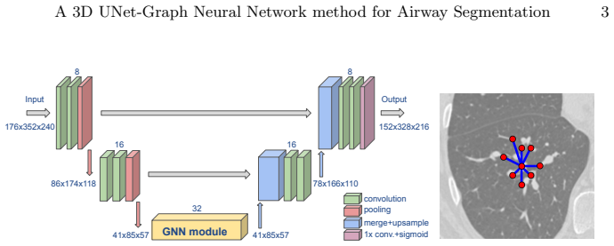
The load-bearing component is the GNN module placed at the deepest level of the 3D UNet, replacing the two convolutional layers of the baseline. The dense feature maps at that level are treated as graph nodes (supervoxels), each carrying a feature vector, with connectivity defined by a binary adjacency matrix $A$; the update rule in Eq. (1) combines each node's own transformed features with the degree-normalized features of its neighbours, and stacking four such layers lets nodes access higher-order neighbourhood information. Two adjacency constructions are tested: a predefined regular 26-neighbour grid and a dynamic adjacency recomputed during training from the $k=26$ nearest neighbours in feature space, constrained to a cube of at most 5 voxels away to fit in GPU memory.
What would settle it
Run the same four models on an independent held-out set of chest CTs with thresholds fixed using only the training and validation ROC curves, and check whether the UNet-GNN models still show significantly lower $d_{FN}$ at 13% volume leakage; if the gain vanishes, it was an artifact of test-set threshold tuning.
Extended reading notes
Core claim
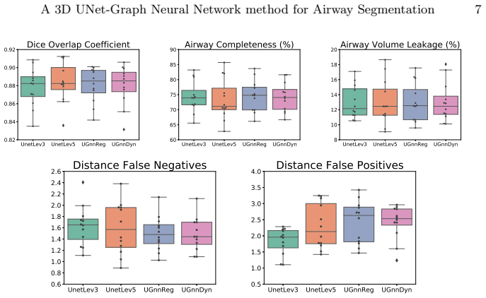
The central claim is that inserting a graph neural network module, consisting of four stacked graph-convolution layers of the form $H^{(l+1)} = \sigma(H^{(l)}W_0^{(l)} + D^{-1}AH^{(l)}W_1^{(l)})$, at the deepest resolution level of a 3D UNet improves airway-tree segmentation in a specific, measurable way. Compared with the baseline 3-level UNet, the UNet-GNN models achieve a significantly lower false-negative centreline distance $d_{FN}$ ($p \le 0.01$ for both adjacency types) at an operating point fixed at 13% volume leakage, while Dice overlap, airway completeness, and leakage show no significant difference. The authors interpret the lower $d_{FN}$ as evidence that the models produce slightly more complete airway trees with more or longer peripheral branches, and they note this comes with fewer trainable parameters (about 50k versus about 90k for the baseline; a deeper 5-level UNet with about 1.4M parameters shows no such gain). The two adjacency choices, fixed 26-neighbour connectivity and a dynamically computed $k$-nearest-neighbour graph in feature space with $k=26$, perform similarly.
Load-bearing premise
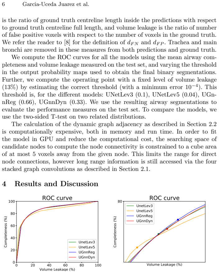
The comparison relies on probability thresholds (0.1, 0.04, 0.66, 0.33) chosen from the test-set ROC curves to fix leakage at 13%, and if those thresholds do not transfer to new scans, the reported improvement in false-negative distance may not reflect real-world performance.
Editorial extensions
If this is right
- Replacing the deepest convolutional block with graph convolutions yields a small but significant reduction in false-negative centreline distance, suggesting more complete airway trees at equal leakage.
- Dice overlap and airway completeness are unchanged, so the gain is specific to branch detection as measured along the centreline rather than to overall volume overlap.
- The UNet-GNN models need about 50k trainable parameters versus about 90k for the baseline and about 1.4M for the 5-level UNet, so graph convolutions can substitute for model capacity in this setting.
- A deeper 5-level UNet does not show the same $d_{FN}$ improvement, suggesting extra resolution depth is not the mechanism behind the gain.
- Both fixed and dynamically computed graph adjacency produce similar results, so the benefit is not sensitive to whether connectivity is predefined or learned in feature space.
Reading between the lines
- Placing GNN modules at multiple resolution levels, not just the deepest, is a testable extension that could amplify the effect if connectivity information helps at several scales; the paper notes this was not attempted due to GPU memory limits.
- The same architecture could transfer to other elongated or tree-like structures in medical imaging, such as vessels, bronchi, or neurons, where peripheral branch detection is the bottleneck and centreline-distance evaluation is the appropriate metric.
- Because the dynamic adjacency is limited to a 5-voxel search cube during training, the model only approximates true long-range connectivity; relaxing that constraint with more memory could either strengthen or weaken the reported gain.
- If test-set threshold tuning is a major source of the improvement, fixed-threshold deployment on new scans would likely narrow the gap; this is worth checking before clinical use.
Signed reviews
Editorial analysis
A structured set of objections, weighed in public.
Referee Report
Summary. The paper proposes a hybrid 3D UNet-GNN architecture for airway segmentation from chest CTs. The two convolutional layers at the deepest encoding level of a 3D UNet are replaced by a module of four graph-convolutional layers operating on a graph whose nodes are derived from the downsampled feature maps. Two adjacency strategies are tested: a fixed 26-neighbour regular grid and a dynamically computed kNN adjacency in feature space. Using 32 low-dose CTs from the Danish Lung Cancer Screening Trial (16 train, 4 validation, 12 test), the authors compare four models: UNetLev3, UNetLev5, UGnnReg, and UGnnDyn. At a fixed 13% volume-leakage operating point, selected by choosing per-model probability thresholds from the test-set ROC curves, the UNet-GNN models show significantly lower false-negative centreline distance dFN and significantly higher false-positive centreline distance dFP relative to UNetLev3, with no significant differences in Dice or airway completeness. The paper concludes that the GNN-based models can segment slightly more complete airway trees with fewer trainable parameters.
Significance. If the reported dFN improvement is a true effect, the paper provides a useful architectural contribution: replacing the deepest UNet convolutions with graph convolutions can reduce false-negative branch detection at fixed leakage, with a smaller parameter count than the baseline. The authors are transparent about the mixed result pattern (lower dFN but higher dFP, no significant change in Dice or completeness) and about the computational constraints of the GNN module. The work is an empirical evaluation rather than a derivation, and the comparison is grounded in held-out test scans. However, the statistical evidence is weakened by the joint selection of operating thresholds and test-set evaluation, and by multiple uncorrected paired t-tests on only 12 scans. The central claim is therefore plausible but not yet securely established.
major comments (3)
- [Section 3.4, Figure 2] The per-model probability thresholds (0.1, 0.04, 0.66, 0.33) are estimated from the test-set ROC curves themselves to achieve a fixed 13% volume leakage, and the resulting segmentations are then used to compute dFN, dFP, Dice, and completeness on the same test set. This procedure selects each model's operating point on the evaluation data, which can systematically favour models with particular ROC curvature and inflate apparent differences between models. The large spread in the selected thresholds also indicates substantially different probability calibrations across models, making it unclear whether the reported dFN gain would generalize to new scans. To support the central claim, the authors should either tune thresholds on a separate validation set (e.g., within cross-validation) or demonstrate that the dFN differences are robust over a range of leakage levels and thresholds rather than only at the single test-selected point.
- [Section 4, Results] The reported significance values (e.g., p = 0.001 for UGnnDyn vs. UNetLev3 in dFN, p < 0.001 for both GNN models vs. UNetLev3 in dFP) come from multiple two-sided paired t-tests performed across four models and several metrics on a test set of only 12 scans. No correction for multiple comparisons is applied, and with this sample size the p-values are sensitive to a few scans. The authors should report effect sizes, confidence intervals, or corrected p-values, and at minimum state how many comparisons were made. As it stands, the p-values do not provide strong evidence for the specific claim that the GNN variants are superior to UNetLev3 in dFN.
- [Section 5, Conclusions] The conclusion that the proposed models "can segment slightly more complete airway trees" is not fully supported by the result pattern: dFN improves significantly, but dFP worsens significantly, while Dice and airway completeness are unchanged. A decrease in false-negative centreline distance cannot be equated with a clinically meaningful gain when it is accompanied by a corresponding increase in false positives. The paper should discuss the trade-off more explicitly and, if the authors wish to claim an overall benefit, justify why reducing false negatives is more desirable than increasing false positives for the airway segmentation task.
minor comments (5)
- [Section 1, Introduction] The sentence "Initial work of combining CNNs and GNNs was by proposed Shin et al." contains a word-order error; it should read "was proposed by Shin et al.".
- [Section 2.2] The dynamic graph adjacency is computed with k = 26 neighbours, the same as the regular 26-neighbour case, but the search space is constrained to a cube 5 voxels away. It would be helpful to clarify whether the self-connection is included in the adjacency and how the degree matrix in Eq. (1) is defined in that case.
- [Section 2.2] The statement that the dynamic graph enables nodes to access "information well beyond their directly connected neighbours" is somewhat undercut by the 5-voxel search-space constraint. Please clarify how much of the long-range benefit actually survives the constraint.
- [Figure 2] In the right-hand detailed view, the markers for the operating points at threshold 0.5 should be clearly described in the caption or legend, as the current figure legend is not fully self-contained.
- [Section 3.3] The loss function in Eq. (3) is written as a single sum over the region of interest, but the dice loss typically involves a sum over classes or a sum over the full image with a masked ground truth. Please clarify the notation so that the masking operation is unambiguous.
Circularity Check
No circularity: the central claim is an empirical comparison measured on held-out test CTs; test-set threshold selection is a fairness concern, not a self-referential derivation.
full rationale
The paper does not derive its headline result from a fitted input or from an author-imported uniqueness claim. The UNet-GNN architecture is compared with the baseline UNet on 12 held-out CT scans; Dice, completeness, leakage, and centreline distances are computed from test-set segmentations. The self-citations ([7] baseline UNet, [8] GNN airway extraction and the dFN/dFP metrics, [10] reference airway segmentation) provide benchmarks, components, and ground truth, but none of them is used to force the reported dFN improvement. The thresholds in Sec. 3.4 (0.1, 0.04, 0.66, 0.33) are chosen from test-set ROC curves to fix leakage at 13%, which means the leakage equality is imposed by construction and threshold choice could affect the fairness of the dFN comparison; however, this is a statistical evaluation issue rather than a circularity, because the dFN/dFP values are independent centreline measurements, not quantities defined by the threshold-fitting procedure. No equation in the paper reduces to its own input, and no load-bearing argument rests solely on a self-citation. Therefore no circularity is present.
Assumptions & free parameters
free parameters (5)
- Per-model probability thresholds for fixed 13% leakage operating point =
UNetLev3: 0.10; UNetLev5: 0.04; UGnnReg: 0.66; UGnnDyn: 0.33
- Number of graph neighbours k =
26
- Number of GNN layers L =
4
- GNN feature expansion factor E = 2F =
E = 2F
- Input patch size =
176 x 352 x 240
assumptions (5)
- domain assumption Reference segmentations from method [10] applied to the union of [11] and [12], then corrected by an expert, represent the true airway lumen.
- domain assumption Excluding the trachea and main bronchi from the loss and evaluation focuses learning on peripheral branches without biasing model comparisons.
- domain assumption The 12 test CTs from DLCST are sufficient and representative for paired t-tests across four models.
- standard math The graph convolution update in Eq. (1), taken from Kipf and Welling [4], is a valid feature-propagation operator for this segmentation task.
- domain assumption Dice loss with a lung mask is an appropriate training objective for airway segmentation.
Cite this review
Pith. "Pith review of A joint 3D UNet-Graph Neural Network-based method for Airway Segmentation from chest CTs." pith.science (2026). https://pith.science/paper/AQPEP2MI
@misc{pith2026190808588,
  author       = {Pith},
  title        = {Pith review of: A joint 3D UNet-Graph Neural Network-based method for Airway Segmentation from chest CTs},
  year         = {2026},
  howpublished = {\url{https://pith.science/paper/AQPEP2MI}},
  note         = {Machine review of arXiv:1908.08588}
}
read the original abstract
We present an end-to-end deep learning segmentation method by combining a 3D UNet architecture with a graph neural network (GNN) model. In this approach, the convolutional layers at the deepest level of the UNet are replaced by a GNN-based module with a series of graph convolutions. The dense feature maps at this level are transformed into a graph input to the GNN module. The incorporation of graph convolutions in the UNet provides nodes in the graph with information that is based on node connectivity, in addition to the local features learnt through the downsampled paths. This information can help improve segmentation decisions. By stacking several graph convolution layers, the nodes can access higher order neighbourhood information without substantial increase in computational expense. We propose two types of node connectivity in the graph adjacency: i) one predefined and based on a regular node neighbourhood, and ii) one dynamically computed during training and using the nearest neighbour nodes in the feature space. We have applied this method to the task of segmenting the airway tree from chest CT scans. Experiments have been performed on 32 CTs from the Danish Lung Cancer Screening Trial dataset. We evaluate the performance of the UNet-GNN models with two types of graph adjacency and compare it with the baseline UNet.
Figures
Reference graph
Works this paper leans on
-
[8]
1st Conference on Medical Imaging with Deep Learning, Amsterdam, 2018
Selvan, R., et al.: Extraction of airways using graph neural networks. 1st Conference on Medical Imaging with Deep Learning, Amsterdam, 2018
work page 2018
-
[9]
Deep Vessel Segmentation By Learning Graphical Connectivity
Shin, S.Y., Lee, S., Yon, I.D., Lee, K.M.: Deep Vessel Segmentation By Learning Graphical Connectivity. ArXiv preprint arXiv:1806.02279, 2018
work page Pith review arXiv 2018
-
[1]
IEEE Conference on Computer Vision and Pattern Recognition (CVPR), 2015
Long, J., Shelhamer, E., Darrell, T.: Fully convolutional networks for semantic seg- mentation. IEEE Conference on Computer Vision and Pattern Recognition (CVPR), 2015
work page 2015
-
[2]
Medical Image Computing and Computer-Assisted Inter- vention MICCAI, pp
Ronneberger, O., Fischer, P., Brox, T.: U-Net: Convolutional networks for biomed- ical image segmentation. Medical Image Computing and Computer-Assisted Inter- vention MICCAI, pp. 234241, 2015
work page 2015
-
[3]
IEEE Transactions on Neural Networks, 20(1):6180, 2009
Scarselli, F., Gori, M., Tsoi, A.C., Hagenbuchner, M., Monfardini, G.: The graph neural network model. IEEE Transactions on Neural Networks, 20(1):6180, 2009
work page 2009
-
[4]
and Welling, M.: Semi-supervised classification with graph convolutional networks
Kipf, T.N. and Welling, M.: Semi-supervised classification with graph convolutional networks. International Conference on Learning Representations, 2017
work page 2017
-
[5]
et al: Extraction of Airways From CT (EXACT09)
Lo, P. et al: Extraction of Airways From CT (EXACT09). IEEE Transactions on Medical Imaging, Vol. 31, No. 11, 2012
work page 2012
-
[6]
Medical Image Computing and Computer-Assisted Intervention MICCAI 2017, pp
Meng, Q., Roth, H.R., Kitasaka, T., Oda, M., Ueno, J., Mori, K.: Tracking and Segmentation of the Airways in Chest CT Using a Fully Convolutional Network. Medical Image Computing and Computer-Assisted Intervention MICCAI 2017, pp. 198-207, 2017
work page 2017
Show all 13 references
-
[7]
Thoracic Image Analysis Workshop, pp 238-250, 2018
Garcia-Uceda Juarez, A., Tiddens, H.A.W.M., de Bruijne, M.: Automatic Airway Segmentation in Chest CT Using Convolutional Neural Networks. Thoracic Image Analysis Workshop, pp 238-250, 2018
2018
-
[10]
Medical image analysis 18, pp
Petersen, J., Nielsen, M., Lo, P., Nordenmark, L.H., Pedersen, J.H., Wille, M.M.W., Dirksen, A., de Bruijne, M.: Optimal surface segmentation using flow lines to quan- tify airway abnormalities in chronic obstructive pulmonary disease. Medical image analysis 18, pp. 531-541, 2014
2014
-
[11]
Medical image analysis 14.4, pp
Lo, P., Sporring, J., Ashraf, H., Pedersen, J.H., de Bruijne, M.: Vessel-guided airway tree segmentation: A voxel classification approach. Medical image analysis 14.4, pp. 527-538, 2010
2010
-
[12]
International Conference on Medical Image Computing and Computer-Assisted Intervention
Lo, P., Sporring, J., Pedersen, J.H., de Bruijne, M.: Airway tree extraction with locally optimal paths. International Conference on Medical Image Computing and Computer-Assisted Intervention. Springer, Berlin, Heidelberg, 2009
2009
-
[13]
Journal of Thoracic Oncology 4.5, pp
Pedersen, J., Jesper, H., et al.: The Danish randomized lung cancer CT screening trial overall design and results of the prevalence round. Journal of Thoracic Oncology 4.5, pp. 608-614, 2009
2009
Reviewed August 14, 2026 · model on record in the stance chip above.
Discussion (0). Continue with ORCID to comment.