REVIEW 3 major objections 4 minor 43 references
Vibrational signatures of diamondoid dimers with large intramolecular London dispersion interactions
T0 review · 3 major / 4 minor · reviewed 2026-08-14 · deepseek-v4-flash
Pith's one-line read Intramolecular dispersion forces in diamondoid dimers shift inward CH2 scissoring and CH stretch vibrations upward by up to 150 cm−1, and the resulting ~3050 cm−1 CH stretch band is proposed as a direct Raman marker of strong…
desk verdict Solid Raman study of diamondoid dimers that identifies new high-frequency CH/CH2 modes and a useful dimer-breathing-mode fingerprint, though the claim that dispersion specifically causes the upshifts is not isolated from geometry effects. read the letter →
The pith
A machine-rendered reading of the paper's core claim, the machinery that carries it, and where it could break.
The reading
What carries the argument
The load-bearing mechanism is intramolecular London dispersion attraction acting across well-aligned, opposing H···H surfaces within a single molecule. These contacts add restoring forces felt by CH bonds pointing toward the opposite diamondoid facet, shifting their stretch and scissor frequencies upward, and they couple the two diamondoid moieties into low-frequency dimer modes. Two named objects carry the spectral analysis: the breathing-like mode (BLM) of the individual diamondoid cages, whose frequency indicates cage size, and the dimer breathing mode (DBM), a collective in-phase elongation of the central carbon-carbon bond that is the strongest low-frequency Raman feature. Computationally, the argument uses the PBE functional with the Grimme-D3 dispersion correction and a factor of 0.976 scaling CH stretch frequencies.
What would settle it
Replace the inward-pointing hydrogens of a large dimer with deuterium and remeasure the Raman spectrum: if the assignment is right, the ~3050 cm−1 modes should shift by roughly the isotopic reduced-mass factor while ordinary CH modes remain, whereas absence of such a shift rules out the inward-CH-stretch interpretation.
Extended reading notes
Core claim
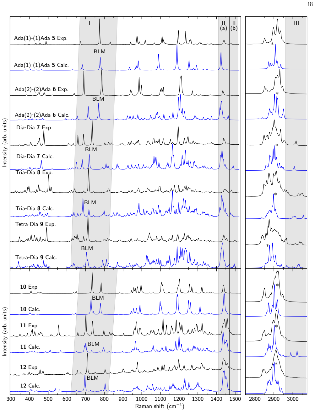
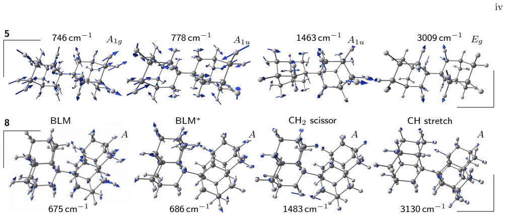
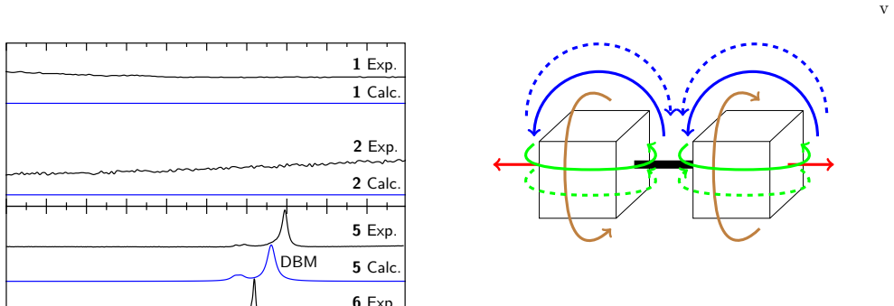
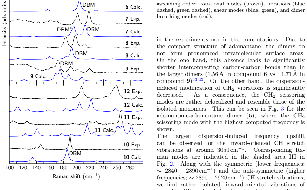
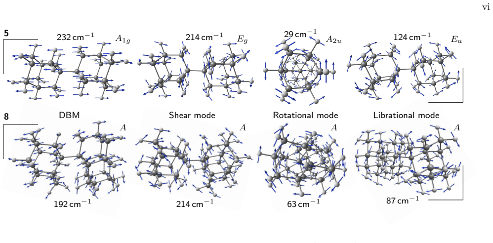
The central discovery is that the same van der Waals attraction that stabilizes the unusually long carbon-carbon single bonds (up to 1.71 Å) also renormalizes the vibrational spectrum. In direct diamondoid dimers, CH2 scissoring modes localized at the inward contact surface appear about 50 cm−1 above the ordinary scissoring modes near 1440 cm−1, while isolated, inward-pointing CH stretch vibrations form a distinct band near 2920-3050 cm−1, up to 150 cm−1 higher than the usual symmetric and antisymmetric CH stretch modes. These high-energy modes are absent in adamantane dimers because their compact structure cannot form extended intramolecular facets. The paper concludes that these high-frequency CH stretch modes can therefore serve as a direct, structure-induced marker of strong intramolecular H···H dispersion contacts. In the low-frequency region, it establishes a ladder of structure-induced dimer modes (rotations, librations, shear modes, and a dimer breathing mode), with the dimer breathing mode carrying the largest Raman intensity and showing an inverse dependence on dimer size.
Load-bearing premise
The load-bearing premise is that an isolated, relaxed PBE-D3 molecule (with a 0.976 CH-stretch scaling factor) reproduces the vibrations of the crystalline samples closely enough for mode assignment; the authors themselves flag that this fails for compound 11 because its relaxed ground-state geometry differs from its van der Waals crystal structure.
Editorial extensions
If this is right
- High-frequency CH stretch modes near 3050 cm−1 can be used as a Raman fingerprint for diamondoid dimers with strong intramolecular H···H contacts, distinguishing them from double-bond-linked diamondoids that lack such contacts.
- The dimer breathing mode frequency provides an inverse size ruler for single-bond diamondoid dimers, and combined with the BLM frequency it can distinguish even homo dimers linked at different positions.
- Dispersion corrections are necessary, not optional, in quantum-chemical calculations of the vibrational properties of these dimers, since the upshifts and dimer modes disappear without them.
- The absence of high-energy CH2 scissoring and CH stretch modes in adamantane dimers follows directly from their inability to form extended intramolecular contact surfaces, so the marker modes are tied to molecular shape, not merely to dimerization.
- The proposed spectral fingerprints could allow identification of specific single-bond diamondoid dimers without relying solely on structural characterization.
Reading between the lines
- Editorial inference: if the ~3050 cm−1 marker depends only on close, well-aligned H···H pairs, it may generalize to other crowded hydrocarbons as a Raman-based reporter of intramolecular dispersion contacts, not just diamondoid dimers.
- Editorial inference: temperature- or pressure-dependent Raman measurements should tune the H···H alignment and therefore shift or broaden the marker modes, a testable prediction the paper does not make.
- Editorial inference: the inverse-size dimer-breathing-mode trend suggests a minimal harmonic model of two rigid cages coupled by a soft central bond, from which an effective force constant for the unusually long C-C bond could be extracted.
- Editorial inference: the authors' explanation for the compound 11 discrepancy implies that a periodic dispersion-corrected calculation of the actual crystal structure should reproduce the missing mode, which would directly test whether the isolated-molecule model is the right comparison.
Signed reviews
Editorial analysis
A structured set of objections, weighed in public.
Referee Report
Summary. The paper presents experimental Raman spectra and dispersion-corrected DFT (PBE-D3/def2-TZVP) calculations for eight diamondoid dimers (compounds 5–12), comparing them with pristine diamondoids. The authors identify new high-frequency modes near 1490 cm^-1 (CH2 scissoring) and 3050 cm^-1 (CH stretch) that they attribute to strong intramolecular dispersion interactions, and low-frequency 'dimer modes' (rotational, librational, shear, and dimer breathing modes) whose most intense feature, the dimer breathing mode, shifts inversely with dimer size. The central claim is that dispersion causes frequency upshifts of up to 150 cm^-1 for inward-oriented CH2/CH vibrations, and that the ~3050 cm^-1 CH stretch modes can serve as a direct marker of strong intramolecular H···H interactions.
Significance. If the causal attribution to dispersion is established, the paper would provide a useful spectroscopic fingerprint for intramolecular London dispersion interactions in diamondoid dimers and a practical way to identify single-bond dimers via the dimer breathing mode. The study benefits from direct visual agreement between experimental and computed Raman spectra, from using standard external benchmarks (PBE-D3, def2-TZVP) rather than parameters fitted to these data, and from clear displacement-vector analysis of the assigned modes. The main weakness is that all calculations already include the D3 dispersion correction, so the specific role of dispersion in producing the upshift is not isolated; the acknowledged exception of compound 11 further complicates the marker claim. These issues are addressable with additional control calculations and more complete uncertainty reporting.
major comments (3)
- [§2 Theoretical details and §3 Results (high-frequency modes)] All vibrational calculations use PBE with the Grimme-D3 dispersion correction, and no dispersion-free control (e.g., PBE without D3) or alternative dispersion treatment is reported. The central claim that dispersion causes the 150 cm^-1 upshift of inward-oriented CH2 and CH stretch vibrations is therefore not actually tested against a scenario in which only the steric/covalent geometry is present. The upshift could be a generic consequence of the crowded intramolecular H···H contacts or an artifact of the D3 parametrization at short H···H distances. I request PBE-without-D3 calculations for compounds 5–9, and ideally a second dispersion-corrected method, to isolate the D3 contribution to the frequencies and to the Hessian.
- [§3 (compound 11 discussion) and Conclusion] The paper states that for compound 11, the computed structure-induced CH stretch upshift is not observed experimentally and attributes this to differences between the relaxed ground-state geometry and the van der Waals crystal structure. This is a load-bearing exception because the conclusion claims the ~3050 cm^-1 modes can serve as a direct marker of strong intramolecular H···H interactions. The authors should either demonstrate that the marker is robust to crystal-environment effects (e.g., through periodic DFT or explicit crystal-structure calculations) or explicitly qualify the marker claim to gas-phase-like isolated molecules, with compound 11 as a documented counterexample.
- [§3 (all experimental spectra)] No experimental frequency uncertainties or measurement precision are reported, despite the quantitative claim of up to 150 cm^-1 shifts and the use of small frequency differences (e.g., ~20 cm^-1 separation between DBM and BLM in compounds 5 and 6) for structural identification. Please provide the spectral resolution and, for at least a few key peaks, the standard deviation of line positions from repeated measurements or from different crystal facets. A table of measured versus computed frequencies for the assigned modes would also clarify the quantitative agreement.
minor comments (4)
- [Fig. 2 caption] The CH stretch scaling factor 0.976 is introduced without justification or reference; please state how this factor was determined (e.g., from a benchmark set) and whether it is applied consistently to all computed CH stretch frequencies.
- [Fig. 4 and text on compound 7] The label 'DBM*' is used for two related dimer breathing modes in compound 7, but neither the figure nor the caption clearly distinguishes the asterisked peaks; consider marking both features explicitly.
- [Conclusion] The conclusion states dimer-mode frequencies between 23 and 232 cm^-1, while the text reports rotational modes from 29 to 77 cm^-1, librations ~90–135 cm^-1, shear modes up to 217 cm^-1, and DBM from 170 to 239 cm^-1; please make the reported range consistent with the body of the paper.
- [§2 Theoretical details] The modeled Raman spectra use a Lorentzian/Gaussian FWHM of 5 cm^-1, but the experimental resolution or broadening is not given; a brief statement of the instrument resolution would aid the visual comparison in Figs. 2 and 4.
Circularity Check
No significant circularity: the DFT calculations are external benchmarks, the CH-stretch scaling is uniform and standard, and the experimental Raman data independently corroborate the assignment of the new modes.
full rationale
The paper's derivation chain is self-contained against external benchmarks. The computed spectra use PBE-D3 with def2-TZVP basis sets, a parameterization fixed independently of the present Raman data; the 0.976 scaling factor applies uniformly to all computed CH stretches and therefore cannot manufacture the claimed relative upshifts of the inward-oriented modes. The experimental Raman spectra are measured independently and show the high-frequency CH2 scissoring and CH stretch features (regions IIb and III) as well as the low-frequency dimer breathing modes, so the mode assignments are corroborated rather than imported solely from prior work. Self-citations to ref. 24 for the concept of dimer modes and for the inverse size dependence of the DBM are not load-bearing in a circular way: the same trend is reproduced here in both experiment and computation for the single-bond dimers. The attribution of the upshifts to intramolecular dispersion is an interpretation supported by the presence of repulsive H...H contacts in the computed geometries, but even if one questions the lack of a dispersion-free control, that is a concern about causal testing, not a reduction of the claimed prediction to its inputs by construction. No equation, fitted parameter, or self-citation chain forces the central result.
Assumptions & free parameters
free parameters (2)
- CH stretch frequency scaling factor =
0.976
- Line broadening FWHM for computed spectra =
5 cm^-1
assumptions (3)
- domain assumption PBE-D3/def2-TZVP yields accurate vibrational frequencies and Raman intensities for these diamondoid dimers.
- domain assumption Raman spectra of crystalline van der Waals samples can be interpreted with isolated-molecule DFT spectra.
- domain assumption The empirical 0.976 scaling factor makes computed CH stretch frequencies directly comparable to experiment.
Cite this review
Pith. "Pith review of Vibrational signatures of diamondoid dimers with large intramolecular London dispersion interactions." pith.science (2026). https://pith.science/paper/AURTA4ZK
@misc{pith2026190805933,
  author       = {Pith},
  title        = {Pith review of: Vibrational signatures of diamondoid dimers with large intramolecular London dispersion interactions},
  year         = {2026},
  howpublished = {\url{https://pith.science/paper/AURTA4ZK}},
  note         = {Machine review of arXiv:1908.05933}
}
abstract
We analyze the vibrational properties of diamondoid compounds via Raman spectroscopy. The compounds are interconnected with carbon-carbon single bonds that exhibit exceptionally large bond lengths up to 1.71 A. Attractive dispersion interactions caused by well-aligned intramolecular H--H contact surfaces determine the overall structures of the diamondoid derivatives. The strong van-der-Waals interactions alter the vibrational properties of the compounds in comparison to pristine diamondoids. Supported by dispersion-corrected density functional theory (DFT) computations, we analyze and explain their experimental Raman spectra with respect to unfunctionalized diamondoids. We find a new set of dispersion-induced vibrational modes comprising characteristic CH/CH$_{2}$ vibrations with exceptionally high energies. Further, we find structure-induced dimer modes that are indicative of the size of the dimers.
Figures

Figures from the paper (3 more)
Reference graph
Works this paper leans on
-
[1]
Landa, S. and Mach\' a c ek, V. Collect. Czech. Commun. 5 , 1--5 (1933)
work page 1933
-
[2]
Dahl, J. E., Liu, S. G., and Carlson, R. M. K. Science 299 (5603), 96--99 (2003)
work page 2003
-
[3]
Balaban, A. T. and Schleyer, P. v. R. Tetrahedron 34 (24), 3599 -- 3609 (1978)
work page 1978
-
[4]
Gund, T. M., Osawa, E., Williams, V. Z., and Schleyer, P. v. R. J. Org. Chem. 39 (20), 2979--2987 (1974)
work page 1974
-
[5]
Tetrahedron 36 (8), 971 -- 992 (1980)
McKervey, M. Tetrahedron 36 (8), 971 -- 992 (1980)
work page 1980
-
[6]
Schwertfeger, H., Fokin, A. A., and Schreiner, P. R. Angew. Chem. Int. Ed. 47 (6), 1022--1036 (2008)
work page 2008
-
[7]
under, K., Dahl, J. E., Carlson, R. M. K., M\
Landt, L., Kl\"under, K., Dahl, J. E., Carlson, R. M. K., M\"oller, T., and Bostedt, C. Phys. Rev. Lett. 103 , 047402 (2009)
work page 2009
-
[8]
Richter, R., Wolter, D., Zimmermann, T., Landt, L., Knecht, A., Heidrich, C., Merli, A., Dopfer, O., Reiß, P., Ehresmann, A., Petersen, J., Dahl, J. E., Carlson, R. M. K., Bostedt, C., Möller, T., Mitrić, R., and Rander, T. Phys. Chem. Chem. Phys. 16 , 3070--3076 (2014)
work page 2014
Show all 43 references
-
[9]
and Saalfrank, P
Banerjee, S. and Saalfrank, P. Phys. Chem. Chem. Phys. 16 , 144--158 (2014)
2014
-
[10]
A., Tkachenko, B
Fokin, A. A., Tkachenko, B. A., Gunchenko, P. A., Gusev, D. V., and Schreiner, P. R. Chem. Eur. J. 11 (23), 7091--7101 (2005)
2005
-
[11]
A., Hierso, J.-C., Poinsot, D., Fokin, A
Gunawan, M. A., Hierso, J.-C., Poinsot, D., Fokin, A. A., Fokina, N. A., Tkachenko, B. A., and Schreiner, P. R. New J. Chem. 38 , 28--41 (2014)
2014
-
[12]
A., Tkachenko, B
Fokina, N. A., Tkachenko, B. A., Merz, A., Serafin, M., Dahl, J. E. P., Carlson, R. M. K., Fokin, A. A., and Schreiner, P. R. Eur. J. Org. Chem. 2007 (28), 4738--4745
2007
-
[13]
R., Fokina, N
Schreiner, P. R., Fokina, N. A., Tkachenko, B. A., Hausmann, H., Serafin, M., Dahl, J. E. P., Liu, S., Carlson, R. M. K., and Fokin, A. A. J. Org. Chem. 71 (18), 6709--6720 (2006)
2006
-
[14]
Mella, M., Freccero, M., Soldi, T., Fasani, E., and Albini, A. J. Org. Chem. 61 (4), 1413--1422 (1996)
1996
-
[15]
P., Balestrieri, C., Becker, J., Hausmann, H., Bodwell, G
Kahl, P., Wagner, J. P., Balestrieri, C., Becker, J., Hausmann, H., Bodwell, G. J., and Schreiner, P. R. Angew. Chem. Int. Ed. 55 (32), 9277--9281 (2016)
2016
-
[16]
E., Carlson, R
Rander, T., Staiger, M., Richter, R., Zimmermann, T., Landt, L., Wolter, D., Dahl, J. E., Carlson, R. M. K., Tkachenko, B. A., Fokina, N. A., Schreiner, P. R., Möller, T., and Bostedt, C. J. Chem. Phys. 138 (2), 024310 (2013)
2013
-
[17]
Landt, L., Bostedt, C., Wolter, D., Möller, T., Dahl, J. E. P., Carlson, R. M. K., Tkachenko, B. A., Fokin, A. A., Schreiner, P. R., Kulesza, A., Mitrić, R., and Bonačić-Koutecký, V. J. Chem. Phys. 132 (14), 144305 (2010)
2010
-
[18]
Banerjee, S., Stüker, T., and Saalfrank, P. Phys. Chem. Chem. Phys. 17 , 19656--19669 (2015)
2015
-
[19]
V\"or\"os, M., Demj\'en, T., Szilv\'asi, T., and Gali, A. Phys. Rev. Lett. 108 , 267401 (2012)
2012
-
[20]
A., Koso, T
Zimmermann, T., Richter, R., Knecht, A., Fokin, A. A., Koso, T. V., Chernish, L. V., Gunchenko, P. A., Schreiner, P. R., Möller, T., and Rander, T. J. Chem. Phys. 139 (8), 084310 (2013)
2013
-
[21]
Rander, T., Bischoff, T., Knecht, A., Wolter, D., Richter, R., Merli, A., and Möller, T. J. Am. Chem. Soc. 139 (32), 11132--11137 (2017)
2017
-
[22]
S., Koso, T., Pashenko, A
Zhuk, T. S., Koso, T., Pashenko, A. E., Hoc, N. T., Rodionov, V. N., Serafin, M., Schreiner, P. R., and Fokin, A. A. J. Am. Chem. Soc. 137 (20), 6577--6586 (2015)
2015
-
[23]
A., Koso, T
Tyborski, C., Meinke, R., Gillen, R., Bischoff, T., Knecht, A., Richter, R., Merli, A., Fokin, A. A., Koso, T. V., Rodionov, V. N., Schreiner, P. R., Möller, T., Rander, T., Thomsen, C., and Maultzsch, J. J. Chem. Phys. 147 (4), 044303 (2017)
2017
-
[24]
A., Koso, T
Tyborski, C., Gillen, R., Fokin, A. A., Koso, T. V., Fokina, N. A., Hausmann, H., Rodionov, V. N., Schreiner, P. R., Thomsen, C., and Maultzsch, J. J. Phys. Chem. C 121 (48), 27082--27088 (2017)
2017
-
[25]
A., Koso, T
Meinke, R., Richter, R., Merli, A., Fokin, A. A., Koso, T. V., Rodionov, V. N., Schreiner, P. R., Thomsen, C., and Maultzsch, J. J. Chem. Phys. 140 (3), 034309 (2014)
2014
-
[26]
A., Schreiner, P
Meinke, R., Richter, R., Möller, T., Tkachenko, B. A., Schreiner, P. R., Thomsen, C., and Maultzsch, J. J. Phys. B 46 (2), 025101 (2013)
2013
-
[27]
and Koenig, J
Tuinstra, F. and Koenig, J. L. J. Chem. Phys. 53 (3), 1126--1130 (1970)
1970
-
[28]
Tyborski, C., Herziger, F., Gillen, R., and Maultzsch, J. Phys. Rev. B 92 , 041401 (2015)
2015
-
[29]
N., Allan, N
Filik, J., Harvey, J. N., Allan, N. L., May, P. W., Dahl, J. E., Liu, S., and Carlson, R. M. Spectrochim. Acta A 64 (3), 681 -- 692 (2006)
2006
-
[30]
Jensen, J. O. Spectrochim. Acta A 60 (8), 1895 -- 1905 (2004)
2004
-
[31]
and Lewis, J
Jenkins, T. and Lewis, J. Spectrochim. Acta A 36 (3), 259 -- 264 (1980)
1980
-
[32]
R., Chernish, L
Schreiner, P. R., Chernish, L. V., Gunchenko, P. A., Tikhonchuk, E. Y., Hausmann, H., Serafin, M., Schlecht, S., Dahl, J. E. P., Carlson, R. M. K., and Fokin, A. A. Nature 477 (308) (2011)
2011
-
[33]
A., Chernish, L
Fokin, A. A., Chernish, L. V., Gunchenko, P. A., Tikhonchuk, E. Y., Hausmann, H., Serafin, M., Dahl, J. E. P., Carlson, R. M. K., and Schreiner, P. R. J. Am. Chem. Soc. 134 (33), 13641--13650 (2012)
2012
-
[34]
Wagner, J. P. and Schreiner, P. R. Angew. Chem. Int. Ed. 54 (42), 12274--12296 (2015)
2015
-
[35]
A., Zhuk, T
Fokin, A. A., Zhuk, T. S., Blomeyer, S., Pérez, C., Chernish, L. V., Pashenko, A. E., Antony, J., Vishnevskiy, Y. V., Berger, R. J. F., Grimme, S., Logemann, C., Schnell, M., Mitzel, N. W., and Schreiner, P. R. J. Am. Chem. Soc. 139 (46), 16696--16707 (2017)
2017
-
[36]
M., Bostedt, C., van Buuren, T., Dahl, J
Willey, T. M., Bostedt, C., van Buuren, T., Dahl, J. E., Liu, S. G., Carlson, R. M. K., Terminello, L. J., and M\"oller, T. Phys. Rev. Lett. 95 , 113401 (2005)
2005
-
[37]
Wires Comp
Neese, F. Wires Comp. Mol. Sci. 2 , 73--78 (2012)
2012
-
[38]
and Maultzsch, J
Gillen, R. and Maultzsch, J. Phys. Rev. B 97 (16), 165306 (2018)
2018
-
[39]
Tornatzky, H., Gillen, R., Uchiyama, H., and Maultzsch, J. Phys. Rev. B 99 , 144309 (2019)
2019
-
[40]
P., Burke, K., and Ernzerhof, M
Perdew, J. P., Burke, K., and Ernzerhof, M. Phys. Rev. Lett. 77 , 3865--3868 (1996)
1996
-
[41]
Grimme, S., Antony, J., Ehrlich, S., and Krieg, H. J. Chem. Phys. 132 , 154104 (2010)
2010
-
[42]
and Ahlrichs, R
Weigend, F. and Ahlrichs, R. Phys. Chem. Chem. Phys. 7 , 3297--3305 (2005)
2005
-
[43]
Bond lengths are computed with a dispersion corrected DFT approach as described in the theoretical details
Reviewed August 14, 2026 · model on record in the stance chip above.
Discussion (0). Continue with ORCID to comment.