Data-Efficient Multimodal Alignment for Histopathology-based Molecular Prediction
Pith reviewed 2026-06-30 04:28 UTC · model grok-4.3
The pith
A lightweight alignment module on frozen models allows gene-set queries of H&E slides to predict molecular pathway activity.
A machine-rendered reading of the paper's core claim, the machinery that carries it, and where it could break.
Core claim
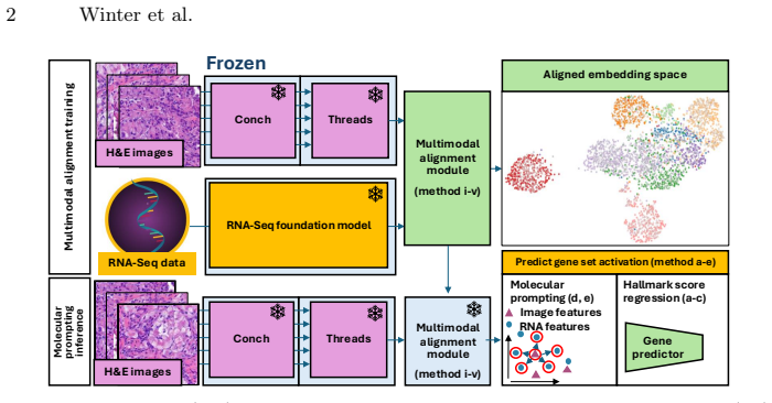
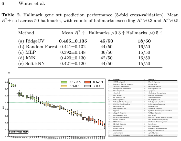
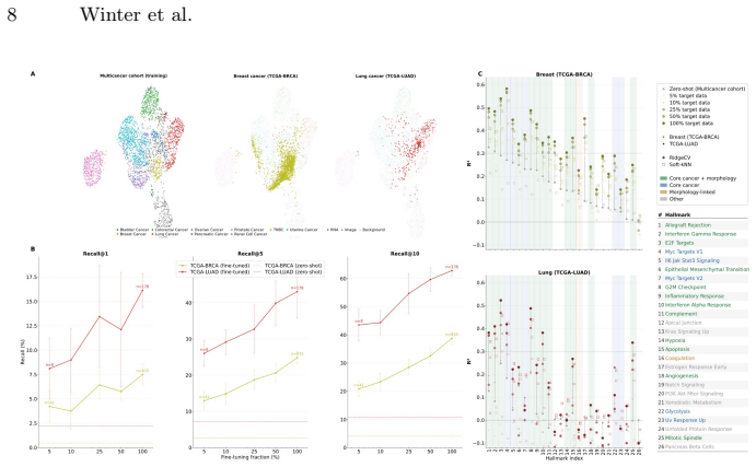
Training a lightweight alignment module atop frozen histopathology and RNA-Seq foundation models enables open-vocabulary molecular prompting by querying H&E slides with gene-set signatures to predict pathway activity without sequencing or end-to-end retraining. Using contrastive learning on a multi-cancer cohort of 1,720 samples yields a 25-fold improvement in retrieval over baselines, with morphologically grounded programs showing high predictability.
What carries the argument
Lightweight alignment module trained via contrastive learning to bridge embeddings from frozen histopathology and RNA-Seq foundation models.
Load-bearing premise
The features from the frozen foundation models are already close enough to molecular signals that a small contrastive alignment on this sample size can connect them effectively.
What would settle it
Failure to achieve similar retrieval gains or clinical correlations when tested on an independent multi-cancer cohort with new gene sets would indicate the alignment does not hold.
Figures
read the original abstract
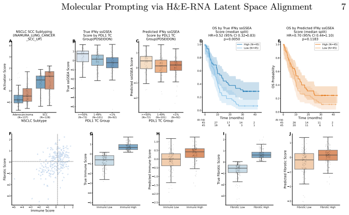
H&E-stained whole-slide images offer cohort-scale availability and rich spatial context but lack molecular specificity, whereas bulk RNA-seq provides transcriptome-wide resolution at high cost with limited archival availability. We show that training a lightweight alignment module atop frozen histopathology and RNA-Seq foundation models enables open-vocabulary molecular prompting -- querying H&E slides with gene-set signatures to predict pathway activity without sequencing or end-to-end retraining. Using contrastive learning on a multi-cancer cohort (N=1,720), we achieve a 25-fold improvement in retrieval over baseline methods. Systematic analysis reveals a graduated predictability spectrum: morphologically grounded programs (cell-cycle programs, immune-related) are most reliably predicted (R^2>0.5), while predicting pathways with no morphological footprint remains challenging as expected. We validate clinical utility on the POSEIDON clinical trial: H&E-predicted squamous cell carcinoma scores recapitulate NSCLC subtype identity and predicted IFN-gamma mirror PD-L1 tumor-cell expression groups. Furthermore, genesets describing immune activation and fibrosis predict known tumor microenvironment archetypes from histology alone. We further validate generalization of our approach across unseen cohorts and demonstrate data-efficient domain adaptation, establishing a slide-native framework for molecular analysis on H&E images.
Editorial analysis
A structured set of objections, weighed in public.
Referee Report
Summary. The paper claims that training a lightweight alignment module atop frozen histopathology and RNA-Seq foundation models with contrastive learning on a multi-cancer cohort (N=1,720) enables open-vocabulary molecular prompting from H&E slides to predict pathway activity without sequencing or end-to-end retraining. It reports a 25-fold retrieval improvement over baseline methods, a graduated predictability spectrum with R²>0.5 for morphologically grounded programs (cell-cycle, immune-related), clinical validation on the POSEIDON trial where predicted squamous cell carcinoma scores and IFN-gamma recapitulate NSCLC subtypes and PD-L1 groups, plus generalization across unseen cohorts and data-efficient domain adaptation.
Significance. If the results hold, the work has clear significance for computational pathology by offering a data-efficient route to molecular inference from routine H&E images that leverages existing foundation models without retraining them. The open-vocabulary gene-set prompting and clinical-trial validation are practical strengths; the graduated predictability spectrum is biologically plausible. The approach avoids the data and compute costs of end-to-end training, which is a genuine advantage if the frozen encoders already carry usable molecular signal.
major comments (2)
- [Abstract] Abstract: the 25-fold retrieval improvement is stated without identifying the baseline methods, their training details, or any statistical significance tests. This is load-bearing for the central claim of substantial superiority.
- [Abstract] Abstract: the central claim presupposes that the frozen foundation models already embed pathway-level structure (e.g., separation of cell-cycle vs. fibrosis signatures) that contrastive learning on N=1,720 can exploit. No zero-shot retrieval baselines, embedding visualizations, or ablation removing one encoder are described to test this precondition; without such evidence the 25-fold gain and R²>0.5 results cannot be confidently attributed to the alignment module rather than to the base encoders.
Simulated Author's Rebuttal
We thank the referee for the constructive comments, which highlight important clarifications needed for the abstract and supporting evidence. We address each point below and will revise the manuscript to strengthen the presentation of results.
read point-by-point responses
-
Referee: [Abstract] Abstract: the 25-fold retrieval improvement is stated without identifying the baseline methods, their training details, or any statistical significance tests. This is load-bearing for the central claim of substantial superiority.
Authors: We agree that the abstract should be more explicit. The baselines are direct cosine similarity between frozen encoders and a linear probe trained on the same N=1,720 cohort; full training details and p-values (Wilcoxon signed-rank tests across 5-fold cross-validation) appear in Section 3.2 and Supplementary Table S2. We will expand the abstract to name the baselines, note their training regime, and report statistical significance for the 25-fold gain. revision: yes
-
Referee: [Abstract] Abstract: the central claim presupposes that the frozen foundation models already embed pathway-level structure (e.g., separation of cell-cycle vs. fibrosis signatures) that contrastive learning on N=1,720 can exploit. No zero-shot retrieval baselines, embedding visualizations, or ablation removing one encoder are described to test this precondition; without such evidence the 25-fold gain and R²>0.5 results cannot be confidently attributed to the alignment module rather than to the base encoders.
Authors: We acknowledge the value of these controls. The current manuscript reports the graduated predictability spectrum and cross-cohort generalization as indirect support, but does not include explicit zero-shot retrieval numbers, t-SNE visualizations of pre-alignment embeddings, or single-encoder ablations. We will add these analyses (zero-shot retrieval near chance, visualizations showing pathway separation only post-alignment, and ablation results) to the revised Methods and Results sections to directly attribute gains to the alignment module. revision: yes
Circularity Check
No significant circularity; empirical alignment results are independent of inputs
full rationale
The paper's central result is an empirical demonstration: contrastive training of a lightweight module on top of frozen encoders yields a 25-fold retrieval gain on N=1720 samples, with graduated predictability for morphologically grounded pathways. No equations, definitions, or claims reduce any prediction to its own fitted parameters or to a self-citation chain; the frozen encoders are treated as external black boxes whose properties are not derived within the paper. Standard contrastive loss is applied without self-referential redefinition or renaming of known results as novel unification. The derivation chain is therefore self-contained against external benchmarks.
Axiom & Free-Parameter Ledger
Reference graph
Works this paper leans on
-
[1]
Cancer cell39(6), 845–865 (2021)
Bagaev, A., Kotlov, N., Nomie, K., Svekolkin, V., Gafurov, A., Isaeva, O., Osokin, N., Kozlov, I., Frenkel, F., Gancharova, O., et al.: Conserved pan-cancer microenvi- ronment subtypes predict response to immunotherapy. Cancer cell39(6), 845–865 (2021)
2021
-
[2]
Nature462(7269), 108–112 (2009)
Barbie, D.A., Tamayo, P., Boehm, J.S., Kim, S.Y., Moody, S.E., Dunn, I.F., Schinzel, A.C., Sandy, P., Meylan, E., et al.: Systematic RNA interference reveals that oncogenic KRAS-driven cancers require TBK1. Nature462(7269), 108–112 (2009)
2009
-
[3]
Nature Medicine30, 850–862 (2024)
Chen, R.J., Ding, T., Lu, M.Y., Williamson, D.F.K., Jaume, G., Song, A.H., Chen, B., Zhang, A., Shao, D., Shaban, M., Williams, M., Oldenburg, L., Weishaupt, L.L., Wang, J.J., Vaidya, A., Le, L.P., Gerber, G., Sahai, S., Williams, W., Mahmood, F.: Towards a general-purpose foundation model for computational pathology. Nature Medicine30, 850–862 (2024)
2024
-
[4]
Nature Methods21, 1470–1480 (2024)
Cui, H., Wang, C., Maan, H., Pang, K., Luo, F., Duan, N., Wang, B.: scGPT: Toward building a foundation model for single-cell multi-omics using generative AI. Nature Methods21, 1470–1480 (2024)
2024
-
[5]
Nature medicine pp
Ding, T., Wagner, S.J., Song, A.H., Chen, R.J., Lu, M.Y., Zhang, A., Vaidya, A.J., Jaume, G., Shaban, M., Kim, A., et al.: A multimodal whole-slide foundation model for pathology. Nature medicine pp. 1–13 (2025)
2025
-
[6]
Nature cancer 1(8), 800–810 (2020)
Fu, Y., Jung, A.W., Torne, R.V., Gonzalez, S., Vöhringer, H., Shmatko, A., Yates, L.R., Jimenez-Linan, M., Moore, L., Gerstung, M.: Pan-cancer computational histopathology reveals mutations, tumor composition and prognosis. Nature cancer 1(8), 800–810 (2020)
2020
-
[7]
Nature Cancer5(9), 1305–1317 (2024)
Hoang, D.T., Dinstag, G., Shulman, E.D., Hermida, L.C., BenHur, A., Kisilev, P., Raspe, E., Vanderstichele, A., Lambrechts, D., Linn, S.C., et al.: A deep- learning framework to predict cancer treatment response from histopathology im- ages through imputed transcriptomics. Nature Cancer5(9), 1305–1317 (2024)
2024
-
[8]
medRxiv (2025), preprint
Howard, F.M., Dolezal, J., Kochanny, S., Khramtsova, G., Vickery, J., Srisuwananukorn, A., Woodard, A., Chen, N., Nanda, R., Olopade, O.I., Huo, D., Pearson, A.T.: Integration of pathology image and gene expression data using a transformer-based multiple instance learning approach for breast cancer. medRxiv (2025), preprint
2025
-
[9]
arXiv preprint arXiv:2601.21560 (2026), published at ICLR 2026
Hu, S., Zeng, Q., Bhasker, N., Kather, J.N., Speidel, S.: HistoPrism: Unlocking functional pathway analysis from pan-cancer histology via gene expression predic- tion. arXiv preprint arXiv:2601.21560 (2026), published at ICLR 2026
-
[10]
Journal of Clinical Oncology41(6), 1213–1227 (2023)
Johnson, M.L., Cho, B., Luft, A., Alatorre-Alexander, J., Geater, S.L., Laktionov, K., Kim, S.W., Ahn, M.J., Carcereny, E., Audigier-Valette, C., et al.: Durvalumab with or without tremelimumab in combination with chemotherapy as first-line therapy for metastatic non-small-cell lung cancer: the phase III POSEIDON study. Journal of Clinical Oncology41(6), ...
2023
-
[11]
BioRxiv pp
Kang, B., Fan, R., Yi, M., Cui, C., Cui, Q.: A large-scale foundation model for bulk transcriptomes. BioRxiv pp. 2025–06 (2025)
2025
-
[12]
Nature Medicine25, 1054–1056 (2019)
Kather, J.N., Charoentong, P., Krisam, J., Renna, T., Hoffmeister, F., Chang- Claude, J., Hoffmeister, M., Brenner, H., Jäger, D., Halama, N.: Deep learning can predict microsatellite instability directly from histology in gastrointestinal cancer. Nature Medicine25, 1054–1056 (2019)
2019
-
[13]
detection of clinically actionable genetic alterations
Kather, J.N., Heij, L.R., Grabsch, H.I., Loeffler, C., Echle, A., Muti, H.S., Krause, J., Niehues, J.M., Sommer, K.A., Bankhead, P., et al.: Pan-cancer image-based 10 Winter et al. detection of clinically actionable genetic alterations. Nature cancer1(8), 789–799 (2020)
2020
-
[14]
PLoS medicine16(1), e1002730 (2019)
Kather, J.N., Krisam, J., Charoentong, P., Luedde, T., Herpel, E., Weis, C.A., Gaiser, T., Marx, A., Valous, N.A., Ferber, D., et al.: Predicting survival from colorectal cancer histology slides using deep learning: A retrospective multicenter study. PLoS medicine16(1), e1002730 (2019)
2019
-
[15]
Liberzon, A., Birger, C., Thorvaldsdottir, H., Ghandi, M., Mesirov, J.P., Tamayo, P.: The molecular signatures database (MSigDB) hallmark gene set collection. Cell Systems1(6), 417–425 (2015).https://doi.org/10.1016/j.cels.2015.12.004
-
[16]
A Multimodal Generative AI Copilot for Human Pathology,
Lu, M.Y., Chen, B., Williamson, D.F.K., Chen, R.J., Liang, I., Ding, T., Jaume, G., Odintsov, I., Le, L.P., Gerber, G., Parwani, A.V., Zhang, A., Mahmood, F.: A multimodal generative AI copilot for human pathology. Nature629, 818–826 (2024).https://doi.org/10.1038/s41586-024-07618-3
-
[17]
Nature Communications15(1), 9858 (2024)
Pizurica, M., Zheng, Y., Carrillo-Perez, F., Noor, H., Yao, W., Wohlfart, C., Vladimirova, A., Marchal, K., Gevaert, O.: Digital profiling of gene expression from histology images with linearized attention. Nature Communications15(1), 9858 (2024)
2024
-
[18]
Radford, A., Kim, J.W., Hallacy, C., Ramesh, A., Goh, G., Agarwal, S., Sastry, G., Askell, A., Mishkin, P., Clark, J., Krueger, G., Sutskever, I.: Learning transferable visual models from natural language supervision. In: Int. Conf. Mach. Learn. pp. 8748–8763 (2021)
2021
-
[19]
Nature Communications11, 3877 (2020)
Schmauch, B., Romagnoni, A., Pronier, E., Saillard, C., Maille, P., Calderaro, J., Kamoun, A., Sefta, M., Toldo, S., Zaslavskiy, M., Clozel, T., Moarii, M., Courtiol, P., Wainrib, G.: A deep learning model to predict RNA-Seq expression of tumours from whole slide images. Nature Communications11, 3877 (2020)
2020
-
[20]
Molecular-driven foundation model for oncologic pathology.arXiv preprint arXiv:2501.16652, 2025
Vaidya, A., Zhang, A., Jaume, G., Song, A.H., Ding, T., Wagner, S.J., Lu, M.Y., Doucet, P., Robertson, H., Almagro-Perez, C., et al.: Molecular-driven foundation model for oncologic pathology. arXiv preprint arXiv:2501.16652 (2025)
-
[21]
arXiv preprint arXiv:2408.09554 (2024)
Wang, Y.K., Tydlitatova, L., Kunz, J.D., Oakley, G., Chow, B.K.B., Godrich, R.A., Lee, M.C., Aghdam, H., Bozkurt, A., Zelechowski, M., et al.: Screen them all: high-throughput pan-cancer genetic and phenotypic biomarker screening from h&e whole slide images. arXiv preprint arXiv:2408.09554 (2024)
-
[22]
Nature Genetics45, 1113–1120 (2013)
Weinstein, J.N., Collisson, E.A., Mills, G.B., Shaw, K.R.M., Ozenberger, B.A., Ellrott, K., Shmulevich, I., Sander, C., Stuart, J.M., The Cancer Genome Atlas Research Network: The cancer genome atlas pan-cancer analysis project. Nature Genetics45, 1113–1120 (2013)
2013
discussion (0)
Sign in with ORCID, Apple, or X to comment. Anyone can read and Pith papers without signing in.