SkelEM: Training-Signal Decoupling of Skeleton and Diffusion for Self-supervised Axial Super-Resolution in Volume Microscopy
Pith reviewed 2026-06-30 06:42 UTC · model grok-4.3
The pith
SkelEM decouples a frozen skeleton network from a diffusion refiner to enable fast, bias-free axial super-resolution from sparse microscopy slices.
A machine-rendered reading of the paper's core claim, the machinery that carries it, and where it could break.
Core claim
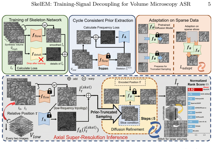
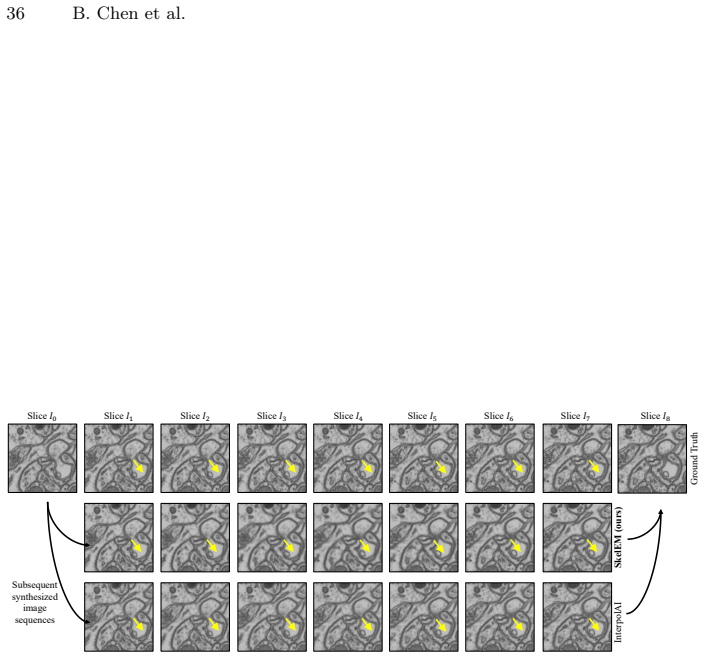
SkelEM achieves axial super-resolution by optimizing a frozen topological network for deterministic skeletons via one objective and a diffusion refiner via a disjoint cycle-consistent objective on sparse input slices, which simultaneously extracts a real-domain residual prior and bidirectionally aligns the refiner so that the reverse diffusion process can be truncated after at most five steps without synthetic bias.
What carries the argument
The training-signal decoupling of skeleton formulation from diffusion refinement, where the skeleton supplies a deterministic low-frequency prior that enables real-domain residual extraction and early truncation of the diffusion reverse process.
If this is right
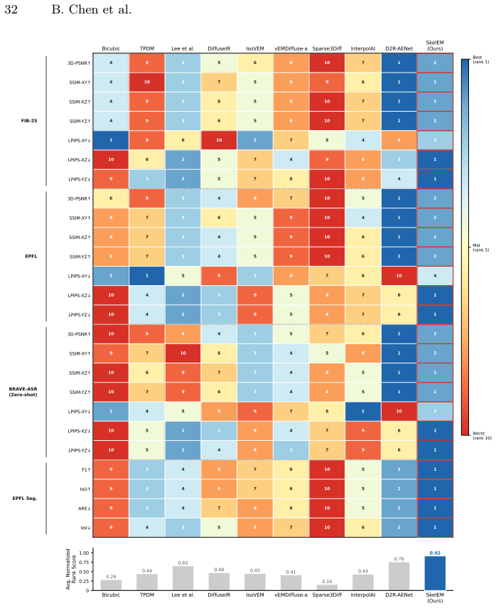
- The method produces the most favorable fidelity-perception balance among self-supervised axial super-resolution approaches on public benchmarks.
- SkelEM delivers state-of-the-art performance on downstream membrane segmentation tasks.
- Zero-shot generalization holds across distinct imaging modalities without retraining.
- Detail restoration remains high-fidelity when the diffusion process is limited to five or fewer steps.
- The BRAVE-ASR benchmark enables rigorous measurement of cross-instrument generalization for future methods.
Where Pith is reading between the lines
- The same cycle-consistent residual extraction could be adapted to other inverse problems where only anisotropic acquisitions are available.
- Truncating diffusion at five steps suggests the skeleton prior captures most of the necessary structural information, which may reduce compute demands in high-throughput volume imaging pipelines.
- If the topological network can be replaced by other deterministic structure extractors, the framework might extend to non-microscopy domains that suffer from directional resolution limits.
Load-bearing premise
A frozen topological network produces a deterministic skeleton that can be used to extract a real-domain residual prior and truncate the reverse diffusion process without introducing synthetic bias or structural hallucinations.
What would settle it
A head-to-head comparison on the BRAVE-ASR benchmark in which SkelEM produces lower membrane segmentation accuracy or more visible structural hallucinations than either pure regression or full-step diffusion baselines would falsify the benefit of the decoupling.
Figures
read the original abstract
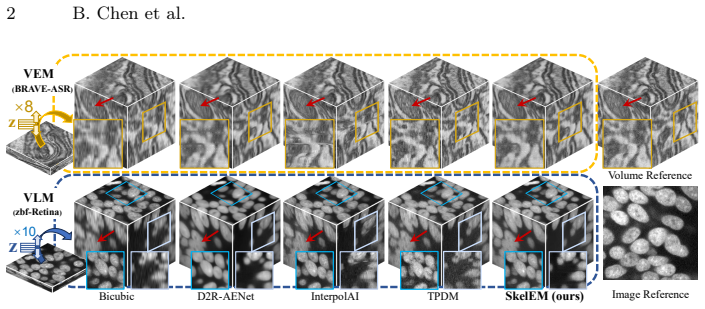
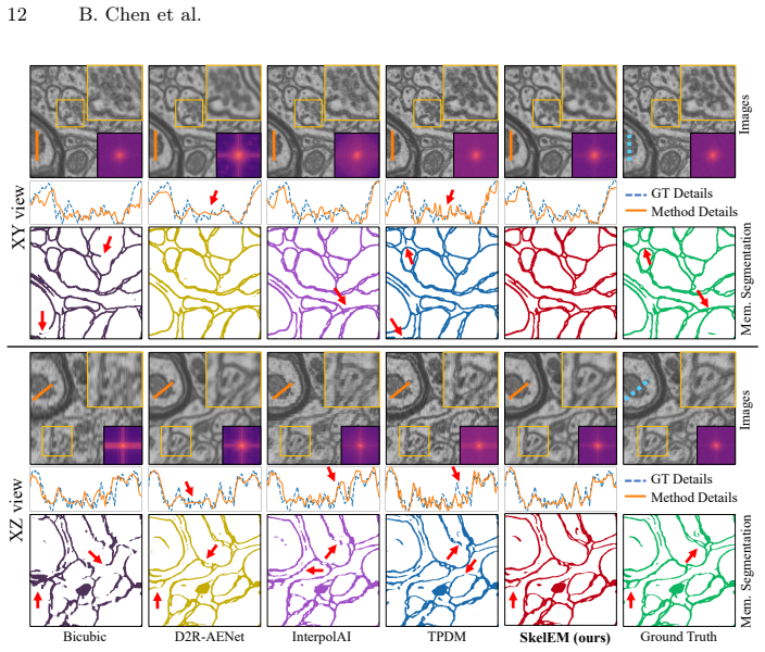
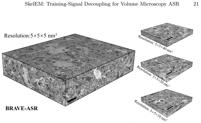
Volume microscopy, including electron and light microscopy, suffers from severe anisotropic resolution due to physical axial sectioning. Existing self-supervised axial super-resolution (ASR) methods face a trilemma bounded by overly smoothed regression textures, structural hallucinations of pure diffusion models, and prohibitive inference latency. In this paper, we propose Skeleton-refinE Microscopy (SkelEM), a self-supervised framework that decouples ASR at the training-signal level: a frozen topological network and a diffusion refiner are optimized by disjoint objectives, separating low-frequency topology formulation from high-frequency detail enhancement. Building on this deterministic skeleton, we exploit a unified cycle-consistent mechanism on input sparse slices to simultaneously extract a real-domain residual prior and bidirectionally align the diffusion refiner, washing away cross-plane artifacts without synthetic bias. By truncating the reverse diffusion process with this physical prior, SkelEM achieves high-fidelity detail restoration in merely $\le 5$ steps. To rigorously assess cross-instrument generalization, we further introduce BRAVE-ASR, a new benchmark of co-aligned anisotropic and isotropic volumes acquired on a Plasma-FIB instrument. Across public benchmarks, SkelEM achieves the most favorable balance across the fidelity-perception trade-off among self-supervised methods, with state-of-the-art downstream membrane segmentation performance and robust zero-shot generalization across distinct modalities.
Editorial analysis
A structured set of objections, weighed in public.
Referee Report
Summary. The paper proposes SkelEM, a self-supervised framework for axial super-resolution in volume microscopy. It decouples the training signal by using a frozen topological network to extract a deterministic skeleton and a diffusion refiner for high-frequency details. A cycle-consistent mechanism on input sparse slices extracts a real-domain residual prior and aligns the refiner bidirectionally, allowing truncation of the reverse diffusion process to ≤5 steps without synthetic bias. The method claims the most favorable fidelity-perception trade-off among self-supervised methods, SOTA downstream membrane segmentation, and robust zero-shot generalization on public benchmarks and the new BRAVE-ASR benchmark.
Significance. If the central claims hold, SkelEM would offer an efficient solution to the trilemma in self-supervised ASR by balancing fidelity and perception while avoiding hallucinations and high latency, with strong performance in downstream tasks and cross-modality generalization. This could have significant impact in volume microscopy applications.
major comments (2)
- [Methods (cycle-consistent mechanism and skeleton extraction)] The central claim relies on the frozen topological network producing a deterministic skeleton from anisotropic sparse slices that enables bias-free residual prior extraction and safe truncation of diffusion at ≤5 steps. However, no explicit validation is provided that the skeleton remains topologically faithful on real low-SNR axial data, nor that the bidirectional alignment eliminates rather than regularizes cross-plane artifacts. This is load-bearing for the 'no synthetic bias' guarantee.
- [Abstract and Results] The abstract states favorable trade-offs and SOTA segmentation performance but provides no quantitative metrics, ablation results, or error analysis to support these claims, making verification of the balance across fidelity-perception trade-off difficult.
minor comments (1)
- [Notation and Methods] Clarify the definition of the residual prior and how it is extracted from the cycle-consistent mechanism to avoid ambiguity in the truncation step.
Simulated Author's Rebuttal
We thank the referee for the constructive comments on our manuscript. We address each major comment point by point below and indicate planned revisions.
read point-by-point responses
-
Referee: [Methods (cycle-consistent mechanism and skeleton extraction)] The central claim relies on the frozen topological network producing a deterministic skeleton from anisotropic sparse slices that enables bias-free residual prior extraction and safe truncation of diffusion at ≤5 steps. However, no explicit validation is provided that the skeleton remains topologically faithful on real low-SNR axial data, nor that the bidirectional alignment eliminates rather than regularizes cross-plane artifacts. This is load-bearing for the 'no synthetic bias' guarantee.
Authors: We agree that direct validation of topological fidelity on low-SNR axial data would strengthen the central claim. In the revised manuscript we will add quantitative evaluation of skeleton accuracy (e.g., topological error metrics and structure-preservation scores) on held-out low-SNR slices from both public datasets and BRAVE-ASR. We will also include an ablation isolating the bidirectional cycle-consistent alignment to demonstrate that it reduces cross-plane artifacts beyond simple regularization, with supporting error analysis. revision: yes
-
Referee: [Abstract and Results] The abstract states favorable trade-offs and SOTA segmentation performance but provides no quantitative metrics, ablation results, or error analysis to support these claims, making verification of the balance across fidelity-perception trade-off difficult.
Authors: The abstract is a high-level summary constrained by length limits. We will revise it to incorporate the key quantitative results already present in the main text (fidelity-perception scores, segmentation accuracies, and latency comparisons) so that the claimed trade-offs are directly supported by numbers. revision: yes
Circularity Check
No significant circularity detected
full rationale
The derivation separates a frozen topological network (producing deterministic skeleton via disjoint objective) from a diffusion refiner, then applies cycle-consistency on input sparse slices to extract residual prior for truncation. No equations, self-citations, or ansatzes are exhibited that reduce any claimed prediction or prior to a fitted input or self-definition by construction. The cycle-consistent extraction is presented as operating on the given anisotropic slices to remove cross-plane artifacts, with the topological network held fixed and objectives explicitly disjoint; this structure is independent of the target super-resolution output. External elements such as the new BRAVE-ASR benchmark and downstream segmentation metrics further anchor the claims outside any internal fit.
Axiom & Free-Parameter Ledger
Reference graph
Works this paper leans on
-
[1]
Aurelien, L., Yunpeng, L., Carlos, B., Pascal, F.:https://www.epfl.ch/labs/ cvlab/data/data-em/(2013), accessed: 2024-08-16
2013
-
[2]
In: Proceedings of the IEEE conference on computer vision and pattern recognition
Blau, Y., Michaeli, T.: The perception-distortion tradeoff. In: Proceedings of the IEEE conference on computer vision and pattern recognition. pp. 6228–6237 (2018)
2018
-
[3]
Journal of the Peripheral Nervous System30(2), e70019 (2025)
Borisovs, V., Bossi, M., Matino, L., Marmiroli, P., Cavaletti, G.: New approaches based on serial-block face electron microscopy to investigate the peripheral nervous system. Journal of the Peripheral Nervous System30(2), e70019 (2025)
2025
-
[4]
In: International Conference on Medical Image Computing and Computer-Assisted Intervention
Chen, B., Zhang, Y., Lv, Y., Han, H., Chen, X.: Self-supervised axial super- resolution for volume microscopy via diffusion-guided structure distillation. In: International Conference on Medical Image Computing and Computer-Assisted Intervention. pp. 467–477. Springer (2025)
2025
-
[5]
In: Proceedings of the AAAI Conference on Artificial Intelligence
Chen, Q., Chen, X., Wang, C., Liu, Y., Xiong, Z., Wu, F.: Learning multimodal volumetric features for large-scale neuron tracing. In: Proceedings of the AAAI Conference on Artificial Intelligence. vol. 38, pp. 1174–1182 (2024) 16 B. Chen et al
2024
-
[6]
In: Med- ical Image Computing and Computer Assisted Intervention–MICCAI 2020: 23rd International Conference, Lima, Peru, October 4–8, 2020, Proceedings, Part V 23
Deng, S., Fu, X., Xiong, Z., Chen, C., Liu, D., Chen, X., Ling, Q., Wu, F.: Isotropic reconstruction of 3d em images with unsupervised degradation learning. In: Med- ical Image Computing and Computer Assisted Intervention–MICCAI 2020: 23rd International Conference, Lima, Peru, October 4–8, 2020, Proceedings, Part V 23. pp. 163–173. Springer (2020)
2020
-
[7]
Advances in neural information processing systems34, 8780–8794 (2021)
Dhariwal, P., Nichol, A.: Diffusion models beat gans on image synthesis. Advances in neural information processing systems34, 8780–8794 (2021)
2021
-
[8]
Current pro- tocols in cytometry92(1), e68 (2020)
Elliott, A.D.: Confocal microscopy: principles and modern practices. Current pro- tocols in cytometry92(1), e68 (2020)
2020
-
[9]
IEEE transactions on image processing17(10), 1737–1754 (2008)
Foi, A., Trimeche, M., Katkovnik, V., Egiazarian, K.: Practical poissonian-gaussian noise modeling and fitting for single-image raw-data. IEEE transactions on image processing17(10), 1737–1754 (2008)
2008
-
[10]
In: Proceedings of the Computer Vision and Pattern Recognition Conference
Hai, Y., Wang, G., Su, T., Jiang, W., Hu, Y.: Hierarchical flow diffusion for ef- ficient frame interpolation. In: Proceedings of the Computer Vision and Pattern Recognition Conference. pp. 22943–22952 (2025)
2025
-
[11]
In: Computer vision: a reference guide, pp
Hasinoff, S.W.: Photon, poisson noise. In: Computer vision: a reference guide, pp. 980–982. Springer (2021)
2021
-
[12]
bioRxiv pp
He, J., Zhang, Y., Sun, W., Yang, G., Sun, F.: Isovem: Isotropic reconstruction for volume electron microscopy based on transformer. bioRxiv pp. 2023–11 (2023)
2023
-
[13]
In: Medical Image Computing and Computer-Assisted Intervention- MICCAI 2017: 20th International Conference, Quebec City, QC, Canada, September 11-13, 2017, Proceedings, Part II 20
Heinrich,L.,Bogovic,J.A.,Saalfeld,S.:Deeplearningforisotropicsuper-resolution from non-isotropic 3d electron microscopy. In: Medical Image Computing and Computer-Assisted Intervention- MICCAI 2017: 20th International Conference, Quebec City, QC, Canada, September 11-13, 2017, Proceedings, Part II 20. pp. 135–143. Springer (2017)
2017
-
[14]
Advances in neural information processing systems30(2017)
Heusel,M.,Ramsauer,H.,Unterthiner,T.,Nessler,B.,Hochreiter,S.:Ganstrained by a two time-scale update rule converge to a local nash equilibrium. Advances in neural information processing systems30(2017)
2017
-
[15]
Advances in neural information processing systems33, 6840–6851 (2020)
Ho, J., Jain, A., Abbeel, P.: Denoising diffusion probabilistic models. Advances in neural information processing systems33, 6840–6851 (2020)
2020
-
[16]
In: European Conference on Computer Vision
Huang, Z., Zhang, T., Heng, W., Shi, B., Zhou, S.: Real-time intermediate flow estimation for video frame interpolation. In: European Conference on Computer Vision. pp. 624–642. Springer (2022)
2022
-
[17]
In: Proceedings of the IEEE/CVF Conference on Computer Vision and Pattern Recognition
Jain, S., Watson, D., Tabellion, E., Poole, B., Kontkanen, J., et al.: Video inter- polation with diffusion models. In: Proceedings of the IEEE/CVF Conference on Computer Vision and Pattern Recognition. pp. 7341–7351 (2024)
2024
-
[18]
In: Proceedings of the Computer Vision and Pat- tern Recognition Conference
Jeong, M.W., Rhee, C.E.: Lc-mamba: Local and continuous mamba with shifted windows for frame interpolation. In: Proceedings of the Computer Vision and Pat- tern Recognition Conference. pp. 17671–17681 (2025)
2025
-
[19]
In: Proceedings of the IEEE/CVF Conference on Computer Vision and Pattern Recognition
Jiang, C., Gedeon, A., Lyu, Y., Landgraf, E., Zhang, Y., Hou, X., Kondepudi, A., Chowdury, A., Lee, H., Hollon, T.: Super-resolution of biomedical volumes with 2d supervision. In: Proceedings of the IEEE/CVF Conference on Computer Vision and Pattern Recognition. pp. 6966–6977 (2024)
2024
-
[20]
In: Proceedings of the IEEE conference on computer vision and pattern recognition
Jiang, H., Sun, D., Jampani, V., Yang, M.H., Learned-Miller, E., Kautz, J.: Super slomo: High quality estimation of multiple intermediate frames for video interpo- lation. In: Proceedings of the IEEE conference on computer vision and pattern recognition. pp. 9000–9008 (2018)
2018
-
[21]
Nature Methods pp
Joshi, S., Forjaz, A., Han, K.S., Shen, Y., Queiroga, V., Selaru, F.A., Gérard, M., Xenes, D., Matelsky, J., Wester, B., et al.: Interpolai: deep learning-based optical flow interpolation and restoration of biomedical images for improved 3d tissue mapping. Nature Methods pp. 1–12 (2025) SkelEM: Training-Signal Decoupling for Volume Microscopy ASR 17
2025
-
[22]
Journal of Mi- croscopy287(3), 114–137 (2022)
Kievits, A.J., Lane, R., Carroll, E.C., Hoogenboom, J.P.: How innovations in methodology offer new prospects for volume electron microscopy. Journal of Mi- croscopy287(3), 114–137 (2022)
2022
-
[23]
Accessed June 29, 2026
Koho, S., Kalinin, A.: MIPLIB: A Python software library for microscopy image restoration, reconstruction and analysis.https://github.com/sakoho81/miplib (2019), version v1.0. Accessed June 29, 2026
2019
-
[24]
Nature communications10(1), 3103 (2019)
Koho, S., Tortarolo, G., Castello, M., Deguchi, T., Diaspro, A., Vicidomini, G.: Fourier ring correlation simplifies image restoration in fluorescence microscopy. Nature communications10(1), 3103 (2019)
2019
-
[25]
In: International Conference on Medical Image Computing and Computer-Assisted Intervention
Lee, H.J., Jo, E., Lim, M., Son, Y.H., Kang, B., Nam, H., Jeong, J.H., Shin, D.H., Kam, T.E.: Sparse3diff: A diffusion framework for 3d reconstruction from sparse 2d slices in volumetric optical imaging. In: International Conference on Medical Image Computing and Computer-Assisted Intervention. pp. 510–519. Springer (2025)
2025
-
[26]
In: International Conference on Medical Image Computing and Computer- Assisted Intervention
Lee, K., Jeong, W.K.: Reference-free isotropic 3d em reconstruction using diffusion models. In: International Conference on Medical Image Computing and Computer- Assisted Intervention. pp. 235–245. Springer (2023)
2023
-
[27]
In: International Conference on Medical Image Computing and Computer-Assisted Intervention
Lee,K.,Jeong,W.K.:Reference-freeaxialsuper-resolutionof3dmicroscopyimages using implicit neural representation with a 2d diffusion prior. In: International Conference on Medical Image Computing and Computer-Assisted Intervention. pp. 593–602. Springer (2024)
2024
-
[28]
In: Proceedings of the IEEE/CVF International Conference on Computer Vision
Lee, S., Chung, H., Park, M., Park, J., Ryu, W.S., Ye, J.C.: Improving 3d imag- ing with pre-trained perpendicular 2d diffusion models. In: Proceedings of the IEEE/CVF International Conference on Computer Vision. pp. 10710–10720 (2023)
2023
-
[29]
Journal of neuroscience methods226, 88–102 (2014)
Liu, T., Jones, C., Seyedhosseini, M., Tasdizen, T.: A modular hierarchical ap- proach to 3d electron microscopy image segmentation. Journal of neuroscience methods226, 88–102 (2014)
2014
-
[30]
In: Computer Vision–ECCV 2020 Workshops: Glasgow, UK, August 23–28, 2020, Proceedings, Part IV 16
Liu, Y., Xie, L., Siyao, L., Sun, W., Qiao, Y., Dong, C.: Enhanced quadratic video interpolation. In: Computer Vision–ECCV 2020 Workshops: Glasgow, UK, August 23–28, 2020, Proceedings, Part IV 16. pp. 41–56. Springer (2020)
2020
-
[31]
Bioinformatics 38(24), 5329–5339 (2022)
Liu, Y., Wang, G., Ascoli, G.A., Zhou, J., Liu, L.: Neuron tracing from light microscopy images: automation, deep learning and bench testing. Bioinformatics 38(24), 5329–5339 (2022)
2022
-
[32]
Nature Communications15(1), 4677 (2024)
Lu, C., Chen, K., Qiu, H., Chen, X., Chen, G., Qi, X., Jiang, H.: Diffusion-based deep learning method for augmenting ultrastructural imaging and volume electron microscopy. Nature Communications15(1), 4677 (2024)
2024
-
[33]
Biology11(9), 1270 (2022)
Ma, H., Chen, J., Deng, Z., Sun, T., Luo, Q., Gong, H., Li, X., Long, B.: Multi- scale analysis of cellular composition and morphology in intact cerebral organoids. Biology11(9), 1270 (2022)
2022
-
[34]
Journal of mul- tivariate analysis98(5), 873–895 (2007)
Meilă, M.: Comparing clusterings—an information based distance. Journal of mul- tivariate analysis98(5), 873–895 (2007)
2007
-
[35]
SDEdit: Guided Image Synthesis and Editing with Stochastic Differential Equations
Meng, C., He, Y., Song, Y., Song, J., Wu, J., Zhu, J.Y., Ermon, S.: Sdedit: Guided image synthesis and editing with stochastic differential equations. arXiv preprint arXiv:2108.01073 (2021)
work page internal anchor Pith review Pith/arXiv arXiv 2021
-
[36]
Auto- matica11(285-296), 23–27 (1975)
Otsu, N., et al.: A threshold selection method from gray-level histograms. Auto- matica11(285-296), 23–27 (1975)
1975
-
[37]
In: International Conference on Medical Image Computing and Computer-Assisted Intervention
Pan, M., Gan, Y., Zhou, F., Liu, J., Zhang, Y., Wang, A., Zhang, S., Li, D.: Diffuseir: Diffusion models for isotropic reconstruction of 3d microscopic images. In: International Conference on Medical Image Computing and Computer-Assisted Intervention. pp. 323–332. Springer (2023) 18 B. Chen et al
2023
-
[38]
Nature Communications13(1), 3297 (2022)
Park, H., Na, M., Kim, B., Park, S., Kim, K.H., Chang, S., Ye, J.C.: Deep learn- ing enables reference-free isotropic super-resolution for volumetric fluorescence mi- croscopy. Nature Communications13(1), 3297 (2022)
2022
-
[39]
Nature Reviews Methods Primers2(1), 51 (2022)
Peddie, C.J., Genoud, C., Kreshuk, A., Meechan, K., Micheva, K.D., Narayan, K., Pape, C., Parton, R.G., Schieber, N.L., Schwab, Y., et al.: Volume electron microscopy. Nature Reviews Methods Primers2(1), 51 (2022)
2022
-
[40]
Schwartz, J., Jiang, Y., Wang, Y., Aiello, A., Bhattacharya, P., Yuan, H., Mi, Z., Bassim, N., Hovden, R.: Removing stripes, scratches, and curtaining with nonre- coverablecompressedsensing.MicroscopyandMicroanalysis25(3),705–710(2019)
2019
-
[41]
Song, J., Meng, C., Ermon, S.: Denoising diffusion implicit models (2020)
2020
-
[42]
Sun, H., Ye, E., Lu, C., Jiang, Z., Bai, W., Ren, S., Wang, C., Zhang, J., Shi, R., Ma, L., et al.: Em generalist: A physics-driven diffusion foundation model for electron microscopy (2025)
2025
-
[43]
Proceedings of the National Academy of Sciences112(44), 13711–13716 (2015)
Takemura, S.y., Xu, C.S., Lu, Z., Rivlin, P.K., Parag, T., Olbris, D.J., Plaza, S., Zhao, T., Katz, W.T., Umayam, L., et al.: Synaptic circuits and their variations within different columns in the visual system of drosophila. Proceedings of the National Academy of Sciences112(44), 13711–13716 (2015)
2015
-
[44]
MICCAI Workshop on Efficient Medical AI (EMA) (2025).https: //doi.org/10.1101/2024.09.07.611785
Troidl, J., Liang, Y., Beyer, J., Tavakoli, M., Danzl, J.G., Hadwiger, M., Pfister, H., Tompkin, J.: niiv: Interactive self-supervised neural implicit isotropic volume reconstruction. MICCAI Workshop on Efficient Medical AI (EMA) (2025).https: //doi.org/10.1101/2024.09.07.611785
-
[45]
In: Medical Image Computing and Computer-Assisted Intervention - MICCAI 2017
Weigert, M., Royer, L., Jug, F., Myers, G.: Isotropic reconstruction of 3d fluores- cence microscopy images using convolutional neural networks. In: Medical Image Computing and Computer-Assisted Intervention - MICCAI 2017. pp. 126–134. Springer International Publishing, Cham (2017)
2017
-
[46]
Nature methods15(12), 1090–1097 (2018)
Weigert, M., Schmidt, U., Boothe, T., Müller, A., Dibrov, A., Jain, A., Wilhelm, B., Schmidt, D., Broaddus, C., Culley, S., et al.: Content-aware image restoration: pushing the limits of fluorescence microscopy. Nature methods15(12), 1090–1097 (2018)
2018
-
[47]
Plos one17(12), e0279005 (2022)
Wu, S., Nakao, M., Imanishi, K., Nakamura, M., Mizowaki, T., Matsuda, T.: Com- puted tomography slice interpolation in the longitudinal direction based on deep learning techniques: To reduce slice thickness or slice increment without dose in- crease. Plos one17(12), e0279005 (2022)
2022
-
[48]
elife6, e25916 (2017)
Xu, C.S., Hayworth, K.J., Lu, Z., Grob, P., Hassan, A.M., García-Cerdán, J.G., Niyogi, K.K., Nogales, E., Weinberg, R.J., Hess, H.F.: Enhanced fib-sem systems for large-volume 3d imaging. elife6, e25916 (2017)
2017
-
[49]
Biomedical Optics Express11(11), 6634–6648 (2020)
Ye, S., Yin, Y., Yao, J., Nie, J., Song, Y., Gao, Y., Yu, J., Li, H., Fei, P., Zheng, W.: Axial resolution improvement of two-photon microscopy by multi-frame re- construction and adaptive optics. Biomedical Optics Express11(11), 6634–6648 (2020)
2020
-
[50]
In: Proceedings of the Computer Vision and Pattern Recognition Con- ference
Yue, Z., Liao, K., Loy, C.C.: Arbitrary-steps image super-resolution via diffusion inversion. In: Proceedings of the Computer Vision and Pattern Recognition Con- ference. pp. 23153–23163 (2025)
2025
-
[51]
In: Proceedings of the IEEE conference on computer vision and pattern recognition
Zhang, R., Isola, P., Efros, A.A., Shechtman, E., Wang, O.: The unreasonable effectiveness of deep features as a perceptual metric. In: Proceedings of the IEEE conference on computer vision and pattern recognition. pp. 586–595 (2018)
2018
-
[52]
In: International Conference on Medical Image Computing and Computer-Assisted Intervention
Zhang, Y., Guo, J., Zhai, H., Liu, J., Han, H.: Segneuron: 3d neuron instance seg- mentation in any em volume with a generalist model. In: International Conference on Medical Image Computing and Computer-Assisted Intervention. pp. 589–600. Springer (2024),https://github.com/yanchaoz/SegNeuron SkelEM: Training-Signal Decoupling for Volume Microscopy ASR 19
2024
-
[53]
In: 2019 international conference on 3D vision (3DV)
Zhou, D., Fang, J., Song, X., Guan, C., Yin, J., Dai, Y., Yang, R.: Iou loss for 2d/3d object detection. In: 2019 international conference on 3D vision (3DV). pp. 85–94. IEEE (2019) 20 B. Chen et al. A Supplementary Materials Overview Thissupplementarymaterialprovidescomprehensivetheoreticalanalyses,imple- mentation details, and extensive experimental eva...
discussion (0)
Sign in with ORCID, Apple, or X to comment. Anyone can read and Pith papers without signing in.