REVIEW 3 major objections 7 minor 25 references
Learning from Anatomy: Supervised Anatomical Pretraining (SAP) for Improved Metastatic Bone Disease Segmentation in Whole-Body MRI
T0 review · 3 major / 7 minor · reviewed 2026-08-06 · deepseek-v4-flash
Pith's one-line read Pretraining a whole-body MRI model to delineate healthy skeletal anatomy, then fine-tuning it to find metastatic bone lesions, substantially outperforms both random initialization and self-supervised pretraining on the same data.
desk verdict Promising and honest paper—supervised pretraining on healthy anatomy beats SSL in a low-data lesion segmentation task—but the threshold/cross-validation description is ambiguous and needs a fix before the superiority claim is solid. read the letter →
The pith
A machine-rendered reading of the paper's core claim, the machinery that carries it, and where it could break.
The reading
What carries the argument
The load-bearing mechanism is a two-stage transfer. In the first stage, a transformer-based segmentation network is trained on 63 whole-body MRI scans from healthy volunteers to delineate nine skeletal regions (cervical, thoracic, and lumbar spine; pelvis; femurs; humeri; scapulae; clavicles; sternum), reaching a whole-skeleton Dice of 0.90 on a held-out validation set. For the downstream task, the encoder and decoder weights are transferred, the multi-class segmentation head is replaced by a binary lesion head, and fine-tuning applies a lower learning rate to the encoder to preserve anatomical features and a higher learning rate to the decoder to adapt to lesions. The authors also inspect the initialized feature space, using a two-dimensional projection and cluster-distance measurements to show that before fine-tuning the anatomical representation forms more compact positive and negative clusters than the SSL representation, which they present as evidence that the inductive bias, not the fine-tuning recipe, drives the improvement.
What would settle it
Have an expert directly annotate the skeleton on a hold-out set of healthy-volunteer scans, compare those annotations with the propagated labels used in pretraining, then retrain the anatomical model on corrected labels and rerun the 7-fold metastatic-lesion evaluation; if the propagated labels deviate substantially from direct expert annotation, or if the retrained model no longer beats SSL on lesion Dice and F2, the central claim is weakened.
Extended reading notes
Core claim
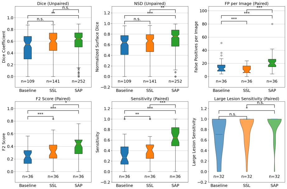
The paper's central claim is that learning bone morphology from healthy anatomy supplies a domain-relevant inductive bias: a network that has first learned to recognize normal skeletal structure finds metastatic lesions more reliably than a network pretrained with generic self-supervised objectives. On 44 prostate-cancer patients with confirmed skeletal metastases, evaluated with 7-fold cross-validation, the method achieves a median Dice coefficient of 0.64 and normalized surface Dice of 0.76, with median lesion-detection sensitivity of 0.64 and an F2 score of 0.44; every value lies above the baseline and the SSL-pretrained model. The authors show that before any fine-tuning, their pretrained representation already separates lesion patches from healthy patches into tighter clusters than the SSL representation does, which supports the interpretation that the anatomical pretraining itself, not the fine-tuning alone, is doing the work. Their stated conclusion is that explicitly learning healthy anatomy is an effective, domain-relevant alternative to generic pretraining in low-data medical imaging.
Load-bearing premise
The load-bearing premise is that the skeletal labels used for anatomical pretraining are accurate: the labels were hand-drawn on one scan per healthy volunteer and then propagated to the other scans by an automatic piecewise registration pipeline, and the reported skeleton Dice of 0.90 is measured against those same propagated labels, so it cannot separate label quality from model quality.
Editorial extensions
If this is right
- For lesions larger than 1 ml, the clinically relevant subpopulation, the method reaches 100% detection sensitivity in 28 of 32 test patients at a mean of 0.47 false positives per image, an operating point close to what a screening-support tool would need.
- Any segmentation task whose pathology is defined against normal anatomy, such as bone lesions, fractures, or organ anomalies, could plausibly be initialized with a supervised anatomical model rather than a generic self-supervised model, at much lower pretraining cost (5.5 GPU hours for anatomical pretraining versus 101 for the SSL baseline).
- The anatomical model's systematic failure to segment bone tissue near metastatic infiltration makes its behavior inspectable: a clinician can see where the anatomical prior breaks down, which the authors argue is an interpretability advantage over black-box SSL.
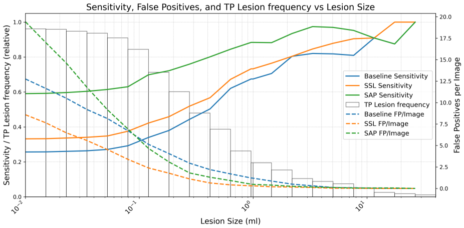
- The stepwise gains from baseline to SSL to anatomical pretraining appear across the full receiver-operating analysis, not at a single threshold, so the ranking is not an artifact of one decision boundary.
Reading between the lines
- Beyond the paper's claims: the reported skeleton Dice of 0.90 is computed against labels propagated by the same registration pipeline that created them, so an independent manual evaluation of those propagated labels is needed to know how much of SAP's success depends on label quality.
- If the inductive-bias explanation is right, the SAP advantage should grow with anatomical diversity and shrink as downstream training data grow large enough to learn bone morphology from lesions alone; that dose-response pattern is testable with the released code and data.
- Because the paper notes that many SAP false positives are clinically acceptable anatomical variations, a metric that counts such detections as neutral rather than false would likely shrink the FPPI gap between SAP and SSL while keeping the sensitivity gain; that is an evaluation-design consequence the authors leave implicit.
- Combining SAP with a self-supervised objective on the healthy dataset, or reusing the same anatomical-pretraining recipe for other anatomical priors such as vasculature or organ boundaries, is a direct extension the paper mentions only implicitly.
Editorial analysis
A structured set of objections, weighed in public.
Referee Report
Summary. The paper proposes Supervised Anatomical Pretraining (SAP), which first trains a Swin UNETR model to segment nine skeletal regions from whole-body MRI of healthy volunteers, then transfers the encoder and decoder weights to a binary metastatic bone disease (MBD) segmentation task in 44 prostate cancer patients. The authors compare SAP against random initialization and a state-of-the-art self-supervised learning (SSL) baseline, reporting improvements in lesion detection F2 score (0.24 to 0.44), Dice (0.55 to 0.64), and Normalized Surface Dice (0.64 to 0.76). They also analyze learned features via t-SNE and report FROC curves, and they emphasize the interpretability and domain relevance of anatomical pretraining. The manuscript is transparent about several limitations, including small cohort size and ground-truth imperfections, and promises public release of code and models.
Significance. If the reported improvements hold up, SAP offers a practical, data-efficient alternative to self-supervised pretraining for medical image segmentation with limited labeled data, and its interpretable inductive bias is a genuine strength. The manuscript is transparent in several respects: the evaluation uses standard Metrics Reloaded, the authors openly discuss limitations, and the promise of public code and models supports reproducibility. However, the strength of the superiority claim is tempered by the small single-center downstream dataset, the lack of external validation, and the ambiguous cross-validation/threshold protocol described in Section 3.4.
major comments (3)
- [3.4] The cross-validation and threshold-selection protocol is internally inconsistent. The text states that a 7-fold cross-validation is used, that in each fold the dataset is divided patient-wise into 6 folds for training and 1 fold for testing, and that 'one split was reserved for determining an optimal decision boundary' with metrics reported on the remaining 6 test splits. A 6+1 split consumes all seven folds, leaving no split for threshold selection, and a 7-fold design has only one test split per fold, not six. If the F2-optimized binarization threshold is selected on the same patients that are later scored, the metrics in Table 4 and the significance stars in Figure 4 are optimistically biased, and the risk is amplified for SAP because its FPPI (20.0) is much higher than that of SSL (8.0) or baseline (11.5). Please clarify the exact split (e.g., nested validation where a portion of each training fold is reserved for threshold selection) and report how many patients were used for threshold selection versus testing. If the threshold was tuned on the test folds, the analysis should be rerun with a properly separated validation split.
- [3.2.2 and 3.2.3] The comparison between SAP and SSL is confounded by the amount of transferred network. Section 3.2.3 states that SAP transfers both the encoder and decoder weights from the skeleton model, while Section 3.2.2 states that the SSL baseline transfers only the encoder and re-initializes the decoder. The downstream gains therefore conflate two differences: the pretraining objective (anatomy vs. self-supervision) and the number of transferred layers. The t-SNE analysis of bottleneck features in Section 4.2 indicates that the SAP encoder is more discriminative, but the segmentation metrics in Table 4 could be driven in part by the decoder initialization. To isolate the contribution of anatomical pretraining, please add an ablation in which the SSL baseline also transfers its decoder (or, conversely, a SAP variant that transfers only the encoder).
- [A.1] The quality of the propagated skeletal labels is not independently assessed. Manual labels were made on one scan per healthy subject and then propagated to the remaining scans via the piecewise registration pipeline described in Section A.1. The skeleton Dice score of 0.90 reported in Section 4.1 is computed on validation scans whose ground-truth labels were generated by this same propagation process, so the evaluation cannot separate registration/labeling errors from model errors. If the propagated labels contain systematic mislabeling, the pretraining signal is corrupted and the claim that the model learns 'bone morphology' is weakened. Please provide an independent estimate of the propagated-label accuracy, for example by comparing a subset of propagated labels against expert manual re-segmentation, or by reporting registration accuracy metrics on held-out scans.
minor comments (7)
- [3.1, 3.3, 5.2] The number of healthy volunteers and scans is inconsistent: Section 3.1 reports 24 volunteers and 72 scans, Section 3.3 reports 24 subjects and 71 scans (with a 63/8 train/validation split), and Section 5.2 reports 23 volunteers. Please reconcile these numbers and ensure the train/validation split sums correctly.
- [4.3.3] Section 4.3.3 reports lesion-level sensitivities for lesions larger than 1 ml (0.68 for baseline, 0.72 for SSL, 0.87 for SAP) that differ from the patient-level medians in Table 4 (0.71, 1.00, 1.00). Please state explicitly whether these are lesion-level or patient-level sensitivities and define how they are computed, so readers can understand the relationship.
- [Figure 4] The significance stars in Figure 4 are not accompanied by p-values in the text. Please report the exact p-values (or Bonferroni-adjusted p-values) for the key comparisons, including SAP versus SSL for F2, Dice, and NSD.
- [Abstract and 4.3.3] The abstract states that SAP achieves 'a detection sensitivity of 100% in 28 out of 32 patients' for lesions larger than 1 ml, while Section 4.3.3 reports a sensitivity of 0.87 for SAP on the same lesion-size subset. Please clarify the relationship (e.g., 28/32 is the fraction of patients with per-patient sensitivity of 1.0, whereas 0.87 is lesion-level sensitivity) so the claims are unambiguous.
- [Table 2 and 4.3.3] The terminology for false positives is inconsistent: Table 2 defines FPPI as 'False Positives per Image,' but the text alternates between 'FP per Image' and 'FPPI.' Please standardize the abbreviation and use it consistently.
- [5.1.2] The computational comparison (101 GPU hours for SSL vs. 5.5 GPU hours for SAP) does not specify whether the times include only pretraining or also the downstream fine-tuning, nor on which GPU hardware. Please clarify these details for reproducibility.
- [References] The reference to Li et al. (2024) is listed as an ICLR paper with an arXiv link (2501.11253) that appears to be from 2025 and may correspond to a different article. Please verify the citation and update if necessary.
Circularity Check
No significant circularity: the central SAP-versus-SSL comparison is empirical and independent; only minor self-citations for data and preprocessing appear.
full rationale
The paper's claimed derivation chain is not circular by construction. SAP pretrains a Swin UNETR on healthy-subject skeleton labels, then fine-tunes on metastatic lesion labels; the pretraining task uses no lesion annotations, and the downstream comparison against a randomly initialized baseline and an external SSL method (Tang et al.) is a measured empirical outcome. A model pretrained on healthy anatomy could plausibly transfer poorly, so the reported gains are not logically forced by the definition of the pretraining task. The healthy dataset (Michoux et al., 2021) and preprocessing pipeline (Ceranka et al., 2023b) are self-citations with overlapping authorship, but they are applied identically to all three training strategies and therefore cannot by themselves determine the reported SAP advantage. The skeleton validation Dice of 0.90 is computed on labels produced by the same manual-labeling-plus-propagation workflow that generated the training labels, so it is best read as an internal consistency check rather than fully independent evidence of anatomical accuracy; this is a limitation but not a circularity in the central claim. The Section 3.4 description of threshold selection is ambiguous, but the text states that metrics are reported on the remaining 6 test splits, so there is no demonstrated reduction of the reported F2 values to a threshold fitted on the scored patients. Overall, the core result is independently grounded and no load-bearing circular step was identified.
Assumptions & free parameters
free parameters (2)
- Segmentation probability threshold per method =
not reported (optimized for F2-score on a reserved split per method)
- SAP differential learning rates =
2e-6 (encoder) and 1e-5 (decoder)
assumptions (4)
- domain assumption Manually segmented and registration-propagated healthy skeletal labels are accurate enough to teach transferable anatomical representations
- domain assumption The healthy volunteer anatomy is representative of the skeletal anatomy of the metastatic prostate cancer patient population
- domain assumption The self-supervised pretraining baseline was given a fair comparison
- ad hoc to paper The Swin UNETR architecture and training hyperparameters are appropriate for both skeleton and lesion segmentation
Cite this review
Pith. "Pith review of Learning from Anatomy: Supervised Anatomical Pretraining (SAP) for Improved Metastatic Bone Disease Segmentation in Whole-Body MRI." pith.science (2026). https://pith.science/paper/BAIP7KA7
@misc{pith2026250619590,
  author       = {Pith},
  title        = {Pith review of: Learning from Anatomy: Supervised Anatomical Pretraining (SAP) for Improved Metastatic Bone Disease Segmentation in Whole-Body MRI},
  year         = {2026},
  howpublished = {\url{https://pith.science/paper/BAIP7KA7}},
  note         = {Machine review of arXiv:2506.19590}
}
read the original abstract
The segmentation of metastatic bone disease (MBD) in whole-body MRI (WB-MRI) is a challenging problem. Due to varying appearances and anatomical locations of lesions, ambiguous boundaries, and severe class imbalance, obtaining reliable segmentations requires large, well-annotated datasets capturing lesion variability. Generating such datasets requires substantial time and expertise, and is prone to error. While self-supervised learning (SSL) can leverage large unlabeled datasets, learned generic representations often fail to capture the nuanced features needed for accurate lesion detection. In this work, we propose a Supervised Anatomical Pretraining (SAP) method that learns from a limited dataset of anatomical labels. First, an MRI-based skeletal segmentation model is developed and trained on WB-MRI scans from healthy individuals for high-quality skeletal delineation. Then, we compare its downstream efficacy in segmenting MBD on a cohort of 44 patients with metastatic prostate cancer, against both a baseline random initialization and a state-of-the-art SSL method. SAP significantly outperforms both the baseline and SSL-pretrained models, achieving a normalized surface Dice of 0.76 and a Dice coefficient of 0.64. The method achieved a lesion detection F2 score of 0.44, improving on 0.24 (baseline) and 0.31 (SSL). When considering only clinically relevant lesions larger than 1~ml, SAP achieves a detection sensitivity of 100% in 28 out of 32 patients. Learning bone morphology from anatomy yields an effective and domain-relevant inductive bias that can be leveraged for the downstream segmentation task of bone lesions. All code and models are made publicly available.
Figures


Figures from the paper (6 more)



Reference graph
Works this paper leans on
- [9]
-
[11]
MONAI: An open-source framework for deep learning in healthcare arXiv:2211.02701. Kim, D.H., Seo, J., Lee, J.H., Jeon, E.T., Jeong, D., Chae, H.D., Lee, E., Kang, J.H., Choi, Y.H., Kim, H.J., Chai, J.W.,
-
[12]
Korean Journal of Radiology 25, 363–373
Automated detection and segmentation of bone metastases on spine mri using u-net: A multicenter study. Korean Journal of Radiology 25, 363–373. URL: https://doi.org/10.3348/kjr.2023.0671, doi:10.3348/kjr.2023.0671. Li, W., Yuille, A., Zhou, Z.,
-
[13]
How well do supervised 3d models transfer to medical imaging tasks?, in: International Conference on Learning Representations (ICLR). URL:https://arxiv. org/abs/2501.11253. Maier-Hein, L., Reinke, A., Godau, P., Tizabi, M.D., Buettner, F., Christodoulou, E., Glocker, B., Isensee, F., Kleesiek, J., Kozubek, M., Reyes, M., Riegler, M.A., Wiesenfarth, M., Ka...
-
[14]
Frontiers in Oncology 11, 772530
Bone metastases are measurable: The role of whole-body mri and positron emission tomography. Frontiers in Oncology 11, 772530. URL: https://www.frontiersin.org/articles/10.3389/ fonc.2021.772530/full, doi:10.3389/fonc.2021.772530. published online 2021-11-19; PMID: 34869009; PMCID: PMC8640187. 23 Padhani, A.R., Lecouvet, F.E., Tunariu, N., Koh, D.M., De K...
-
[17]
Tustison, N.J., Avants, B.B., Cook, P.A., Zheng, Y., Egan, A., Yushkevich, P.A., Gee, J.C.,
Self- Supervised Pre-Training of swin transformers for 3D medical image analysis arXiv:2111.14791. Tustison, N.J., Avants, B.B., Cook, P.A., Zheng, Y., Egan, A., Yushkevich, P.A., Gee, J.C.,
-
[19]
Journal of Magnetic Resonance Imaging 55, 653–680
Whole-body magnetic resonance imaging for prostate cancer assessment: Current status and future directions. Journal of Magnetic Resonance Imaging 55, 653–680. doi: 10.1002/jmri.27485, arXiv:2020 Dec
-
[20]
arXiv preprint arXiv:2412.17041 URL: https://arxiv.org/abs/2412.17041
An openmind for 3d medical vision self-supervised learning. arXiv preprint arXiv:2412.17041 URL: https://arxiv.org/abs/2412.17041. Wasserthal, J., Meyer, M., Breit, H.C., Cyriac, J., Yang, S., Segeroth, M.,
Show all 25 references
-
[21]
TotalSegmentator: robust segmentation of 104 anatomical structures in CT images arXiv:2208.05868. 24 Wennmann, M., Klein, A., Bauer, F., Chmelik, J., Gr¨ ozinger, M., Uhlenbrock, C., Lochner, J., Nonnenmacher, T., Rotkopf, L.T., Sauer, S., Hielscher, T., G¨ otz, M., Floca, R.O...
-
[25]
Medical Im- age Analysis 67, 101840
Models genesis. Medical Im- age Analysis 67, 101840. URL: https://www.sciencedirect.com/science/article/pii/ S1361841520302048, doi:https://doi.org/10.1016/j.media.2020.101840. 25
2020
-
[159]
pMID: 36194301; PMCID: PMC9525241
URL: https://doi.org/10.1186/s13244-022-01287-4 , doi:10.1186/s13244-022-01287-4 . pMID: 36194301; PMCID: PMC9525241. Eisenhauer, E.A., Therasse, P., Bogaerts, J., Schwartz, L.H., Sargent, D., Ford, R., Dancey, J., Arbuck, S., Gwyther, S., Mooney, M., Rubinstein, L., Shankar, ...
-
[1945]
Biometrics Bulletin 1, 80–83
Individual comparisons by ranking methods. Biometrics Bulletin 1, 80–83. URL: https://doi.org/10.2307/3001968, doi:10.2307/3001968. Yushkevich, P.A., Gao, Y., Gerig, G.,
-
[1994]
Springer Netherlands, Dor- drecht
Anisotropic Diffusion. Springer Netherlands, Dor- drecht. pp. 73–92. URL: https://doi.org/10.1007/978-94-017-1699-4_3 , doi: 10.1007/ 978-94-017-1699-4_3 . Reinke, A., Eisenmann, M., Tizabi, M., Sudre, C., Radsch, T., Antonelli, M., Arbel, T., Bakas, S., Cardoso, M., Cheplygin...
-
[2010]
IEEE Transactions on Medical Imaging 29, 1310–1320
N4itk: improved n3 bias correction. IEEE Transactions on Medical Imaging 29, 1310–1320. doi:10.1109/TMI.2010.2046908. epub 2010 Apr
2010
-
[2012]
Magnetic Resonance Imaging 30, 1323–1341
3d slicer as an image computing platform for the quantitative imaging network. Magnetic Resonance Imaging 30, 1323–1341. doi: 10.1016/j.mri.2012.05.001. Gibson, E., Giganti, F., Hu, Y., Bonmati, E., Bandula, S., Gurusamy, K., Davidson, B., Pereira, S.P., Clarkson, M.J., Barratt, D.C.,
2012 doi
-
[2015]
Radiology 275, 155–166
Whole-body 3d t1-weighted mr imaging in patients with prostate cancer: feasibility and evaluation in screening for metastatic disease. Radiology 275, 155–166. doi: 10.1148/radiol.14141242, arXiv:2014 Dec
2014 doi
-
[2016]
3342–3345
ITK-SNAP: An interactive tool for semi-automatic seg- mentation of multi-modality biomedical images, in: 2016 38th Annual International Conference of the IEEE Engineering in Medicine and Biology Society (EMBC), pp. 3342–3345. Zhang, C., Zheng, H., Gu, Y.,
2016
-
[2018]
URL: https://doi.org/10.5281/zenodo.1169361, doi:10.5281/zenodo.1169361
Multi-organ abdominal ct reference standard segmenta- tions (1.0). URL: https://doi.org/10.5281/zenodo.1169361, doi:10.5281/zenodo.1169361. Hatamizadeh, A., Nath, V., Tang, Y., Yang, D., Roth, H., Xu, D.,
-
[2019]
Magnetic Resonance in Medicine 82, 1872–1884
Multi-atlas segmentation of the skeleton from whole-body mri: Impact of atlas selection on segmentation accuracy. Magnetic Resonance in Medicine 82, 1872–1884. doi: 10.1002/mrm.27879. Ceranka, J., Lecouvet, F., Mey, J.D., Vandemeulebroucke, J.,
-
[2020]
(Eds.), Medical Imaging 2020: Computer-Aided Diagnosis, SPIE
Computer-aided detection of focal bone metastases from whole-body multi-modal MRI, in: Hahn, H.K., Mazurowski, M.A. (Eds.), Medical Imaging 2020: Computer-Aided Diagnosis, SPIE. Ceranka, J., Lecouvet, F., Michoux, N., de Mey, J., Raeymaekers, H., Metens, T., Vandemeule- brouck...
2020
-
[2021]
Physiological Reviews 101, 797–855
Bone metastasis: mechanisms, therapies, and biomarkers. Physiological Reviews 101, 797–855. URL: https://doi.org/10.1152/physrev.00012.2019, doi:10.1152/physrev. 00012.2019. epub 2020 Dec
2019
-
[2022]
He, K., Chen, X., Xie, S., Li, Y., Doll´ ar, P., Girshick, R.B.,
Swin UNETR: Swin transformers for semantic segmentation of brain tumors in MRI images arXiv:2201.01266. He, K., Chen, X., Xie, S., Li, Y., Doll´ ar, P., Girshick, R.B.,
-
[2023]
Medical Image Analysis 89, 102879
Dive into the details of self-supervised learning for medical im- age analysis. Medical Image Analysis 89, 102879. doi: 10.1016/j.media.2023.102879. available online 30 June
2023
-
[2024]
arXiv preprint arXiv:2401.12974 URL: https://arxiv.org/abs/2401.12974
Segment any bone: A data-efficient framework for universal bone segmentation in radiographic imaging. arXiv preprint arXiv:2401.12974 URL: https://arxiv.org/abs/2401.12974. 22 Jorge Cardoso, M., Li, W., Brown, R., Ma, N., Kerfoot, E., Wang, Y., Murrey, B., Myronenko, A., Zhao,...
-
[4128]
Armstrong, R.A.,
URL: https://doi.org/10.1038/ s41467-022-30695-9 , doi:10.1038/s41467-022-30695-9 . Armstrong, R.A.,
Reviewed August 6, 2026 · model on record in the stance chip above.
Discussion (0). Continue with ORCID to comment.