REVIEW 3 major objections 4 minor 13 references
Multimodal Volume-Aware Detection and Segmentation for Brain Metastases Radiosurgery
T0 review · 3 major / 4 minor · reviewed 2026-08-14 · deepseek-v4-flash
Pith's one-line read A multimodal ensemble plus a size-weighted loss lifts brain-metastasis segmentation recall by 24 percent.
desk verdict A sensible multimodal segmentation system whose headline gains look inflated because the reweighting loss was chosen on the test set; the method is worth engaging but the reported numbers shouldn't be trusted as-is. read the letter →
The pith
A machine-rendered reading of the paper's core claim, the machinery that carries it, and where it could break.
The reading
What carries the argument
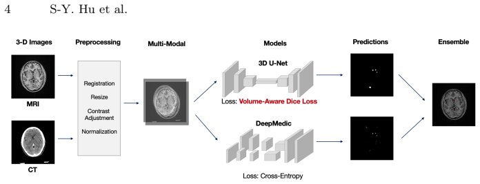
The central mechanism is the volume-aware Dice loss, a modification of the standard Dice loss in which the ground-truth vector $g$ is weighted by a diagonal matrix $W$ with entries $W_{ii} = (\lambda/\mathrm{volume}(i))^{1/2}$ for voxels inside a tumor and $0$ otherwise, together with a normalization constant $C$ that keeps the loss maximum at one. The paper tests constant reweighting, where $\lambda$ is a fixed hyperparameter, and batch reweighting, where $\lambda$ is the largest tumor volume in the batch; batch reweighting avoids hand-tuning $\lambda$ and gives the best balance of precision and recall. This loss makes the optimization sensitive to small lesions that standard Dice would ignore. The second mechanism is the ensemble: a full-volume 3D U-Net trained with the volume-aware Dice loss and a patch-based DeepMedic model trained with cross-entropy, whose probability maps are averaged at test time.
What would settle it
Re-delineate the same test scans with an independent protocol aimed at true lesion boundaries, or use follow-up imaging as the reference, and recompute the volume-aware loss's small-tumor recall; if the gap between standard Dice and volume-aware Dice narrows sharply or disappears, the measured benefit is an artifact of the treatment-contour ground truth.
Extended reading notes
Core claim
The central claim is that a volume-aware Dice loss combined with a multimodal ensemble segmentation system outperforms prior methods on brain metastases, and that the improvement concentrates where clinicians need it most: small lesions. The authors show that replacing the standard Dice loss with a reweighted version in which each voxel's contribution is weighted by the inverse square root of its tumor's volume raises recall on small tumors from 0.466 to 0.633 pixel-wise and from 0.619 to 0.672 metastasis-wise, while leaving large-tumor performance nearly unchanged. Combining a full-volume 3D U-Net and a patch-based DeepMedic network, trained with different objectives and input strategies, and averaging their probability maps raises the Dice score further, to 0.740. The paper interprets these results as evidence that an AI-assisted system can support SRS treatment planning for multiple brain metastases.
Load-bearing premise
The evaluation assumes the physician-drawn SRS treatment contours used as ground truth match the true tumor boundaries, but the paper itself notes these contours can be more aggressive or conservative than the lesion, ignore previously treated tumors, and include highly subjective cases; if those contours deviate systematically, the reported accuracy and the measured benefit of the volume-aware loss could reflect contouring style rather than true tumor extent.
Editorial extensions
If this is right
- If the reported performance holds, automated contouring could take over a large part of the manual delineation burden in SRS, since the system uses the same MRI and CT inputs clinicians already work with.
- Because the volume-aware Dice loss is an architecture-agnostic change to the objective, its small-lesion recall gain should transfer to other segmentation networks trained on the same data.
- With metastasis-wise recall on small lesions improved from 0.619 to 0.672, the system could act as a safety net that draws attention to lesions a human contourer might miss before radiosurgery.
- The ensemble result implies that combining a full-context model and a patch-based model is enough to exploit both global brain anatomy and fine local detail in this task.
Reading between the lines
- A natural extension beyond the paper is to apply batch-reweighted volume-aware Dice to other size-imbalanced medical segmentation tasks, such as lung nodules, lymph nodes, or liver lesions, where standard Dice is likewise dominated by large objects.
- Because the ground truth in this study is treatment-planning contour rather than pathology, the true clinical sensitivity to small lesions remains unknown; a prospective comparison against a second expert contouring pass or follow-up MRI would settle it.
- The batch-reweighting rule makes the loss self-adjusting to the largest lesion in each batch, which suggests it may be robust across institutions with different tumor-size distributions, but that robustness is not demonstrated here.
Signed reviews
Editorial analysis
A structured set of objections, weighed in public.
Referee Report
Summary. This manuscript proposes a volume-aware Dice loss for brain metastasis detection and segmentation in SRS treatment planning, and combines it with a multimodal (MRI+CT) ensemble of a 3D U-Net and DeepMedic. On a single-center cohort of 305 patients plus an additional 36-patient test set, the authors report that the batch-reweighted volume-aware loss improves DSC by 8.57% and recall by 24.14% over a 3D U-Net baseline, and that the final ensemble reaches DSC 0.740 and recall 0.803. The paper also reports size-stratified results showing the largest recall gains on small tumors (pixel-wise recall from 0.466 to 0.633), and it acknowledges that the ground-truth contours are treatment-planning contours rather than true lesion boundaries.
Significance. The underlying idea of weighting small lesions more heavily in a Dice-style loss is clinically motivated and the arithmetic in Section 2.1 is internally consistent, including the normalization constant C in Eq. (2). If the reported gains survived independent evaluation, the system would be a useful contribution to SRS contouring, where small metastases are frequently missed. I credit the authors for evaluating two network architectures and multiple reweighting schemes, for reporting precision and recall separately, and for explicitly discussing the treatment-contour limitation. The evidence as presented, however, does not yet support the headline claims, because the best configuration was selected on the test set and no comparison against prior work is included.
major comments (3)
- [§3.1, Table 1] In Section 3.1 the authors compare constant reweighting at λc = 500, 1000, 2500, 5000 with batch reweighting and select batch reweighting because it gives the best performance on the combination of the two test sets; the final ensemble in Section 3.2 is then also chosen using these same test sets. The reported improvements of +8.57% DSC and +24.14% recall are therefore the maximum of a set of configurations evaluated on the test data, not the performance of a configuration selected on an independent validation split, and the magnitude of any selection bias cannot be estimated because no confidence intervals or repeated splits are given. Please re-run the selection using the validation set (or nested cross-validation) and report the performance of the pre-specified final configuration on a held-out test set that was never used for any model or hyperparameter choice.
- [§3.2, Table 3 / Abstract] The abstract and Section 3.2 state that the system 'surpasses current benchmark levels,' but no quantitative comparison with the prior brain-metastases segmentation methods cited as benchmarks ([1,6]) is provided; the only comparators in Table 3 are the authors' own 3D U-Net and DeepMedic. Because prior work used different data, modalities, and evaluation protocols, the benchmark claim is not supported. Add a direct comparison on a common dataset or clearly reframe the claim as an internal comparison.
- [§3.3] The limitation section correctly states that the ground truth is the SRS treatment contour rather than the true tumor extent, and that these contours can be more aggressive or conservative, ignore previously treated tumors, and be highly subjective. This is not merely a caveat: the reported absolute DSC and the size-stratified recall gains in Table 2 may partly reflect agreement with a contouring protocol rather than true tumor detection, and no analysis quantifies the sensitivity of the results to contour variability. At minimum, the conclusions should be worded in terms of replicating treatment-planning contours, and ideally the authors should provide a contour-agreement or multi-reader analysis.
minor comments (4)
- [§2.1, Eq. (2)] Because W appears only in the ground-truth terms (g^T W p and g^T W g) and not in p^T p, the proposed loss is not symmetric between prediction and ground truth; please state explicitly that this is an intentional recall-oriented weighting and discuss the expected effect on precision.
- [Table 3] The table reports median (std) for apparently repeated runs, but the number of runs and the rule for selecting the reported model (e.g., best validation epoch) are not stated; adding this information would help interpret the small standard deviations.
- [§3.1] The cutoff of 1500 mm3 for small versus large tumors is introduced without justification; state whether this threshold was chosen a priori or based on the data.
- [§2.2 / §3.2] The ensemble confidence map is described as the average of the two models' probability maps, but no details are given for how the final binary mask is thresholded; specify the threshold selection procedure.
Circularity Check
The headline gains are selected on the test set rather than independently predicted, a partial circularity in the reported improvement.
-
fitted input called prediction
[Section 3.1, Table 1; final-system selection in Section 3.2, Table 3]
"We tested the λc of 500, 1000, 2500, 5000 for the CR strategy. ... Table 1 lists the results of applying 𝓁vol-dice relative to the baseline. Overall, using the 𝓁vol-dice with BR (λl) yields the best performance, improving 8.57% of DSC and 24.14% of recall compared to the baseline."
The reweighting strategy and λ value (BR vs CR with λc = 500, 1000, 2500, 5000) are chosen because they give the best numbers on the combined test sets, and the same test-set numbers are then reported as evidence that the volume-aware loss works. Thus the headline 8.57% DSC and 24.14% recall improvements are the maximum over configurations evaluated on the test data, not an independent prediction. The same issue occurs in Section 3.2, where the final ensemble and loss configuration is selected from test-set comparisons in Table 3. The loss formula itself is not definitionally circular; the circularity is in treating test-set-selected performance as a predictive result.
full rationale
The volume-aware Dice loss is a newly proposed objective whose weights depend on ground-truth tumor volume; it is not derived from the reported DSC/recall results, so the loss definition itself is not circular. There are no load-bearing self-citations and no imported uniqueness theorems. The main circularity concern is statistical: the paper evaluates multiple reweighting schemes (BR and CR with several λ values) on the same two test sets, selects the best performer, and then reports that selected performance as the central evidence for the method. Consequently, the claimed improvements are selected estimates and may be inflated by test-set overfitting; a held-out or externally validated dataset would be needed to confirm the improvement. The acknowledged ground-truth contour limitation is a validity issue rather than a circularity issue, since it affects baseline and proposed methods alike. Overall, the contribution retains independent content, but the reported headline gains reduce, in part, to test-set model selection rather than to an unbiased prediction.
Assumptions & free parameters
free parameters (4)
- lambda_c (constant reweight) =
500, 1000, 2500, 5000
- lambda_l (batch reweight) =
dynamic max tumor volume in batch
- exponent in weight formula =
1/2
- small/large tumor cutoff =
1500 mm3
assumptions (4)
- domain assumption Rigid registration between CT and MRI provides accurate spatial alignment for multimodal input.
- domain assumption Ground truth contours delineated for SRS treatment planning are a valid proxy for true tumor extent.
- domain assumption DeepMedic trained with cross-entropy and 3D U-Net trained with Dice loss are complementary for ensemble.
- domain assumption The 80/10/10 random split yields a representative test set.
Cite this review
Pith. "Pith review of Multimodal Volume-Aware Detection and Segmentation for Brain Metastases Radiosurgery." pith.science (2026). https://pith.science/paper/BOZ6BOFW
@misc{pith2026190805418,
  author       = {Pith},
  title        = {Pith review of: Multimodal Volume-Aware Detection and Segmentation for Brain Metastases Radiosurgery},
  year         = {2026},
  howpublished = {\url{https://pith.science/paper/BOZ6BOFW}},
  note         = {Machine review of arXiv:1908.05418}
}
read the original abstract
Stereotactic radiosurgery (SRS), which delivers high doses of irradiation in a single or few shots to small targets, has been a standard of care for brain metastases. While very effective, SRS currently requires manually intensive delineation of tumors. In this work, we present a deep learning approach for automated detection and segmentation of brain metastases using multimodal imaging and ensemble neural networks. In order to address small and multiple brain metastases, we further propose a volume-aware Dice loss which optimizes model performance using the information of lesion size. This work surpasses current benchmark levels and demonstrates a reliable AI-assisted system for SRS treatment planning for multiple brain metastases.
Figures


Reference graph
Works this paper leans on
-
[1]
Computers in biology and medicine 95, 43--54 (2018)
Charron, O., Lallement, A., Jarnet, D., Noblet, V., Clavier, J.B., Meyer, P.: Automatic detection and segmentation of brain metastases on multimodal mr images with a deep convolutional neural network. Computers in biology and medicine 95, 43--54 (2018)
work page 2018
-
[2]
In: International conference on medical image computing and computer-assisted intervention
C i c ek, \"O ., Abdulkadir, A., Lienkamp, S.S., Brox, T., Ronneberger, O.: 3d u-net: learning dense volumetric segmentation from sparse annotation. In: International conference on medical image computing and computer-assisted intervention. pp. 424--432. Springer (2016)
work page 2016
-
[3]
Frontiers in oncology 8, 154 (2018)
Hartgerink, D.E., van der Heyden, B., De Ruysscher, D., Hoeben, A., Terhaag, K., Lambin, P., Postma, A., Jochems, A., Dekker, A., Schoenmaekers, J., et al.: Stereotactic radiosurgery in the management of patients with brain metastases of non-small cell lung cancer; indications, decision tools and future directions. Frontiers in oncology 8, 154 (2018)
work page 2018
-
[4]
Medical image analysis 36, 61--78 (2017)
Kamnitsas, K., Ledig, C., Newcombe, V.F., Simpson, J.P., Kane, A.D., Menon, D.K., Rueckert, D., Glocker, B.: Efficient multi-scale 3d cnn with fully connected crf for accurate brain lesion segmentation. Medical image analysis 36, 61--78 (2017)
work page 2017
-
[5]
Journal of clinical oncology 33(30), 3475 (2015)
Lin, X., DeAngelis, L.M.: Treatment of brain metastases. Journal of clinical oncology 33(30), 3475 (2015)
work page 2015
-
[6]
PloS one 12(10), e0185844 (2017)
Liu, Y., Stojadinovic, S., Hrycushko, B., Wardak, Z., Lau, S., Lu, W., Yan, Y., Jiang, S.B., Zhen, X., Timmerman, R., et al.: A deep convolutional neural network-based automatic delineation strategy for multiple brain metastases stereotactic radiosurgery. PloS one 12(10), e0185844 (2017)
work page 2017
-
[7]
IEEE transactions on medical imaging 34(10), 1993--2024 (2015)
Menze, B.H., Jakab, A., Bauer, S., Kalpathy-Cramer, J., Farahani, K., Kirby, J., Burren, Y., Porz, N., Slotboom, J., Wiest, R., et al.: The multimodal brain tumor image segmentation benchmark (brats). IEEE transactions on medical imaging 34(10), 1993--2024 (2015)
work page 2015
-
[8]
In: 2016 Fourth International Conference on 3D Vision (3DV)
Milletari, F., Navab, N., Ahmadi, S.A.: V-net: Fully convolutional neural networks for volumetric medical image segmentation. In: 2016 Fourth International Conference on 3D Vision (3DV). pp. 565--571. IEEE (2016)
work page 2016
Show all 13 references
-
[9]
BioMed research international 2014 (2014)
Pereira, G.C., Traughber, M., Muzic, R.F.: The role of imaging in radiation therapy planning: past, present, and future. BioMed research international 2014 (2014)
2014
-
[10]
Practical radiation oncology 2(3), 210--225 (2012)
Tsao, M.N., Rades, D., Wirth, A., Lo, S.S., Danielson, B.L., Gaspar, L.E., Sperduto, P.W., Vogelbaum, M.A., Radawski, J.D., Wang, J.Z., et al.: Radiotherapeutic and surgical management for newly diagnosed brain metastasis (es): An american society for radiation oncology eviden...
2012
-
[11]
Radiotherapy and Oncology 121(2), 169--179 (2016)
Vinod, S.K., Jameson, M.G., Min, M., Holloway, L.C.: Uncertainties in volume delineation in radiation oncology: a systematic review and recommendations for future studies. Radiotherapy and Oncology 121(2), 169--179 (2016)
2016
-
[12]
, " * write output.state after.block = add.period write
ENTRY address author booktitle chapter doi edition editor eid howpublished institution journal key month note number organization pages publisher school series title type url volume year label INTEGERS output.state before.all mid.sentence after.sentence after.block FUNCTION in...
-
[13]
write newline
" write newline "" before.all 'output.state := FUNCTION n.dashify 't := "" t empty not t #1 #1 substring "-" = t #1 #2 substring "--" = not "--" * t #2 global.max substring 't := t #1 #1 substring "-" = "-" * t #2 global.max substring 't := while if t #1 #1 substring * t #2 gl...
Reviewed August 14, 2026 · model on record in the stance chip above.
Discussion (0). Continue with ORCID to comment.