Trustworthy MRI Reconstruction via Bayesian Uncertainty Quantification with Sparsity Prior Models
Pith reviewed 2026-06-27 02:59 UTC · model grok-4.3
The pith
Bayesian inference with sparsity priors in transform domains reconstructs MRI images more accurately than optimization methods while supplying uncertainty maps that track actual errors.
A machine-rendered reading of the paper's core claim, the machinery that carries it, and where it could break.
Core claim
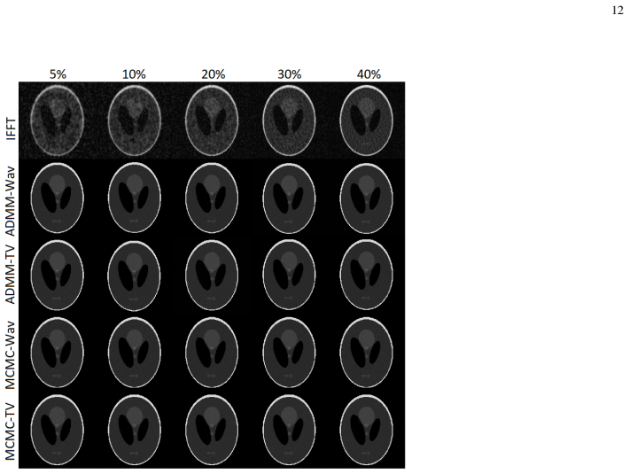
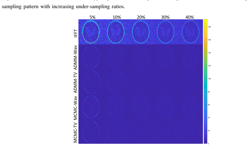
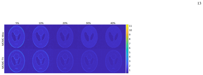
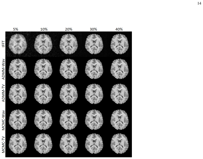
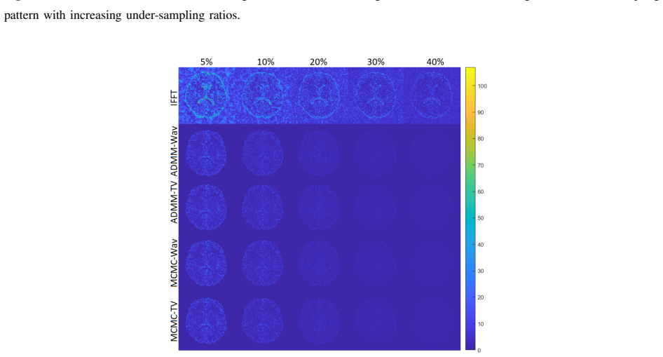
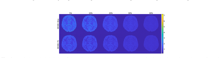
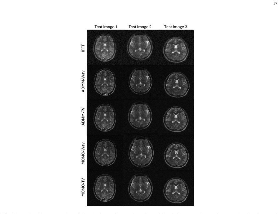
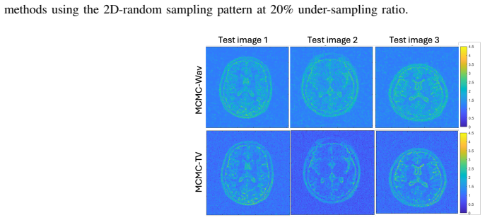
Assigning sparsity priors in total-variation or wavelet domains, then performing Bayesian inference with a split-and-augmented Gibbs sampler and proximal MCMC, produces both higher-quality image reconstructions and uncertainty maps whose values align closely with true reconstruction errors, outperforming optimization-based baselines and deep-learning uncertainty estimators on single-coil and multi-coil datasets.
What carries the argument
Split-and-augmented Gibbs sampler that draws from non-differentiable conditionals via proximal Markov chain Monte Carlo under sparsity priors in a chosen transform domain.
If this is right
- Bayesian reconstructions consistently exceed the image quality of optimization-based counterparts across acceleration factors and sampling trajectories.
- Posterior uncertainty maps correlate strongly with true reconstruction errors on both single-coil and multi-coil data.
- The same uncertainty maps outperform deep-learning uncertainty estimators in correlation with ground-truth errors.
- The framework works for any sparsifying transform once the proximal operator for the associated regularizer is available.
Where Pith is reading between the lines
- The uncertainty maps could be used to adaptively acquire additional k-space lines only in regions flagged as uncertain during an ongoing scan.
- Because the method supplies a full posterior rather than a point estimate, it can be inserted into downstream tasks such as segmentation or registration to propagate reconstruction uncertainty.
- Replacing the fixed transform with a learned dictionary while retaining the Bayesian sampler would test whether the performance gain is due to the inference procedure or the choice of sparsity model.
Load-bearing premise
The unknown image is sparse in a chosen transform domain so that prior distributions can be placed on its coefficients.
What would settle it
On a new multi-coil dataset the posterior variance maps show no correlation with pixel-wise absolute reconstruction error, or the Bayesian reconstructions yield lower PSNR than total-variation minimization under identical sampling.
Figures
read the original abstract
We propose a novel Bayesian framework for joint image reconstruction and uncertainty quantification from compressed sensing magnetic resonance imaging data. The problem is formulated as a linear inverse problem, where prior distributions are assigned to the unknown image parameters. Specifically, the image is assumed to be sparse in a given transform domain. We develop a general framework applicable to any sparsifying transform and demonstrate its performance using (1) a total variation transform based on image spatial gradients and (2) a wavelet-domain transform. Bayesian inference is performed using a split-and-augmented Gibbs sampler, while the resulting non-differentiable conditional distributions are efficiently sampled using a proximal Markov chain Monte Carlo method. The proposed algorithms are validated on both single-coil and multi-coil datasets using various k-space sampling patterns and acceleration factors. The results demonstrate that the proposed Bayesian methods consistently outperform their optimisation-based counterparts in image reconstruction while providing uncertainty estimates for the reconstructed images. Furthermore, the estimated uncertainty maps show a strong correlation with the true reconstruction errors and substantially outperformed deep learning-based uncertainty estimation methods.
Editorial analysis
A structured set of objections, weighed in public.
Referee Report
Summary. The manuscript proposes a Bayesian framework for compressed-sensing MRI reconstruction and uncertainty quantification. Sparsity priors are placed on the image in a chosen transform domain (total variation or wavelet); inference uses a split-and-augmented Gibbs sampler whose non-differentiable conditionals are handled by proximal MCMC. The central claims are that the Bayesian reconstructions outperform optimization-based baselines, that the resulting uncertainty maps correlate strongly with ground-truth reconstruction error, and that these uncertainty estimates substantially outperform deep-learning uncertainty methods.
Significance. If the posterior samples are shown to be reliable, the work would supply a principled, non-deep-learning route to calibrated uncertainty in clinical MRI, which is a recognized need. The generality of the framework to arbitrary sparsifying transforms and the explicit handling of non-differentiable priors via proximal MCMC are technical strengths that could be leveraged beyond the two transforms demonstrated.
major comments (2)
- [Bayesian inference / proximal MCMC description] The reliability of the uncertainty maps (and therefore both the correlation claim and the outperformance over DL uncertainty methods) rests on the quality of the posterior samples produced by the split-and-augmented Gibbs sampler with proximal MCMC. No convergence diagnostics—trace plots, autocorrelation times, effective sample size, or Gelman–Rubin statistics—are reported for the high-dimensional image-space chains. This omission directly weakens the trustworthiness assertions in the abstract.
- [Experimental validation section] The experimental claims of consistent superiority across single-coil and multi-coil data, multiple sampling patterns, and acceleration factors are presented without statistical significance tests, confidence intervals, or error bars on the quantitative metrics. This makes it impossible to judge whether the reported gains are robust or could be explained by post-hoc hyper-parameter choices.
minor comments (2)
- Notation for the proximal operator and the augmented variables in the Gibbs sampler could be introduced more explicitly with a short table of symbols to aid readability.
- The abstract states that uncertainty maps 'substantially outperformed' DL methods but does not name the DL baselines or the quantitative metric used for that comparison.
Simulated Author's Rebuttal
We thank the referee for their thoughtful and constructive comments, which highlight important aspects for strengthening the manuscript. We address each major comment below and will revise the paper accordingly.
read point-by-point responses
-
Referee: [Bayesian inference / proximal MCMC description] The reliability of the uncertainty maps (and therefore both the correlation claim and the outperformance over DL uncertainty methods) rests on the quality of the posterior samples produced by the split-and-augmented Gibbs sampler with proximal MCMC. No convergence diagnostics—trace plots, autocorrelation times, effective sample size, or Gelman–Rubin statistics—are reported for the high-dimensional image-space chains. This omission directly weakens the trustworthiness assertions in the abstract.
Authors: We agree that convergence diagnostics are necessary to substantiate the quality of the posterior samples and the resulting uncertainty maps. The proximal MCMC approach is intended to handle the non-differentiable conditionals arising from the sparsity priors, but we omitted explicit diagnostics in the initial submission. In the revised manuscript we will report Gelman–Rubin statistics across multiple independent chains, effective sample sizes, autocorrelation times, and representative trace plots for image-domain variables. These additions will directly support the trustworthiness claims. revision: yes
-
Referee: [Experimental validation section] The experimental claims of consistent superiority across single-coil and multi-coil data, multiple sampling patterns, and acceleration factors are presented without statistical significance tests, confidence intervals, or error bars on the quantitative metrics. This makes it impossible to judge whether the reported gains are robust or could be explained by post-hoc hyper-parameter choices.
Authors: We concur that quantitative claims benefit from measures of variability and statistical testing. The original results were obtained by averaging over multiple test cases, yet standard deviations and formal significance tests were not included. In the revision we will add error bars (standard deviation across test images or repeated runs) to all reported metrics and include paired statistical tests (e.g., Wilcoxon signed-rank or t-tests) with p-values to evaluate whether the observed improvements over baselines are statistically significant. This will allow readers to assess the robustness of the performance gains. revision: yes
Circularity Check
No circularity in derivation; empirical validation independent of inputs
full rationale
The paper sets up a standard Bayesian linear inverse problem with sparsity priors (TV or wavelet), derives a split-and-augmented Gibbs sampler using proximal MCMC for the non-differentiable conditionals, and reports empirical outperformance plus uncertainty-error correlation on held-out single- and multi-coil MRI datasets with various sampling patterns. No equation reduces a reported prediction or uncertainty map to a quantity fitted on the same evaluation data; no load-bearing uniqueness theorem or ansatz is imported via self-citation; the central claims rest on external benchmarks rather than self-definition. This is the normal case of a self-contained methodological paper whose results are not forced by its own construction.
Axiom & Free-Parameter Ledger
axioms (1)
- domain assumption The image is assumed to be sparse in a given transform domain
Reference graph
Works this paper leans on
-
[1]
Sparse mri: The application of compressed sensing for rapid mr imaging,
M. Lustig, D. Donoho, and J. M. Pauly, “Sparse mri: The application of compressed sensing for rapid mr imaging,”Magnetic Resonance in Medicine: An Official Journal of the International Society for Magnetic Resonance in Medicine, vol. 58, no. 6, pp. 1182–1195, 2007. 20
2007
-
[2]
Advances in sensitivity encoding with arbitrary k-space trajectories,
K. P. Pruessmann, M. Weiger, P. B ¨ornert, and P. Boesiger, “Advances in sensitivity encoding with arbitrary k-space trajectories,”Magnetic Resonance in Medicine: An Official Journal of the International Society for Magnetic Resonance in Medicine, vol. 46, no. 4, pp. 638–651, 2001
2001
-
[3]
A complex quasi-newton proximal method for image reconstruction in compressed sensing mri,
T. Hong, L. Hernandez-Garcia, and J. A. Fessler, “A complex quasi-newton proximal method for image reconstruction in compressed sensing mri,”IEEE Transactions on Computational Imaging, 2024
2024
-
[4]
Plug-and-play methods for magnetic resonance imaging: Using denoisers for image recovery,
R. Ahmad, C. A. Bouman, G. T. Buzzard, S. Chan, S. Liu, E. T. Reehorst, and P. Schniter, “Plug-and-play methods for magnetic resonance imaging: Using denoisers for image recovery,”IEEE signal processing magazine, vol. 37, no. 1, pp. 105–116, 2020
2020
-
[5]
Fast iteratively reweighted least squares algorithms for analysis-based sparse reconstruction,
C. Chen, L. He, H. Li, and J. Huang, “Fast iteratively reweighted least squares algorithms for analysis-based sparse reconstruction,”Medical image analysis, vol. 49, pp. 141–152, 2018
2018
-
[6]
Image reconstruction of compressed sensing mri using graph-based redundant wavelet transform,
Z. Lai, X. Qu, Y . Liu, D. Guo, J. Ye, Z. Zhan, and Z. Chen, “Image reconstruction of compressed sensing mri using graph-based redundant wavelet transform,”Medical image analysis, vol. 27, pp. 93–104, 2016
2016
-
[7]
Modl: Model-based deep learning architecture for inverse problems,
H. K. Aggarwal, M. P. Mani, and M. Jacob, “Modl: Model-based deep learning architecture for inverse problems,”IEEE transactions on medical imaging, vol. 38, no. 2, pp. 394–405, 2018
2018
-
[8]
Learning a variational network for reconstruction of accelerated mri data,
K. Hammernik, T. Klatzer, E. Kobler, M. P. Recht, D. K. Sodickson, T. Pock, and F. Knoll, “Learning a variational network for reconstruction of accelerated mri data,”Magnetic resonance in medicine, vol. 79, no. 6, pp. 3055–3071, 2018
2018
-
[9]
Mr image reconstruction using deep density priors,
K. C. Tezcan, C. F. Baumgartner, R. Luechinger, K. P. Pruessmann, and E. Konukoglu, “Mr image reconstruction using deep density priors,”IEEE transactions on medical imaging, vol. 38, no. 7, pp. 1633–1642, 2018
2018
-
[10]
Mri reconstruction using deep bayesian estimation,
G. Luo, N. Zhao, W. Jiang, E. S. Hui, and P. Cao, “Mri reconstruction using deep bayesian estimation,”Magnetic resonance in medicine, vol. 84, no. 4, pp. 2246–2261, 2020
2020
-
[11]
Unsupervised mri reconstruction via zero-shot learned adversarial transformers,
Y . Korkmaz, S. U. Dar, M. Yurt, M. ¨Ozbey, and T. Cukur, “Unsupervised mri reconstruction via zero-shot learned adversarial transformers,” IEEE Transactions on Medical Imaging, vol. 41, no. 7, pp. 1747–1763, 2022
2022
-
[12]
Radial magnetic resonance image reconstruction with a deep unrolled projected fast iterative soft-thresholding network,
B. Qu, J. Zhang, T. Kang, J. Lin, M. Lin, H. She, Q. Wu, M. Wang, and G. Zheng, “Radial magnetic resonance image reconstruction with a deep unrolled projected fast iterative soft-thresholding network,”Computers in Biology and Medicine, vol. 168, p. 107707, 2024
2024
-
[13]
Hierarchical neural architecture search with adaptive global–local feature learning for magnetic resonance image reconstruction,
C. Cao, W. Huang, F. Hu, and X. Gao, “Hierarchical neural architecture search with adaptive global–local feature learning for magnetic resonance image reconstruction,”Computers in Biology and Medicine, vol. 168, p. 107774, 2024
2024
-
[14]
A systematic review and identification of the challenges of deep learning techniques for undersampled magnetic resonance image reconstruction,
M. B. Hossain, R. K. Shinde, S. Oh, K.-C. Kwon, and N. Kim, “A systematic review and identification of the challenges of deep learning techniques for undersampled magnetic resonance image reconstruction,”Sensors, vol. 24, no. 3, p. 753, 2024
2024
-
[15]
Mip-enhanced uncertainty-aware network for fast 7t time-of-flight mra reconstruction,
K. Sun, C. Duan, X. Lou, and D. Shen, “Mip-enhanced uncertainty-aware network for fast 7t time-of-flight mra reconstruction,”IEEE Transactions on Medical Imaging, vol. 44, no. 5, pp. 2270–2282, 2025
2025
-
[16]
Dropout as a bayesian approximation: Representing model uncertainty in deep learning,
Y . Gal and Z. Ghahramani, “Dropout as a bayesian approximation: Representing model uncertainty in deep learning,” ininternational conference on machine learning. PMLR, 2016, pp. 1050–1059
2016
-
[17]
Weight uncertainty in neural network,
C. Blundell, J. Cornebise, K. Kavukcuoglu, and D. Wierstra, “Weight uncertainty in neural network,” inInternational conference on machine learning. PMLR, 2015, pp. 1613–1622
2015
-
[18]
Simple and scalable predictive uncertainty estimation using deep ensembles,
B. Lakshminarayanan, A. Pritzel, and C. Blundell, “Simple and scalable predictive uncertainty estimation using deep ensembles,”Advances in neural information processing systems, vol. 30, 2017
2017
-
[19]
Optimization methods for magnetic resonance image reconstruction: Key models and optimization algorithms,
J. A. Fessler, “Optimization methods for magnetic resonance image reconstruction: Key models and optimization algorithms,”IEEE signal processing magazine, vol. 37, no. 1, pp. 33–40, 2020
2020
-
[20]
Sparsity averaging for compressive imaging,
R. E. Carrillo, J. D. McEwen, D. Van De Ville, J.-P. Thiran, and Y . Wiaux, “Sparsity averaging for compressive imaging,”IEEE Signal Processing Letters, vol. 20, no. 6, pp. 591–594, 2013
2013
-
[21]
Patch-based sparse representation for bacterial detection,
A. K. Eldaly, Y . Altmann, A. Akram, A. Perperidis, and S. McLaughlin, “Patch-based sparse representation for bacterial detection,” in IEEE International Symposium on Biomedical Imaging (ISBI), Venice, Italy, April 2019, pp. 1–5
2019
-
[22]
Deconvolution and restoration of optical endomicroscopy images,
A. K. Eldaly, Y . Altmann, A. Perperidis, N. Krstajic, T. R. Choudhary, K. Dhaliwal, and S. McLaughlin, “Deconvolution and restoration of optical endomicroscopy images,”IEEE Trans. Comput. Imag., vol. 4, no. 2, pp. 194–205, June 2018
2018
-
[23]
Bayesian bacterial detection using irregularly sampled optical endomicroscopy images,
A. K. Eldaly, Y . Altmann, A. Akram, P. McCool, A. Perperidis, K. Dhaliwal, and S. McLaughlin, “Bayesian bacterial detection using irregularly sampled optical endomicroscopy images,”Medical Image Analysis, 2019
2019
-
[24]
Bayesian activity estimation and uncertainty quantification of spent nuclear fuel using passive gamma emission tomography,
A. K. Eldaly, M. Fang, A. Di Fulvio, S. McLaughlin, M. E. Davies, Y . Altmann, and Y . Wiaux, “Bayesian activity estimation and uncertainty quantification of spent nuclear fuel using passive gamma emission tomography,”Journal of Imaging, vol. 7, no. 10, p. 212, 2021
2021
-
[25]
Bayesian magnetic resonance image reconstruction and uncertainty quantification,
A. Karam Eldaly and D. C. Alexander, “Bayesian magnetic resonance image reconstruction and uncertainty quantification,” inISMRM & SMRT Virtual Conference & Exhibition, Singapore, May 2024. 21
2024
-
[26]
Robust compressed sensing mri with deep generative priors,
A. Jalal, M. Arvinte, G. Daras, E. Price, A. G. Dimakis, and J. Tamir, “Robust compressed sensing mri with deep generative priors,” Advances in Neural Information Processing Systems, vol. 34, pp. 14 938–14 954, 2021
2021
-
[27]
Bayesian uncertainty-aware mri reconstruction,
A. K. Eldaly, M. Figini, and D. C. Alexander, “Bayesian uncertainty-aware mri reconstruction,” inICASSP 2026-2026 IEEE International Conference on Acoustics, Speech and Signal Processing (ICASSP). IEEE, 2026, pp. 6–10
2026
-
[28]
Multi-contrast reconstruction with bayesian compressed sensing,
B. Bilgic, V . K. Goyal, and E. Adalsteinsson, “Multi-contrast reconstruction with bayesian compressed sensing,”Magnetic resonance in medicine, vol. 66, no. 6, pp. 1601–1615, 2011
2011
-
[29]
Bayesian sparse regularization for parallel mri reconstruction using complex bernoulli–laplace mixture priors,
S. Chaabene, L. Chaari, and A. Kallel, “Bayesian sparse regularization for parallel mri reconstruction using complex bernoulli–laplace mixture priors,”Signal, Image and Video Processing, vol. 14, pp. 445–453, 2020
2020
-
[30]
Parameter estimation in spike and slab variational inference for blind image deconvolution,
J. G. Serra, J. Mateos, R. Molina, and A. K. Katsaggelos, “Parameter estimation in spike and slab variational inference for blind image deconvolution,” inIEEE European Signal Processing Conference (EUSIPCO), Greece Kos island, Aug 2017, pp. 1495–1499
2017
-
[31]
Oedipus: An experiment design framework for sparsity-constrained mri,
J. P. Haldar and D. Kim, “Oedipus: An experiment design framework for sparsity-constrained mri,”IEEE transactions on medical imaging, vol. 38, no. 7, pp. 1545–1558, 2019
2019
-
[32]
Maximum likelihood estimation of regularization parameters in high-dimensional inverse problems: An empirical bayesian approach part i: Methodology and experiments,
A. F. Vidal, V . De Bortoli, M. Pereyra, and A. Durmus, “Maximum likelihood estimation of regularization parameters in high-dimensional inverse problems: An empirical bayesian approach part i: Methodology and experiments,”SIAM Journal on Imaging Sciences, vol. 13, no. 4, pp. 1945–1989, 2020
1945
-
[33]
An algorithm for total variation minimization and applications,
A. Chambolle, “An algorithm for total variation minimization and applications,”Journal of Mathematical imaging and vision, vol. 20, no. 1-2, pp. 89–97, 2004
2004
-
[34]
Nonlinear total variation based noise removal algorithms,
L. I. Rudin, S. Osher, and E. Fatemi, “Nonlinear total variation based noise removal algorithms,”Physica D: nonlinear phenomena, vol. 60, no. 1-4, pp. 259–268, 1992
1992
-
[35]
Split-and-augmented gibbs sampler—application to large-scale inference problems,
M. V ono, N. Dobigeon, and P. Chain ´ais, “Split-and-augmented gibbs sampler—application to large-scale inference problems,”IEEE Transactions on Signal Processing, vol. 67, no. 6, pp. 1648–1661, 2019
2019
-
[36]
Efficient bayesian computation by proximal markov chain monte carlo: when langevin meets moreau,
A. Durmus, E. Moulines, and M. Pereyra, “Efficient bayesian computation by proximal markov chain monte carlo: when langevin meets moreau,”SIAM Journal on Imaging Sciences, vol. 11, no. 1, pp. 473–506, 2018
2018
-
[37]
Robert,The Bayesian choice: from decision-theoretic foundations to computational implementation
C. Robert,The Bayesian choice: from decision-theoretic foundations to computational implementation. Springer Science & Business Media, 2007
2007
-
[38]
Concentration of the information in data with log-concave distributions,
S. Bobkov, M. Madimanet al., “Concentration of the information in data with log-concave distributions,”Annals of Probability, vol. 39, no. 4, pp. 1528–1543, 2011
2011
-
[39]
Advances in diffusion mri acquisition and processing in the human connectome project,
S. N. Sotiropoulos, S. Jbabdi, J. Xu, J. L. Andersson, E. J. Auerbach, M. Hernandez, G. Sapiro, M. Jenkinsonet al., “Advances in diffusion mri acquisition and processing in the human connectome project,”Neuroimage, vol. 80, pp. 125–143, 2013
2013
-
[40]
M4raw: A multi-contrast, multi-repetition, multi-channel mri k-space dataset for low-field mri research,
M. Lyu, L. Mei, S. Huang, S. Liu, Y . Li, K. Yang, Y . Liu, Y . Dong, L. Dong, and E. X. Wu, “M4raw: A multi-contrast, multi-repetition, multi-channel mri k-space dataset for low-field mri research,”Scientific Data, vol. 10, no. 1, p. 264, 2023
2023
-
[41]
Image reconstruction with low-rankness and self-consistency of k-space data in parallel mri,
X. Zhang, D. Guo, Y . Huang, Y . Chen, L. Wang, F. Huang, Q. Xu, and X. Qu, “Image reconstruction with low-rankness and self-consistency of k-space data in parallel mri,”Medical image analysis, vol. 63, p. 101687, 2020
2020
discussion (0)
Sign in with ORCID, Apple, or X to comment. Anyone can read and Pith papers without signing in.