REVIEW 3 major objections 6 minor 13 references
GLENDA: Gynecologic Laparoscopy Endometriosis Dataset
T0 review · 3 major / 6 minor · reviewed 2026-08-05 · deepseek-v4-flash
Pith's one-line read This paper releases GLENDA, a 25,682-frame laparoscopy dataset annotated with 520 expert-drawn endometriosis lesion regions, described as the first public region-based endometriosis dataset.
desk verdict First public region-annotated endometriosis laparoscopy dataset; a solid resource paper, but annotation quality is asserted, not demonstrated. read the letter →
The pith
A machine-rendered reading of the paper's core claim, the machinery that carries it, and where it could break.
The reading
What carries the argument
The central object is the GLENDA dataset itself, structured as paired frame images and binary annotation images whose file names encode video ID, sequence range, frame ID, class, and annotation ID, allowing frames to be mapped to their regions by partial path matching. The annotation scheme carries the argument: every endometriosis region is a closed freehand drawing, polygon, or rectangle drawn in the ECAT tool, tied either to a single frame or to a keyframe of a video segment. The load-bearing design choice is that keyframes are selected only where camera motion is minimal, so a region drawn on one frame is claimed to remain visible for all frames in the segment, enabling tracking-based au
What would settle it
Collect a sample of GLENDA's video segments and have independent endometriosis specialists draw regions on non-keyframe frames, then measure overlap with the keyframe annotation propagated by tracking; alternatively, biopsy-confirm a subset of annotated lesions. If propagated regions consistently miss the lesion (low IoU) or if confirmed false-positive annotations are frequent, the ground-truth and tracking assumptions are falsified.
Extended reading notes
Core claim
The authors' central claim is that GLENDA is 'the first of its kind': the first public endometriosis dataset whose annotations are regions, not just image-level or video-level labels. To establish this, they introduce a dataset of 25,682 frames from over 400 gynecologic laparoscopy videos, including more than 12,000 positive frames with visible endometriosis and more than 13,000 negative frames without visible endometriosis. A total of 520 annotations were hand-drawn by leading endometriosis experts on 302 frames, distributed across four location-based categories—peritoneum (402 annotations), ovary (51), uterus (14), and deep infiltrating endometriosis (53)—using the ECAT annotation tool wit
Load-bearing premise
The value of GLENDA rests on the assumption that an expert's hand-drawn region on a keyframe is an accurate label for endometriosis presence and location, and that this label stays correct for every frame of the video segment because camera motion is minimal.
Editorial extensions
If this is right
- Binary pathology-versus-no-pathology classification can be trained directly on the 25K+ frames without using region labels.
- Multi-label classification and localization can be trained on the 520 expert regions across four endometriosis locations.
- Because 138 sequences are annotated at keyframes with minimal camera motion, tracking algorithms can propagate regions to neighboring frames, multiplying the effective number of labeled samples.
- The file-naming convention lets users map every frame to its binary annotation masks and convert them to bounding boxes or polygons as needed.
- Planned extensions—additional categories, lesion severities, and an 'endometriosis suspicion' class—aim to improve classifier robustness on visually ambiguous cases.
Reading between the lines
- Beyond the paper's stated plans, a validation study comparing tracked keyframe regions against fresh expert annotations on non-keyframe frames would test whether the minimal-camera-motion assumption actually holds; without such a check, tracking-based augmentation could silently propagate annotation errors.
- The uterus class, with only 14 annotations on 8 frames, is likely too small for standalone deep-learning training, so the dataset's practical utility for rare classes depends on augmentation, class fusion, or future expansion.
- Because no-pathology segments carry no region annotations, negative regions cannot serve as hard negatives in detection training; detectors trained on GLENDA can only learn positive regions and frame-level negatives.
- The paper's warning about near-duplicate sequential frames implies that an independent benchmark built on GLENDA should split by video or sequence rather than by random frame, which would give a more realistic estimate of model performance.
Signed reviews
Editorial analysis
A structured set of objections, weighed in public.
Referee Report
Summary. The paper introduces GLENDA, a dataset of gynecologic laparoscopy images with region-level annotations for endometriosis. It contains 25,682 frames from 138 video sequences, of which 520 hand-drawn region annotations are provided on 302 frames across four pathological classes (peritoneum, ovary, uterus, DIE) plus a no-pathology class. The authors describe the data collection process using the ECAT tool, the dataset structure, and intended uses for classification, detection, localization, and tracking. The main claim is that GLENDA is the first publicly released region-annotated endometriosis laparoscopy dataset.
Significance. If the annotations are accurate, GLENDA fills a genuine gap: no other public dataset offers region-level endometriosis annotations from laparoscopy. The dataset has a plausible scale (25K+ frames, 520 regions) and the authors are transparent about practical limitations such as class imbalance and temporal redundancy. The paper does not include any experiments or benchmarks, but for a dataset release that is not an intrinsic flaw. The decisive issue is whether the ground-truth labels can be trusted for training and evaluation; the manuscript currently provides no inter-annotator agreement, adjudication procedure, or histopathologic confirmation, and it relies on an unquantified keyframe-to-sequence propagation assumption. These are load-bearing because the contribution is precisely the correctness of the 520 expert regions and the usability of the video sequences for annotation augmentation.
major comments (3)
- [Section 2 (Dataset Creation) and Section 4 (Limitations)] The central claim that GLENDA is a usable ground-truth dataset is not supported by any quantitative annotation-reliability evidence. Section 2 states only that 'leading medical experts' created the annotations; it does not report the number of annotators, the number of cases annotated by more than one expert, inter-annotator agreement (e.g., Dice, IoU, Cohen's kappa), or an adjudication protocol. Section 4 and the Conclusion further acknowledge that even specialists sometimes cannot classify endometriosis without further inspection, and the authors propose a future 'suspicion' class. This self-admitted uncertainty is not reflected in the released binary region labels. Without any reliability measure, the reader cannot distinguish systematic label noise from accurate ground truth. This is the core weakness of the paper's central claim and should be addressed by adding annotation statistic
- [Section 2 (Dataset Creation)] The assertion that keyframe annotations remain valid throughout video segments because 'camera motion is kept at a minimal level' is not quantified. No motion metric, threshold, or per-sequence verification is provided. Since most of the 12K+ positive frames are unannotated frames that would be augmented by tracking keyframe masks, propagation error could substantially affect any downstream localization or tracking evaluation. The authors should either provide per-sequence camera-motion statistics (e.g., frame differencing, optical flow magnitude), or release only the annotated keyframes for evaluation and clearly mark propagated annotations as weak labels.
- [Section 3, Table 1] The class distribution is extremely imbalanced, with peritoneum having 402 of 520 annotations and uterus only 14 annotations on 8 frames. The paper discusses this in Section 4, but it remains a significant limitation for any model trained on GLENDA as-is. The authors should provide explicit guidance on recommended evaluation protocols (e.g., class-wise AP, sequence-level splits, excluding or reweighting under-represented classes) and ideally include a benchmark or baseline experiment to demonstrate that the dataset is actually usable for the claimed detection/localization tasks. Without such a sanity check, the dataset's practical value is uncertain.
minor comments (6)
- [Section 2] Typo: 'a a complete dataset' should be 'a complete dataset'.
- [Section 3.2] Typo: 'addionally' should be 'additionally'.
- [Section 5] The acronym expansion 'Gynecologic Laparoscy ENdometriosis DAtaset' contains a typo ('Laparoscy') and does not match the spelled-out form used in the title.
- [Table 1] The 'T otal' row uses an unusual space; also 'max. cat.: 3' is not explained in the table caption or surrounding text. Please clarify whether the maximum of three categories per frame is a property of the annotation protocol or of the data.
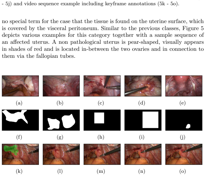
- [Section 3.1, Figure captions] The figures show examples of each class, but the captions do not specify whether the green overlay corresponds to the keyframe annotation or the propagated mask. The caption in Figure 3 mentions 'keyframe annotations (green overlay)', while Figures 4–6 do not repeat this; please standardize.
- [Section 4] The recommendation to split training/validation/test by video sequence is sound, but the sentence 'yielding a perfect classification score' is colloquial and could be misread as a claim about the dataset's difficulty. Rephrase to make clear this is a warning about train/test leakage.
Circularity Check
No circular derivation: GLENDA is a self-contained dataset contribution; the central claims are counts and first-of-its-kind status, not predictions derived from fitted inputs.
full rationale
This is a dataset paper with no equations, fitted parameters, or predictive claims, so there is no derivation chain to collapse. The contribution is the dataset itself: 25,682 frames, 520 expert region annotations, and 138 sequences (Table 1). These numbers are definitional properties of the released artifact, not outputs derived from inputs. The only self-references are to the authors' own ECAT annotation tool [6], the LapGyn4 dataset [4], and an Enzian classification paper by co-author J. Keckstein [3]; none is load-bearing for the dataset's existence, contents, or first-of-its-kind assertion. The paper's acknowledged limitations (class imbalance, small uterus class, need for sequence-based splits, and the future 'endometriosis suspicion' class reflecting expert difficulty) are validity/correctness concerns about label noise and generalizability, not circular reasoning. Therefore no circularity is present.
Assumptions & free parameters
assumptions (3)
- domain assumption Expert hand-drawn annotations are accurate ground truth for endometriosis
- domain assumption The rASRM and Enzian classifications define meaningful endometriosis categories for image annotation
- domain assumption Keyframe annotations remain valid for entire video segments because camera motion is minimal
Cite this review
Pith. "Pith review of GLENDA: Gynecologic Laparoscopy Endometriosis Dataset." pith.science (2026). https://pith.science/paper/CO3GINHJ
@misc{pith2026250821398,
  author       = {Pith},
  title        = {Pith review of: GLENDA: Gynecologic Laparoscopy Endometriosis Dataset},
  year         = {2026},
  howpublished = {\url{https://pith.science/paper/CO3GINHJ}},
  note         = {Machine review of arXiv:2508.21398}
}
read the original abstract
Gynecologic laparoscopy as a type of minimally invasive surgery (MIS) is performed via a live feed of a patient's abdomen surveying the insertion and handling of various instruments for conducting treatment. Adopting this kind of surgical intervention not only facilitates a great variety of treatments, the possibility of recording said video streams is as well essential for numerous post-surgical activities, such as treatment planning, case documentation and education. Nonetheless, the process of manually analyzing surgical recordings, as it is carried out in current practice, usually proves tediously time-consuming. In order to improve upon this situation, more sophisticated computer vision as well as machine learning approaches are actively developed. Since most of such approaches heavily rely on sample data, which especially in the medical field is only sparsely available, with this work we publish the Gynecologic Laparoscopy ENdometriosis DAtaset (GLENDA) - an image dataset containing region-based annotations of a common medical condition named endometriosis, i.e. the dislocation of uterine-like tissue. The dataset is the first of its kind and it has been created in collaboration with leading medical experts in the field.
Figures


Figures from the paper (5 more)




Reference graph
Works this paper leans on
-
[1]
Fertility and sterility 43(3), 351–352 (1985)
Andrews, W., Buttram Jr, V., Behrman, S., Carmichael, E., Cohen, M., Dmowski, P., Eward, R., Guzick, D., Jones Jr, H., Kistner, R., et al.: Revised american fertility society classification of endometriosis: 1985. Fertility and sterility 43(3), 351–352 (1985)
work page 1985
-
[2]
Canis, M., Donnez, J., Guzick, D., Halme, J., Rock, J., Schenken, R., Vernon, M.: Revised american society for reproductive medicine classification of endometriosis:
-
[3]
coloproctology 39(2), 121–133 (mar 2017)
Keckstein, J.: Endometriosis in the Intestinal Tract – Important Facts for Diagnosis and Therapy. coloproctology 39(2), 121–133 (mar 2017). https://doi.org/10.1007/s00053-017-0144-5
-
[4]
Leibetseder, A., Petscharnig, S., Primus, M.J., Kletz, S., M¨ unzer, B., Schoeffmann, K., Keckstein, J.: Lapgyn4: a dataset for 4 automatic content analysis problems in the domain of laparoscopic gynecology. In: Proc. of the 9th ACM Multimedia Systems Conf. pp. 357–362. ACM (2018)
work page 2018
-
[5]
Medical image analysis 42, 60–88 (2017)
Litjens, G., Kooi, T., Bejnordi, B.E., Setio, A.A.A., Ciompi, F., Ghafoorian, M., Van Der Laak, J.A., Van Ginneken, B., S´ anchez, C.I.: A survey on deep learning in medical image analysis. Medical image analysis 42, 60–88 (2017)
work page 2017
- [6]
-
[7]
Multimedia Tools and Applications (jan 2017)
M¨ unzer, B., Schoeffmann, K., B¨ osz¨ ormenyi, L.: Content-based processing and anal- ysis of endoscopic images and videos: A survey. Multimedia Tools and Applications (jan 2017). https://doi.org/10.1007/s11042-016-4219-z
-
[8]
Multimedia Tools and Applications 77(13), 16813–16832 (jul 2018)
Schoeffmann, K., Husslein, H., Kletz, S., Petscharnig, S., Muenzer, B., Beecks, C.: Video retrieval in laparoscopic video recordings with dynamic content de- scriptors. Multimedia Tools and Applications 77(13), 16813–16832 (jul 2018). https://doi.org/10.1007/s11042-017-5252-2
Show all 13 references
-
[9]
arXiv preprint arXiv:1610.09278 (2016)
Stauder, R., Ostler, D., Kranzfelder, M., Koller, S., Feußner, H., Navab, N.: The tum lapchole dataset for the m2cai 2016 workflow challenge. arXiv preprint arXiv:1610.09278 (2016)
2016 arXiv
-
[10]
Surgical endoscopy 27(7), 2253–2257 (2013)
Tsui, C., Klein, R., Garabrant, M.: Minimally invasive surgery: national trends in adoption and future directions for hospital strategy. Surgical endoscopy 27(7), 2253–2257 (2013)
2013
-
[11]
IEEE Transactions on Medical Imaging 36(1), 86–97 (jan 2017)
Twinanda, A.P., Shehata, S., Mutter, D., Marescaux, J., de Mathelin, M., Padoy, N.: EndoNet: A Deep Architecture for Recognition Tasks on Laparo- scopic Videos. IEEE Transactions on Medical Imaging 36(1), 86–97 (jan 2017). https://doi.org/10.1109/TMI.2016.2593957
2017
-
[12]
Medical image analysis 30, 144–157 (2016)
Ye, M., Giannarou, S., Meining, A., Yang, G.Z.: Online tracking and retargeting with applications to optical biopsy in gastrointestinal endoscopic examinations. Medical image analysis 30, 144–157 (2016)
2016
-
[1996]
https://doi.org/10.1016/S0015- 0282(97)81391-X
Fertility and Sterility67(5), 817–821 (1997). https://doi.org/10.1016/S0015- 0282(97)81391-X
1997 doi
Reviewed August 5, 2026 · model on record in the stance chip above.
Discussion (0). Continue with ORCID to comment.