REVIEW 4 major objections 5 minor 43 references
SFD-Mamba2Net: Structure-Guided Frequency-Enhanced Dual-Stream Mamba2 Network for Coronary Artery Segmentation
T0 review · 4 major / 5 minor · reviewed 2026-08-04 · deepseek-v4-flash
Pith's one-line read SFD-Mamba2Net claims that joining a Hessian-based structural prior, a dual-axis bidirectional Mamba2 bottleneck, and wavelet high-frequency refinement yields the best coronary-artery segmentation and stenosis detection among eight methods o
desk verdict A reasonable incremental Mamba2-vessel-segmentation paper whose headline claim is contradicted by its own Table 2, and whose dataset description has an unresolved count mismatch; worth engaging but needs real revision. read the letter →
The pith
A machine-rendered reading of the paper's core claim, the machinery that carries it, and where it could break.
The reading
What carries the argument
The architecture rests on three named modules. CASE (Curvature-Aware Structural Enhancement) is a parameter-free multi-scale Hessian vesselness filter applied to the input to amplify slender tubular structures and suppress background. AA-DS Mamba2 (Axial-Alternating Dual-Stream Mamba2) is a bottleneck module that flattens features along width and height in two alternating paths, each containing forward and backward Mamba2 sequences under the State Space Duality (SSD) framework, to capture long-range spatial dependencies with linear complexity. PHFP (Progressive High-Frequency Perception) is a decoder module that recursively applies Haar wavelet decomposition, enhances high-frequency sub-band
What would settle it
Run the same eight methods under a strictly patient-external split (all frames of a given patient placed entirely in training or entirely in testing) on the exact dataset reported in Table 1, and check whether SFD-Mamba2Net still leads VM-UNet by the reported margins; a drop of Dice or stenosis metrics to within one standard deviation, or evidence that training and test frames share the same patients, would falsify the generalization claim.
Extended reading notes
Core claim
The central claim is that SFD-Mamba2Net, by explicitly injecting geometric vesselness priors, bidirectional state-space modeling along orthogonal axes, and progressive wavelet high-frequency refinement, produces more complete and boundary-accurate coronary vessel segmentations than existing U-Net, Transformer, state-space, and frequency-based models. The paper reports the best Dice (88.10%), F1 (89.22%), IoU (80.81%), Accuracy (98.80%), and Specificity (99.50%) among eight compared methods, and in a centerline-based stenosis-detection pipeline it reports the highest true positive rate (0.60) and positive predictive value (0.64), with lower absolute and relative severity errors than the other
Load-bearing premise
The central claim rests on the unstated assumption that the reported metrics come from a fair, patient-independent train/test split on a correctly described dataset; the paper gives no split procedure and its abstract counts (187 LCA + 127 RCA) disagree with Table 1 (403 LCA + 213 RCA), so leakage could inflate all results.
Editorial extensions
If this is right
- If the reported results are correct, automated ICA analysis can localize and grade coronary stenoses with fewer misses and false positives than the seven comparison models, including for moderate and severe lesions.
- The ablation results indicate each module contributes and the full combination is best, so the proposed recipe of geometric priors, state-space long-range modeling, and wavelet frequency refinement is a workable template for low-contrast tubular-structure segmentation.
- Because the CASE module is parameter-free, it can be attached to other backbones during inference without adding learnable parameters, providing structural guidance at no training cost.
- The stratified stenosis results imply the pipeline can distinguish minimal, mild, moderate, and severe narrowing, supporting its stated clinical value for risk stratification and interventional planning.
Reading between the lines
- A strict patient-independent split is the decisive next test: the paper never describes the train/test split or whether all frames from the same patient were kept together, so if nearby frames from the same angiography run appear in both training and testing, the reported margins could shrink under external validation.
- The wavelet high-frequency refinement idea is likely transferable to other thin-structure segmentation problems, such as retinal vessels, airways, or road networks, since PHFP is not coronary-specific.
- The paper's own Section 5.4 acknowledges that 2D ICA is a projection of 3D arteries; this sets a geometric upper bound on stenosis grading accuracy, suggesting that multi-view fusion or 3D reconstruction should be the next priority rather than further single-image accuracy gains.
- The dataset-count inconsistency (314 vs 616 images) should be resolved and the metrics recomputed on the clarified set before accepting the headline comparison.
- The parameter-free vesselness prior could also serve as a self-supervision signal, not just a concatenated input, in settings where manual annotations are scarce.
Editorial analysis
A structured set of objections, weighed in public.
Referee Report
Summary. The paper proposes SFD-Mamba2Net, an encoder-decoder network for coronary artery segmentation in invasive coronary angiography (ICA), integrating a Hessian-based Curvature-Aware Structure Enhancement (CASE/MASE) module, an Axial-Alternating Dual-Stream Mamba2 (AA-DS Mamba2) bottleneck, and a Progressive High-Frequency Perception (PHFP) decoder module based on multi-level wavelet decomposition. The authors evaluate segmentation on a private ICA dataset against seven published models using eight metrics, and also report stenosis detection metrics obtained by feeding segmentation outputs into a centerline-based stenosis-detection algorithm. The abstract and conclusion claim consistent superiority across all eight segmentation metrics and best stenosis-detection performance.
Significance. If the reported results are reliable, the architecture offers a useful combination of structural priors, bidirectional state-space modeling, and frequency-domain refinement for vascular imaging, and the public code release is a practical strength. The paper is an empirical architecture comparison rather than a theoretical contribution, so its value rests on the validity and clarity of the experimental evaluation. The stenosis-detection evaluation reuses the authors' prior algorithm with several hand-set thresholds, but because the same pipeline is applied to every segmentation model, the relative comparisons are not circular. However, the headline claim of consistent superiority is contradicted by the paper's own quantitative results, and the private dataset and missing split details limit the generalizability claims.
major comments (4)
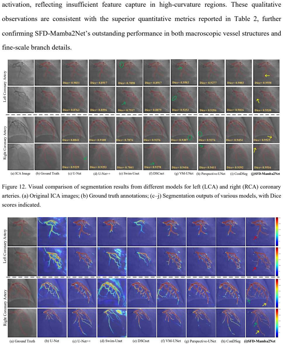
- [Abstract and §4.2, Table 2] The abstract states that SFD-Mamba2Net 'consistently outperformed state-of-the-art methods across eight segmentation metrics.' This is not supported by Table 2. SFD-Mamba2Net ranks first only on Dice, Acc, F1, IoU, and Spec; it is second on Sens (87.48 vs. DSCNet 91.84), third on HD95 (7.57 vs. VM-UNet 5.64), and second on ASSD (1.25 vs. ConDSeg 1.17). Section 5.1 concedes these exceptions, but the abstract and conclusion still make the unqualified claim. This is a load-bearing inconsistency that must be fixed by either rewriting the claim to 'best on 5 of 8 metrics' or providing evidence that the differences on the three remaining metrics are not meaningful.
- [§4.1 and Table 1] The dataset description is internally inconsistent. Section 4.1 says 'The dataset comprises 187 left coronary artery (LCA) images and 127 right coronary artery (RCA) images,' while Table 1 lists totals of 403 LCA and 213 RCA images. The paper never specifies the train/test split procedure, whether it is patient-independent, or how many images/patients are in each split. Section 4.4 mentions '132 test images' without explaining how this number follows from the stated totals. If multiple frames from the same patient appear in both training and test sets, the reported performance ranking could reflect memorization rather than generalization. This concerns the central comparative claim and must be resolved with exact split details and patient-level separation.
- [§4.3, Table 3] The ablation results do not show that the full model is uniformly best. In Table 3, the U+A+P variant achieves HD95 = 7.10 ± 1.41, while the full SFD-Mamba2Net has HD95 = 7.57 ± 0.35; the full model is also not best on ASSD relative to U+M+A (1.25 vs. 1.51, so it is best) but the HD95 regression is unexplained. The text in §4.3 says the full model 'achieves the best performance across Dice, Acc, F1, IoU, and Specificity,' which is accurate, but the qualitative statement that the full model integrates the modules 'to yield an accurate segmentation map' glosses over the HD95 anomaly. The authors should either explain the HD95 behavior or temper the claim that the full model is the optimal combination.
- [§4.4, Algorithm 3 and Table 4] The stenosis detection evaluation depends on several threshold parameters (detection radius r = 10, D_thresh = 1.8 mm, L_thresh = 20, b_thresh = 0.1, plus second-derivative heuristics). No sensitivity analysis is provided, and the same pipeline is used for all models, so the relative ranking is fair. However, the absolute TPR/PPV numbers (0.60/0.64) are claimed as clinically relevant while the detection ratio is only 44.74% and 1,212 false negatives remain in 132 test images. The clinical-applicability conclusion in §5.2 is stronger than the evidence supports; the authors should moderate it or add a failure analysis.
minor comments (5)
- [Title and throughout] The title has a typo: 'Strcture-Guided' should be 'Structure-Guided.' Also, the module is called CASE in the abstract and Section 3.2, but Figure 2 and Section 4.3 call it MASE; please unify the nomenclature.
- [§4.1, Eq. (15)] The IoU formula in Eq. (15) is written as TP / (TN + FP + FN), which is not the standard IoU definition (TP / (TP + FP + FN)). If this is a typo, correct it; if not, the metric is misdefined and the reported IoU values are suspect.
- [Table 2 header] The column header says 'Swim-Unet' but the model and references use 'Swin-Unet.' Please correct the typo.
- [§4.3, Table 3] The ablation variant names are inconsistent with the module names: MASE in the table versus CASE in the text. Also, the standard deviations in Table 3 (e.g., HD95 = 7.10 ± 1.41 for U+A+P) overlap substantially with those of the full model (7.57 ± 0.35), so the discussion of 'further optimization' should acknowledge this overlap.
- [§5.1, Figure 11 reference] The text says 'Visualizations of difference maps (Figure 11)' but Figure 11 shows PHFP Grad-CAM heatmaps; the difference maps are in Figure 17. Please correct the cross-reference.
Circularity Check
No circularity found; empirical architecture comparison against external baselines, with a minor non-load-bearing self-citation in stenosis evaluation.
full rationale
The paper is an empirical architecture study and contains no theoretical derivation chain that could collapse into its inputs. The segmentation model is assembled from existing building blocks (conv-BN-LeakyReLU encoder/decoder, Hessian-based vesselness, Mamba2 SSD, Haar wavelet transforms) and is compared directly with seven external baselines (U-Net, U-Net++, Swin-Unet, DSCNet, VM-UNet, Perspective-UNet, ConDSeg) using the same metrics and protocol (Tables 2-3). The stenosis evaluation in Section 4.4 does reuse the authors' own prior detection algorithm [39], and Chen Zhao and Zhihui Xu are co-authors of both works, so a self-citation is present. However, it is not load-bearing in a circular sense: the algorithm is a fixed, externally published pipeline with hand-set thresholds (r=10 pixels, L_thresh=20, D_thresh=1.8 mm, b_thresh=0.1), and it is applied identically to every model's predicted vessel contours. Relative rankings in Table 4 are therefore not forced by construction, and no parameter is fitted to the proposed model and then renamed as a prediction. The paper's internal contradictions (e.g., the abstract claims superiority on all eight segmentation metrics while Table 2 shows DSCNet, VM-UNet, and ConDSeg lead on Sensitivity, HD95, and ASSD; the dataset counts in the abstract vs. Table 1 differ) are correctness and reporting concerns, not circularity. The Limitations section (5.4) also explicitly acknowledges dataset scale and static-view limitations, which further indicates the authors' claims are empirical rather than derivational. Because the central comparison is self-contained against external benchmarks, the circularity score is 0.
Assumptions & free parameters
free parameters (5)
- Vesselness hyperparameters beta and c in Eq. (7) =
not specified
- Gaussian scale set sigma in CASE module =
1, 2, 3
- Stenosis detection radius r =
10 pixels
- Stenosis detection thresholds =
D_thresh=1.8 mm, L_thresh=20 px, b_thresh=0.1
- PHFP wavelet decomposition levels n =
not specified
assumptions (5)
- domain assumption Hessian eigenvalue analysis correctly distinguishes tubular vessels from background in low-contrast ICA images.
- standard math Haar wavelet transforms are linearly additive, so high-frequency subbands can be summed before inverse transformation.
- domain assumption Mamba2's SSD chunked parallel scan captures long-range spatial dependencies when sequences are flattened along width and height.
- domain assumption Ground-truth annotations by interventional cardiologists are consistent and correct for both segmentation and stenosis severity.
- domain assumption The stenosis detection algorithm from [39] is a valid and unbiased clinical metric when applied to predicted contours.
Cite this review
Pith. "Pith review of SFD-Mamba2Net: Structure-Guided Frequency-Enhanced Dual-Stream Mamba2 Network for Coronary Artery Segmentation." pith.science (2026). https://pith.science/paper/D35LJLHZ
@misc{pith2026250908934,
  author       = {Pith},
  title        = {Pith review of: SFD-Mamba2Net: Structure-Guided Frequency-Enhanced Dual-Stream Mamba2 Network for Coronary Artery Segmentation},
  year         = {2026},
  howpublished = {\url{https://pith.science/paper/D35LJLHZ}},
  note         = {Machine review of arXiv:2509.08934}
}
read the original abstract
Background: Coronary Artery Disease (CAD) is one of the leading causes of death worldwide. Invasive Coronary Angiography (ICA), regarded as the gold standard for CAD diagnosis, necessitates precise vessel segmentation and stenosis detection. However, ICA images are typically characterized by low contrast, high noise levels, and complex, fine-grained vascular structures, which pose significant challenges to the clinical adoption of existing segmentation and detection methods. Objective: This study aims to improve the accuracy of coronary artery segmentation and stenosis detection in ICA images by integrating multi-scale structural priors, state-space-based long-range dependency modeling, and frequency-domain detail enhancement strategies. Methods: We propose SFD-Mamba2Net, an end-to-end framework tailored for ICA-based vascular segmentation and stenosis detection. In the encoder, a Curvature-Aware Structural Enhancement (CASE) module is embedded to leverage multi-scale responses for highlighting slender tubular vascular structures, suppressing background interference, and directing attention toward vascular regions. In the decoder, we introduce a Progressive High-Frequency Perception (PHFP) module that employs multi-level wavelet decomposition to progressively refine high-frequency details while integrating low-frequency global structures. Results and Conclusions: SFD-Mamba2Net consistently outperformed state-of-the-art methods across eight segmentation metrics, and achieved the highest true positive rate and positive predictive value in stenosis detection.
Figures


Figures from the paper (9 more)



Reference graph
Works this paper leans on
-
[1]
Okrainec, D.K
K. Okrainec, D.K. Banerjee, M.J. Eisenberg, Coronary artery disease in the developing world, American heart journal, 148 (2004) 7-15
2004
-
[2]
Z. Li, Y. Zhang, G. Liu, H. Shao, W. Li, X. Tang, A robust coronary artery identification and centerline extraction method in angiographies, Biomedical Signal Processing and Control, 16 (2015) 1-8
2015
-
[3]
Kim, A new computerized measurement approach of carotid artery stenosis on tomographic image sequence, Academic radiology, (2010)
D.Y. Kim, A new computerized measurement approach of carotid artery stenosis on tomographic image sequence, Academic radiology, (2010)
2010
-
[4]
Otsu, A threshold selection method from gray -level histograms, Automatica, 11 (1975) 23 - 27
N. Otsu, A threshold selection method from gray -level histograms, Automatica, 11 (1975) 23 - 27
1975
-
[5]
Soille, L.M
P. Soille, L.M. Vincent, Determining watersheds in digital pictures via flooding simulations, Visual Communications and Image Processing'90: Fifth in a Series, SPIE, 1990, pp. 240-250
1990
-
[6]
Ronneberger, P
O. Ronneberger, P. Fischer, T. Brox, U -net: Convolutional networks for biomedical image segmentation, International Conference on Medical image computing and computer -assisted intervention, Springer, 2015, pp. 234-241
2015
-
[7]
D. Bahdanau, K. Cho, Y. Bengio, Neural machine translation by jointly learning to align and translate, arXiv preprint arXiv:1409.0473, (2014)
arXiv 2014
-
[8]
Vaswani, N
A. Vaswani, N. Shazeer, N. Parmar, J. Uszkoreit, L. Jones, A.N. Gomez, Ł. Kaiser, I. Polosukhin, Attention is all you need, Advances in neural information processing systems, 30 (2017)
2017
Show all 43 references
-
[9]
J. Chen, Y. Lu, Q. Yu, X. Luo, E. Adeli, Y. Wang, L. Lu, A.L. Yuille, Y. Zhou, Transunet: Transformers make strong encoders for medical image segmentation, arXiv preprint arXiv:2102.04306, (2021)
2021 arXiv
-
[10]
A. Gu, K. Goel, C. Ré, Efficiently modeling long sequences with structured state spaces, arXiv preprint arXiv:2111.00396, (2021)
2021 arXiv
-
[11]
T. Dao, A. Gu, Transformers are ssms: Generalized models and efficient algorithms through structured state space duality, arXiv preprint arXiv:2405.21060, (2024)
2024 arXiv
-
[12]
A. Gu, T. Dao, Mamba: Linear -time sequence modeling with selective state spaces, arXiv preprint arXiv:2312.00752, (2023)
2023 arXiv
-
[13]
J. Ruan, J. Li, S. Xiang, Vm -unet: Vision mamba unet for medical image segmentation, arXiv preprint arXiv:2402.02491, (2024)
2024 arXiv
-
[14]
Soares, J.J
J.V. Soares, J.J. Leandro, R.M. Cesar, H.F. Jelinek, M.J. Cree, Retinal vessel segmentation using the 2-D Gabor wavelet and supervised classification, IEEE Transactions on medical Imaging, 25 (2006) 1214-1222
2006
-
[15]
Huang, X.-H
D.-X. Huang, X.-H. Zhou, X.-L. Xie, S.-Q. Liu, S.-Y. Wang, Z.-Q. Feng, M.-J. Gui, H. Li, T.- Y. Xiang, B. -X. Yao, Spironet: Spatial -frequency learning and topological channel interaction network for vessel segmentation, arXiv preprint arXiv:2406.19749, (2024)
2024 arXiv
-
[16]
W. Wang, J. Wang, C. Chen, J. Jiao, L. Sun, Y. Cai, S. Song, J. Li, Fremae: Fourier transform meets masked autoencoders for medical image segmentation, arXiv preprint arXiv:2304.10864, 4 (2023)
2023 arXiv
-
[17]
Frangi, W.J
A.F. Frangi, W.J. Niessen, K.L. Vincken, M.A. Viergever, Multiscale vessel enhancement filtering, International conference on medical image computing and computer -assisted intervention, Springer, 1998, pp. 130-137
1998
-
[18]
LeCun, L
Y. LeCun, L. Bottou, Y. Bengio, P. Haffner, Gradient -based learning applied to document recognition, Proceedings of the IEEE, 86 (2002) 2278-2324
2002
-
[19]
H. Wang, S. Xie, L. Lin, Y. Iwamoto, X.-H. Han, Y.-W. Chen, R. Tong, Mixed transformer u- net for medical image segmentation, ICASSP 2022 -2022 IEEE international conference on acoustics, speech and signal processing (ICASSP), IEEE, 2022, pp. 2390-2394
2022
-
[20]
J. Hu, S. Chen, Z. Pan, S. Zeng, W. Yang, Perspective+ unet: Enhancing segmentation with bi- path fusion and efficient non-local attention for superior receptive fields, International Conference on Medical Image Computing and Computer-Assisted Intervention, Springer, 2024, pp. 499-509
2024
-
[21]
M. Lei, H. Wu, X. Lv, X. Wang, Condseg: A general medical image segmentation framework via contrast -driven feature enhancement, Proceedings of the AAAI Conference on Artificial Intelligence, 2025, pp. 4571-4579
2025
-
[22]
Wang, J.-Q
Z. Wang, J.-Q. Zheng, Y. Zhang, G. Cui, L. Li, Mamba-unet: Unet-like pure visual mamba for medical image segmentation, arXiv preprint arXiv:2402.05079, (2024)
2024 arXiv
-
[23]
L. Kong, J. Dong, J. Ge, M. Li, J. Pan, Efficient frequency domain-based transformers for high- quality image deblurring, Proceedings of the IEEE/CVF Conference on Computer Vision and Pattern Recognition, 2023, pp. 5886-5895
2023
-
[24]
N. Mu, Z. Lyu, X. Zhang, R. McBane, A.S. Pandey, J. Jiang, Exploring a frequency -domain attention-guided cascade U-Net: Towards spatially tunable segmentation of vasculature, Computers in biology and medicine, 167 (2023) 107648
2023
-
[25]
R. Azad, A. Bozorgpour, M. Asadi -Aghbolaghi, D. Merhof, S. Escalera, Deep frequency re - calibration u -net for medical image segmentation, Proceedings of the IEEE/CVF international conference on computer vision, 2021, pp. 3274-3283
2021
-
[26]
L. Chen, L. Gu, L. Li, C. Yan, Y. Fu, Frequency Dynamic Convolution for Dense Image Prediction, Proceedings of the Computer Vision and Pattern Recognition Conference, 2025, pp. 30178-30188
2025
-
[27]
J. -H. Nam, N.S. Syazwany, S.J. Kim, S. -C. Lee, Modality -agnostic domain generalizable medical image segmentation by multi -frequency in multi -scale attention, Proceedings of the IEEE/CVF conference on computer vision and pattern recognition, 2024, pp. 11480-11491
2024
-
[28]
Ioffe, C
S. Ioffe, C. Szegedy, Batch normalization: Accelerating deep network training by reducing internal covariate shift, International conference on machine learning, pmlr, 2015, pp. 448 -456
2015
-
[29]
Maas, A.Y
A.L. Maas, A.Y. Hannun, A.Y. Ng, Rectifier nonlinearities improve neural network acoustic models, Proc. icml, Atlanta, GA, 2013, pp. 3
2013
-
[30]
Srivastava, G
N. Srivastava, G. Hinton, A. Krizhevsky, I. Sutskever, R. Salakhutdinov, Dropout: a simple way to prevent neural networks from overfitting, The journal of machine learning research, 15 (2014) 1929-1958
2014
-
[31]
Selvaraju, M
R.R. Selvaraju, M. Cogswell, A. Das, R. Vedantam, D. Parikh, D. Batra, Grad -cam: Visual explanations from deep networks via gradient -based localization, Proceedings of the IEEE international conference on computer vision, 2017, pp. 618-626
2017
-
[32]
Ramachandran, B
P. Ramachandran, B. Zoph, Q.V. Le, Searching for activation functions, arXiv preprint arXiv:1710.05941, (2017)
2017 arXiv
-
[33]
Zhang, R
B. Zhang, R. Sennrich, Root mean square layer normalization, Advances in neural information processing systems, 32 (2019)
2019
-
[34]
Mallat, A theory for multiresolution signal decomposition: the wavelet representation, IEEE transactions on pattern analysis and machine intelligence, 11 (2002) 674-693
S.G. Mallat, A theory for multiresolution signal decomposition: the wavelet representation, IEEE transactions on pattern analysis and machine intelligence, 11 (2002) 674-693
2002
-
[35]
Kingma, J
D.P. Kingma, J. Ba, Adam: A method for stochastic optimization, arXiv preprint arXiv:1412.6980, (2014)
2014 arXiv
-
[36]
Zhou, M.M
Z. Zhou, M.M. Rahman Siddiquee, N. Tajbakhsh, J. Liang, Unet++: A nested u-net architecture for medical image segmentation, International workshop on deep learning in medical image analysis, Springer, 2018, pp. 3-11
2018
-
[37]
H. Cao, Y. Wang, J. Chen, D. Jiang, X. Zhang, Q. Tian, M. Wang, Swin -unet: Unet-like pure transformer for medical image segmentation, European conference on computer vision, Springer, 2022, pp. 205-218
2022
-
[38]
Y. Qi, Y. He, X. Qi, Y. Zhang, G. Yang, Dynamic snake convolution based on topological geometric constraints for tubular structure segmentation, Proceedings of the IEEE/CVF international conference on computer vision, 2023, pp. 6070-6079
2023
-
[39]
C. Zhao, A. Vij, S. Malhotra, J. Tang, H. Tang, D. Pienta, Z. Xu, W. Zhou, Automatic extraction and stenosis evaluation of coronary arteries in invasive coronary angiograms, Computers in biology and medicine, 136 (2021) 104667
2021
-
[40]
Arbab -Zadeh, J
A. Arbab -Zadeh, J. Hoe, Quantification of coronary arterial stenoses by multidetector CT angiography in comparison with conventional angiography: methods, caveats, and implications, JACC: Cardiovascular Imaging, 4 (2011) 191-202
2011
-
[41]
Zir, S.W
L.M. Zir, S.W. Miller, R.E. Dinsmore, J. Gilbert, J. Harthorne, Interobserver variability in coronary angiography, Circulation, 53 (1976) 627-632
1976
-
[42]
Galbraith, M.L
J.E. Galbraith, M.L. Murphy, N. de Soyza, Coronary angiogram interpretation: interobserver variability, Jama, 240 (1978) 2053-2056
1978
-
[43]
Galassi, M
F. Galassi, M. Alkhalil, R. Lee, P. Martindale, R.K. Kharbanda, K.M. Channon, V. Grau, R.P. Choudhury, 3D reconstruction of coronary arteries from 2D angiographic projections using non - uniform rational basis splines (NURBS) for accurate modelling of c oronary stenoses, PloS ...
2018
Reviewed August 4, 2026 · model on record in the stance chip above.
Discussion (0). Sign in to comment.