REVIEW 5 major objections 5 minor 30 references
Mixtures of Neural Cellular Automata: A Stochastic Framework for Growth Modelling and Self-Organization
T0 review · 5 major / 5 minor · reviewed 2026-08-06 · deepseek-v4-flash
Pith's one-line read Mixing several learned local update rules, selected cell by cell through a probability network, lets neural cellular automata represent stochastic tissue growth, recover from image perturbations, and segment cells without labels.
desk verdict Genuine but incremental extension of NCA with a learned rule selector and noise; the synthetic tissue results look good, but the biological claims outrun the evidence. read the letter →
The pith
A machine-rendered reading of the paper's core claim, the machinery that carries it, and where it could break.
The reading
What carries the argument
The carrying mechanism is the mixture update rule. At each cell $i$ a rule-selector network $\pi$ maps the current state $s_i^t$ to a categorical distribution over $K$ local transition networks $\phi_k$. The active rule index $z$ is sampled from that distribution through the Gumbel-Softmax relaxation, and the next state is $s_i^{t+1}=s_i^t+\prod_{k=1}^K \phi_k(s_i^t,\{s_j^t\}_{j\in\mathcal{N}(i)},\mathbf{x}_k)^{z_k}$, where $\mathbf{x}_k\sim\mathcal{N}(0,1)$ is injected Gaussian noise in the noisy variant. The categorical selector imposes a soft clustering of cells into rule regimes, which is what produces segmentation and interpretability, while the sampling and noise turn the update into a stochastic transition kernel that can represent rare and cell-specific events.
What would settle it
Train an MNCA and a single-rule NCA of matched parameter count on time-lapse images of real growing tissue with stochastic lineage decisions, for instance organoid cultures tracked cell by cell; if the MNCA's KL divergence on cell-type composition and Wasserstein distances on tissue shape are not clearly better than the NCA's, the paper's central advantage claim falls. Alternatively, ablate the mixture by forcing all K rules to share identical weights: if perturbation recovery remains as good as a full mixture, then the mixture itself is not the mechanism.
Extended reading notes
Core claim
Read on its own terms, the paper's discovery is that replacing the single update rule of a neural cellular automaton with a stochastic mixture of K rules changes both what the model can represent and what can be read out of it. In the tissue-growth setup the MNCA lowers the KL divergence of cell-type proportions from 2.057 (NCA) to 0.018 (MNCA) and improves Wasserstein distances for tissue size and border structure; in morphogenesis the mixture recovers the target image after patch deletion, pixel removal, and Gaussian noise more accurately than the deterministic NCA and the Gaussian NCA baseline; and in microscopy images the learned rule probabilities correlate with a cell-shape parameter and can be re-weighted during inference to steer the population toward different morphologies. The authors interpret these outcomes as showing that MNCAs reproduce stochastic growth dynamics, provide interpretable rule segmentation, and are more stable under perturbations.
Load-bearing premise
The biological half of the central claim is tested only against a hand-written computer simulation of tissue growth, and the paper assumes that simulation is a faithful stand-in for real tissues; if that assumption fails, the claim that MNCAs recapitulate real biological growth patterns is unsupported.
Editorial extensions
If this is right
- A trained MNCA can act as a black-box surrogate for a stochastic agent-based tissue model, reproducing cell-type proportions and spatial borders without hand-specified kinetic rates or ABC inference.
- The perturbation results imply that mixture updates confer stability to learned morphogenesis without adversarial training, because no single rule determines the whole recovery trajectory.
- Rule-probability maps are an emergent unsupervised segmentation of the input, and constraining rule probabilities at inference time steers the final phenotype.
- The number of mixture rules is a capacity knob: fidelity improves with more rules but saturates, so practitioners can trade cost against divergence.
Reading between the lines
- If the Lipschitz-averaging explanation is right, adding a categorical mixture of K copies of an already trained deterministic NCA should improve perturbation recovery even when the rules are not learned from biological data; this is a testable, biology-free prediction.
- The paper's noise-tail analysis suggests the injected Gaussian channel acts as an interpretable latent variable for rare events; one could try to read out event types from the learned noise partition and use it as a controllable randomness knob in other self-organizing models.
- Because the biological evaluation is synthetic, a natural next test is fitting MNCA to real time-lapse organoid or tumor imaging with lineage data; only that would settle whether the claimed recapitulation of real growth patterns holds outside the simulator.
Editorial analysis
A structured set of objections, weighed in public.
Referee Report
Summary. The paper proposes Mixture of Neural Cellular Automata (MNCA), an extension of NCA in which each cell's update is produced by one of K learned transition rules, selected probabilistically through a Gumbel-Softmax categorical sampler, optionally augmented with per-rule Gaussian noise. The framework is evaluated on three tasks: a synthetic tissue-growth simulator, emoji image morphogenesis under perturbations, and microscopic cell-image segmentation from synthetic high-content screening data. The authors claim that MNCAs are more robust to perturbations, better recapitulate biological growth patterns, and yield interpretable rule assignments, and they provide an appendix with an ABC comparison and a Lipschitz-based robustness argument. The core idea is clean and the experimental appendix is extensive, but several of the headline claims are broader than the reported evidence.
Significance. If the central claims hold after capacity controls and more careful evaluation, MNCAs are a useful extension of NCAs for stochastic self-organizing systems, with potential value in computational biology and generative modeling. The paper deserves credit for including an ABC baseline, a rule-number ablation, and CIFAR-10 generalization experiments in the appendices. The main tissue experiment shows a large improvement in cell-type distribution KL divergence relative to the single-rule NCA baseline. However, the robustness claim is contradicted in part of Table 3, the biological-dynamics claim is not supported by the marginal metrics used, and the mixture mechanism is not isolated from a parameter-count confound. These issues need to be resolved before the headline conclusions are acceptable.
major comments (5)
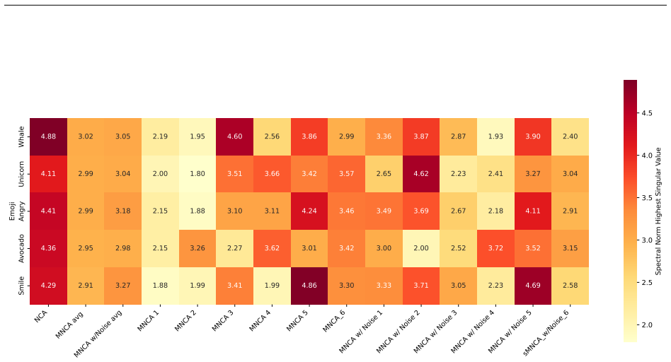
- [Section 4.3, Table 3] The blanket claim in Section 4.3 that 'Mixture-based NCA vastly outperforms the single-rule baseline across all perturbation scenarios' is contradicted by the first emoji block of Table 3: for Gaussian noise at 10% and 25%, the basic MNCA reports MSE 0.071±0.002 and 0.079±0.004, which are worse than the NCA's 0.049±0.007 and 0.052±0.010. Only MNCA with internal noise outperforms NCA in those conditions. Please report per-condition statistics and qualify the robustness claim accordingly.
- [Section 4.2, Appendix A and C, Section 5] The claim that MNCAs 'reproduce the stochastic dynamics observed in biological processes' is not supported by the reported metrics. The evaluation in Section 4.2 compares only marginal summaries—cell-type proportion KL, tissue-size Wasserstein, and border-size Wasserstein—of a hand-specified agent-based simulator (Algorithm 1). These marginals do not constrain transition probabilities, temporal correlations, or per-step stochastic behavior, even though the models are trained to reconstruct the state at each time step. Moreover, Section 5's reference to 'real spatial transcriptomic data' is unsupported: the microscopy experiment uses synthetic BBBC031 images, not real spatial transcriptomic data. Please add trajectory-level or transition-level metrics and real data, or temper the claims to synthetic surrogates.
- [Section 4.1, Table 2, Table 3] The MNCA baseline has K=5 update networks plus a rule selector, whereas the NCA baseline has a single update network, so part of the improvement in Tables 2 and 3 may reflect parameter count rather than the mixture mechanism. A matched-capacity control is needed—for example, a single NCA with proportionally wider layers, or an MNCA with K=1—before attributing the gains to probabilistic rule selection.
- [Appendix F] The theoretical robustness argument is not applied to the model actually evaluated. Equation (15) defines a convex-combination update F(s)=Σ π_k(s) f_k(s), but Eqs. (4b) and (5c) implement a hard categorical selection via the Gumbel-Softmax. The Lipschitz bound on the averaged update does not directly bound the stochastic selection process. Either revise the argument to address the sampled update or explicitly present it as a heuristic for why averaging may help.
- [Section 4.4, Figure 6C] The interpretable-segmentation claim rests on a visual 'clear correlation' between inferred rule assignments and the Cell Shape Parameter, but no quantitative correlation, statistical test, or agreement metric is reported. Since rule interpretability is one of the headline contributions, please quantify this relationship (e.g., correlation coefficient, adjusted Rand index against known phenotypes).
minor comments (5)
- [Section 3.1, Eq. (4)] The symbols z∼Cat(π(s_i,η)) and z_k are inconsistent: if z is a categorical value, z_k is undefined; if z is a one-hot vector, it should be defined as such. Please clarify the notation.
- [Section 4.1] The text lists 'Visium spatial transcriptomics' as one of the three experiments, but Section 4.4 uses BBBC031 microscopy images; please align the naming.
- [Table 2] The baseline is labeled 'GNCA' in Table 2 but 'GCA' in the text; please unify the notation.
- [Table 1] The table caption states 'k stands for 103'; this should be '10^3' or 'k=10^3'.
- [Appendix E, Table 4] The statement that ABC results are 'comparable' to the MNCA approach is not fully supported by Table 4: the ABC proportion-based model has Border-W 0.054 versus 0.184 for MNCA, so the comparison is mixed.
Circularity Check
No significant circularity: the MNCA derivation is self-contained; the only mild circular element is the K=5 rule count chosen to match the five known cell types in the synthetic tissue study, which partially pre-specifies the 'emergent' rule segmentation claim.
-
fitted input called prediction
[Section 4.2, rule-assignment paragraph near Figure 3]
"When then looking at the rules assignment for the MNCAs, by plotting the rule assignment probability for a given state in time, we find that the model correctly gets that approximately each cell type has a different rule. This has also been our main rationale for setting the number of rules to 5."
The number of mixture rules K is set to 5 because the synthetic tissue was generated with exactly five cell types (STEM, INT1, INT2, DIFF1, DIFF2). The paper then reports as an emergent result that approximately each cell type has a different rule. With five mixture components and five label classes, the coarse one-rule-per-cell-type alignment is a capacity choice rather than an independent discovery; the model cannot discover a different number of rule segments unless components are left unused or shared. The specific grouping (stem and DIFF2 sharing a rule, empty space owning a rule) is learned and not forced, so the circularity is partial and mild.
full rationale
The central MNCA formulation (Eq. 4-5) is a straightforward extension of NCAs with a categorical rule selector and Gaussian noise; no fitted parameter is renamed as a prediction. The robustness experiments in Section 4.3 and Appendix G train on clean targets and then test on unseen perturbations, so the improved recovery is an emergent external benchmark, not a self-fulfilling fit. The microscopy experiment in Section 4.4 correlates unsupervised rule assignments with the independent BBBC031 Cell Shape Parameter, providing external evidence for the segmentation claim. The only mild circular element is the choice K=5 in the synthetic tissue study, which matches the known five cell types and therefore partially pre-specifies the granularity of the claimed interpretable rule segmentation; however, the actual learned groupings deviate from the labels, so the observation retains unsupervised content. Appendix E explicitly acknowledges that the ABC baseline is evaluated on a quantity tied to its fitting statistic, an honest limitation of the comparison rather than a circularity in the MNCA derivation. Finally, the Conclusion's mention of 'real spatial transcriptomic data' is not supported by any experiment in the manuscript (only synthetic BBBC031 microscopy images are used); this is an evidence and reporting gap, not a circular reasoning step. Overall, the paper's core contributions are independently testable and not circular, with only minor pre-specification issues, so a low score is appropriate.
Assumptions & free parameters
free parameters (3)
- Number of mixture rules K =
5 for tissue and microscopy, 6 for emoji
- Gumbel-Softmax temperature =
not reported
- Gaussian noise injection distribution =
unit normal N(0,1)
assumptions (4)
- standard math Gumbel-Softmax provides a differentiable approximation to sampling from the categorical rule distribution
- domain assumption The mixture update's Lipschitz constant is bounded by the weighted sum of component Lipschitz constants under mild regularity on the mixture weights
- domain assumption The induced Markov chain over states is ergodic, or stochasticity helps escape attractors
- domain assumption The synthetic agent-based tissue model in Appendix A is a valid proxy for real biological growth
Cite this review
Pith. "Pith review of Mixtures of Neural Cellular Automata: A Stochastic Framework for Growth Modelling and Self-Organization." pith.science (2026). https://pith.science/paper/DNKXNCZM
@misc{pith2026250620486,
  author       = {Pith},
  title        = {Pith review of: Mixtures of Neural Cellular Automata: A Stochastic Framework for Growth Modelling and Self-Organization},
  year         = {2026},
  howpublished = {\url{https://pith.science/paper/DNKXNCZM}},
  note         = {Machine review of arXiv:2506.20486}
}
read the original abstract
Neural Cellular Automata (NCAs) are a promising new approach to model self-organizing processes, with potential applications in life science. However, their deterministic nature limits their ability to capture the stochasticity of real-world biological and physical systems. We propose the Mixture of Neural Cellular Automata (MNCA), a novel framework incorporating the idea of mixture models into the NCA paradigm. By combining probabilistic rule assignments with intrinsic noise, MNCAs can model diverse local behaviors and reproduce the stochastic dynamics observed in biological processes. We evaluate the effectiveness of MNCAs in three key domains: (1) synthetic simulations of tissue growth and differentiation, (2) image morphogenesis robustness, and (3) microscopy image segmentation. Results show that MNCAs achieve superior robustness to perturbations, better recapitulate real biological growth patterns, and provide interpretable rule segmentation. These findings position MNCAs as a promising tool for modeling stochastic dynamical systems and studying self-growth processes.
Figures


Figures from the paper (13 more)





Reference graph
Works this paper leans on
-
[1]
Cellular automata models for vegetation dynamics
Heiko Balzter, Paul W Braun, and Wolfgang K \"o hler. Cellular automata models for vegetation dynamics. Ecological modelling, 107 0 (2-3): 0 113--125, 1998
work page 1998
-
[2]
Cell fate specification and differentiation in the adult mammalian intestine
Joep Beumer and Hans Clevers. Cell fate specification and differentiation in the adult mammalian intestine. Nature reviews Molecular cell biology, 22 0 (1): 0 39--53, 2021
work page 2021
-
[3]
Calibrating agent-based models to tumor images using representation learning
Colin G Cess and Stacey D Finley. Calibrating agent-based models to tumor images using representation learning. PLoS Computational Biology, 19 0 (4): 0 e1011070, 2023
work page 2023
-
[4]
Cellular automata approaches to biological modeling
G Bard Ermentrout and Leah Edelstein-Keshet. Cellular automata approaches to biological modeling. Journal of theoretical Biology, 160 0 (1): 0 97--133, 1993
work page 1993
-
[5]
Cellular automata as convolutional neural networks
William Gilpin. Cellular automata as convolutional neural networks. Physical Review E, 100 0 (3): 0 032402, 2019
work page 2019
-
[6]
Learning graph cellular automata
Daniele Grattarola, Lorenzo Livi, and Cesare Alippi. Learning graph cellular automata. Advances in Neural Information Processing Systems, 34: 0 20983--20994, 2021
work page 2021
-
[7]
Neural cellular automata manifold
Alejandro Hernandez, Armand Vilalta, and Francesc Moreno-Noguer. Neural cellular automata manifold. In Proceedings of the IEEE/CVF Conference on Computer Vision and Pattern Recognition, pp.\ 10020--10028, 2021
work page 2021
-
[8]
Categorical reparameterization with gumbel-softmax
Eric Jang, Shixiang Gu, and Ben Poole. Categorical reparameterization with gumbel-softmax. arXiv preprint arXiv:1611.01144, 2016
arXiv 2016
Show all 30 references
-
[9]
Parameter-efficient diffusion with neural cellular automata
John Kalkhof, Arlene K \"u hn, Yannik Frisch, and Anirban Mukhopadhyay. Parameter-efficient diffusion with neural cellular automata. npj Unconventional Computing, 2 0 (1): 0 1--9, 2025
2025
-
[10]
Adam: A method for stochastic optimization
Diederik P Kingma. Adam: A method for stochastic optimization. arXiv preprint arXiv:1412.6980, 2014
2014 arXiv
-
[11]
Computational framework for simulating fluorescence microscope images with cell populations
Antti Lehmussola, Pekka Ruusuvuori, Jyrki Selinummi, Heikki Huttunen, and Olli Yli-Harja. Computational framework for simulating fluorescence microscope images with cell populations. IEEE transactions on medical imaging, 26 0 (7): 0 1010--1016, 2007
2007
-
[12]
State-dependent evolutionary models reveal modes of solid tumour growth
Maya A Lewinsohn, Trevor Bedford, Nicola F M \"u ller, and Alison F Feder. State-dependent evolutionary models reveal modes of solid tumour growth. Nature Ecology & Evolution, 7 0 (4): 0 581--596, 2023
2023
-
[13]
Annotated high-throughput microscopy image sets for validation
Vebjorn Ljosa, Katherine L Sokolnicki, and Anne E Carpenter. Annotated high-throughput microscopy image sets for validation. Nature methods, 9 0 (7): 0 637, 2012
2012
-
[14]
Latent neural cellular automata for resource-efficient image restoration
Andrea Menta, Alberto Archetti, and Matteo Matteucci. Latent neural cellular automata for resource-efficient image restoration. arXiv preprint arXiv:2403.15525, 2024
2024 arXiv
-
[15]
Spectral normalization for generative adversarial networks
Takeru Miyato, Toshiki Kataoka, Masanori Koyama, and Yuichi Yoshida. Spectral normalization for generative adversarial networks. arXiv preprint arXiv:1802.05957, 2018
2018 arXiv
-
[16]
Growing neural cellular automata
Alexander Mordvintsev, Ettore Randazzo, Eyvind Niklasson, and Michael Levin. Growing neural cellular automata. Distill, 5 0 (2): 0 e23, 2020
2020
-
[17]
Spatial structure governs the mode of tumour evolution
Robert Noble, Dominik Burri, C \'e cile Le Sueur, Jeanne Lemant, Yannick Viossat, Jakob Nikolas Kather, and Niko Beerenwinkel. Spatial structure governs the mode of tumour evolution. Nature ecology & evolution, 6 0 (2): 0 207--217, 2022
2022
-
[18]
Variational neural cellular automata
Rasmus Berg Palm, Miguel Gonz \'a lez-Duque, Shyam Sudhakaran, and Sebastian Risi. Variational neural cellular automata. arXiv preprint arXiv:2201.12360, 2022
2022 arXiv
-
[19]
Advanced cell classifier: user-friendly machine-learning-based software for discovering phenotypes in high-content imaging data
Filippo Piccinini, Tamas Balassa, Abel Szkalisity, Csaba Molnar, Lassi Paavolainen, Kaisa Kujala, Krisztina Buzas, Marie Sarazova, Vilja Pietiainen, Ulrike Kutay, et al. Advanced cell classifier: user-friendly machine-learning-based software for discovering phenotypes in high-...
2017
-
[20]
Self-classifying mnist digits
Ettore Randazzo, Alexander Mordvintsev, Eyvind Niklasson, Michael Levin, and Sam Greydanus. Self-classifying mnist digits. Distill, 5 0 (8): 0 e00027--002, 2020
2020
-
[21]
Learning spatio-temporal patterns with neural cellular automata
Alex D Richardson, Tibor Antal, Richard A Blythe, and Linus J Schumacher. Learning spatio-temporal patterns with neural cellular automata. PLOS Computational Biology, 20 0 (4): 0 e1011589, 2024
2024
-
[22]
Morphogen gradients: from generation to interpretation
Katherine W Rogers and Alexander F Schier. Morphogen gradients: from generation to interpretation. Annual review of cell and developmental biology, 27 0 (1): 0 377--407, 2011
2011
-
[23]
Spatial biology of cancer evolution
Zaira Seferbekova, Artem Lomakin, Lucy R Yates, and Moritz Gerstung. Spatial biology of cancer evolution. Nature Reviews Genetics, 24 0 (5): 0 295--313, 2023
2023
-
[24]
Cancer stem cell tumor model reveals invasive morphology and increased phenotypical heterogeneity
Andrea Sottoriva, Joost JC Verhoeff, Tijana Borovski, Shannon K McWeeney, Lev Naumov, Jan Paul Medema, Peter MA Sloot, and Louis Vermeulen. Cancer stem cell tumor model reveals invasive morphology and increased phenotypical heterogeneity. Cancer research, 70 0 (1): 0 46--56, 2010
2010
-
[25]
Quantification of spatial subclonal interactions enhancing the invasive phenotype of pediatric glioma
Haider Tari, Ketty Kessler, Nick Trahearn, Benjamin Werner, Maria Vinci, Chris Jones, and Andrea Sottoriva. Quantification of spatial subclonal interactions enhancing the invasive phenotype of pediatric glioma. Cell Reports, 40 0 (9), 2022
2022
-
[26]
Attention-based neural cellular automata
Mattie Tesfaldet, Derek Nowrouzezahrai, and Chris Pal. Attention-based neural cellular automata. Advances in Neural Information Processing Systems, 35: 0 8174--8186, 2022
2022
-
[27]
Theory of self-reproducing automata
John Von Neumann, Arthur Walter Burks, et al. Theory of self-reproducing automata. 1966
1966
-
[28]
Learning to generate 3d shapes with generative cellular automata
Dongsu Zhang, Changwoon Choi, Jeonghwan Kim, and Young Min Kim. Learning to generate 3d shapes with generative cellular automata. arXiv preprint arXiv:2103.04130, 2021
2021 arXiv
-
[29]
Outdoor scene extrapolation with hierarchical generative cellular automata
Dongsu Zhang, Francis Williams, Zan Gojcic, Karsten Kreis, Sanja Fidler, Young Min Kim, and Amlan Kar. Outdoor scene extrapolation with hierarchical generative cellular automata. In Proceedings of the IEEE/CVF Conference on Computer Vision and Pattern Recognition, pp.\ 20145--...
2024
-
[30]
write newline
" write newline "" before.all 'output.state := FUNCTION n.dashify 't := "" t empty not t #1 #1 substring "-" = t #1 #2 substring "--" = not "--" * t #2 global.max substring 't := t #1 #1 substring "-" = "-" * t #2 global.max substring 't := while if t #1 #1 substring * t #2 gl...
Reviewed August 6, 2026 · model on record in the stance chip above.
Discussion (0). Continue with ORCID to comment.