REVIEW 5 major objections 5 minor 1 cited by
Viscoelastic Characterization of Melanoma Cells Using Brillouin Spectroscopy
T0 review · 5 major / 5 minor · reviewed 2026-08-06 · deepseek-v4-flash
Pith's one-line read Confocal Brillouin spectroscopy distinguishes cultured melanoma cells from their surroundings by spectral markers of stiffness and viscosity, and prolonged laser exposure visibly destroys the cells.
desk verdict Careful instrumentation and an honest photodamage note, but the paper's central viscoelastic claim is not supported by the reported data. read the letter →
The pith
A machine-rendered reading of the paper's core claim, the machinery that carries it, and where it could break.
The reading
What carries the argument
The central mechanism is the Brillouin spectrum itself: the Brillouin shift, the frequency displacement of the Stokes and anti-Stokes lines converted from CCD pixels via a VIPA dispersion curve, and the FWHM of those peaks. The shift carries stiffness-like information and the FWHM carries viscosity-like information, and the custom single-stage VIPA spectrometer uses an iodine vapor cell plus polarization filtering to separate the faint Brillouin signal from the elastic background. A confocal pinhole below one Airy unit provides optical sectioning so the spectral values can be mapped cell-by-cell.
What would settle it
A decisive test would be to image the same cells with an independent mechanical probe, such as atomic force microscopy or a rheometer, while also swapping the culture medium for index-matched fluids; if the spectral differences vanish or fail to track the mechanical readings, the central claim would not hold.
Extended reading notes
Core claim
The central claim is that a custom confocal Brillouin microspectrometer, with an iodine vapor cell and VIPA disperser to reject elastic scattering, yields reproducible spectral differences between cultured melanoma cells and the background: higher Brillouin shift in cell regions and broader Brillouin FWHM, with consistent Stokes and anti-Stokes trends. The shift is read as a proxy for longitudinal stiffness and the FWHM as a proxy for viscosity and structural heterogeneity, so the paper asserts that the cells are mechanically distinct from their environment. A secondary finding is that high-power 532 nm exposure induces progressive morphological degeneration ending in apparent membrane rupture, while lower dwell times preserve viability; this connects laser dosing to mechanical and cytotoxic effects.
Load-bearing premise
The load-bearing premise is that the measured differences in Brillouin shift and linewidth between cells and background reflect mechanical differences in stiffness and viscosity, not optical effects such as refractive-index mismatch, water content, medium composition, or spectral drift; no independent mechanical measurement is supplied.
Editorial extensions
If this is right
- If the spectral readouts really track cell mechanics, Brillouin microscopy becomes a contact-free, label-free way to map stiffness and viscosity of melanoma cells and compare them with surrounding tissue.
- The consistent Stokes and anti-Stokes trends across 11 runs mean the cell-versus-background contrast is reproducible enough to serve as a candidate imaging biomarker.
- The photodamage timeline, from rounding and loss of protrusions to membrane rupture, gives a concrete threshold for safe laser exposure in live-cell imaging and could guide dwell-time and power choices in other cell types.
- Because a confocal pinhole sections the sample, the method can map mechanical properties within a single cell rather than only averaging over a whole population.
Reading between the lines
- The paper does not rule out that part of the cell-versus-background contrast comes from refractive-index and water-content differences rather than stiffness; a control experiment with index-matched media would separate these effects.
- The linewidth changes could partly reflect local heating or spectrometer resolution rather than viscosity, so correlating FWHM with independent temperature measurements would test the viscosity interpretation.
- The photodamage results suggest that Brillouin imaging parameters may need to be tuned per cell line, and that spectral changes during exposure could themselves serve as an early-warning readout of cell stress.
Signed reviews
Editorial analysis
A structured set of objections, weighed in public.
Referee Report
Summary. The manuscript reports a Brillouin microspectroscopy study of cultured melanoma cells, claiming that cells exhibit higher Brillouin shifts and broader FWHM values than the surrounding medium and interpreting these as markers of increased stiffness and viscosity. It also describes a photodamage effect at high laser exposure. The paper presents the experimental setup, acquisition protocol, and a data-analysis pipeline, with representative heatmaps and histograms, but it provides no quantitative shift or FWHM values, no error bars, and no statistical test statistics. The central claim that the data constitute a 'detailed viscoelastic profile' of melanoma cells therefore rests on unreported numbers and on an unexamined assumption that optical contrasts (notably refractive index mismatch) do not dominate the measured spectral differences.
Significance. If properly substantiated, the ability to distinguish melanoma cells from their surroundings without extrinsic labels using Brillouin-derived spectral markers would be of genuine interest for cancer biology and diagnostic development. The manuscript has concrete strengths: a detailed description of a custom-built confocal Brillouin microspectrometer, explicit pinhole calibration, the use of both Stokes and anti-Stokes components to check consistency, and a clear observation of a phototoxic endpoint. However, the current version is qualitative: the claimed reproducible cell/background contrast is not quantified, and the mechanical interpretation is not separated from refractive-index effects. As a result, the significance of the work cannot yet be assessed beyond that of a technical feasibility demonstration.
major comments (5)
- [Section 4.3 and Figures 3-5] The central claim of 'significant distinctions' and a 'reproducible pattern across all 11 imaging runs' is not supported by any reported quantitative values, uncertainties, or test statistics. The manuscript never states the mean Brillouin shift or FWHM for cells versus background, the number of spectra per cell or per background region, the run-to-run variation, or the statistical test used. Please add a table or supplementary material with per-run and pooled means, standard deviations, and the outcome of an appropriate test (e.g., a mixed-effects model accounting for repeated cells/runs).
- [Section 4.1 and Section 2.1.4] The interpretation of higher Brillouin shift as a mechanical stiffness difference is confounded by refractive index. In the backscattering geometry, the shift is proportional to n·v_s, and cells (n ≈ 1.38-1.40) immersed in culture medium (n ≈ 1.33-1.34) would exhibit a shift increase of roughly 4% even with no change in mechanical modulus. The manuscript provides no per-pixel refractive index measurement, no index correction, and no independent mechanical validation. Please either correct the shifts using measured or literature-based index maps, or explicitly state the assumption that the index contrast is negligible and quantify the resulting systematic uncertainty in the inferred stiffness contrast.
- [Section 2 versus Section 4.4] There is an inconsistency in the reported exposure conditions: Section 2 states that the exposure time for each measurement was maintained between 30 and 40 ms, whereas Section 4.4 describes the high-density damaging run with 'exposure (laser dwell time) equal to 40 µs'. These values differ by three orders of magnitude and affect both the viability claim and the photodamage interpretation. Please reconcile the two values and report the total illumination dose per cell (dwell time per spectrum, number of spectra per cell, and laser power) so that the damage threshold can be assessed.
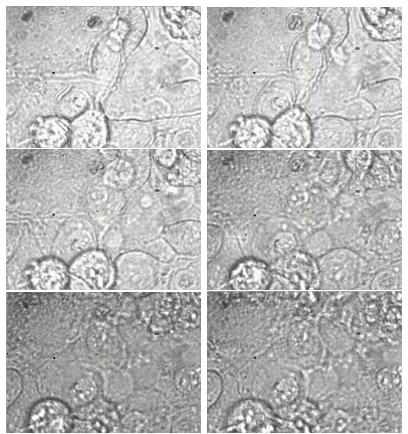
- [Section 4.4] The photodamage section is framed as a mechanical effect of laser-induced damage, but no Brillouin shift or FWHM data are reported during the cell degradation; only bright-field morphology is shown. The title and abstract promise a viscoelastic characterization, but the damage narrative is not connected to any spectral markers. Please either add time-lapse Brillouin metrics accompanying the morphological changes, or clearly label this section as an incidental observation that is not part of the central claim.
- [Section 3.1, Step 1] The Savitzky-Golay filter (window 7, polynomial order 3) is applied before extracting FWHM values, yet the effect of the smoothing parameters on the reported FWHM is not quantified. Since FWHM is the basis for the viscosity claim, filter-induced broadening could produce cell/background differences that are not mechanical. Please add a calibration test (e.g., on a stable reference spectrum or simulated Lorentzian) showing that the smoothing does not differentially affect the extracted FWHM in cells versus background.
minor comments (5)
- [Abstract] The phrase 'detailed viscoelastic profile' overstates the content: the paper does not report quantitative mechanical moduli, loss tangents, or spatially resolved profiles beyond qualitative heatmaps.
- [Section 4.4] The sentence beginning 'After the exposure the cell has been observed...' is followed by 'shown As shown in Figure 6' containing a doubled phrase; please edit for clarity.
- [Section 4.3] The line '11 imagings (30 cells) in total' is a sentence fragment and does not explain how the 30 cells were distributed across the 11 runs or how cellular regions were segmented from background for the histograms.
- [Section 1] The phrase 'the authors observed' in the introduction is redundant and should be replaced with a direct statement of the observation.
- [Figure captions] Figure 3 and Figure 4 captions would benefit from explicit definitions of 'cell group #4' and of how the background region was selected, since these definitions are necessary to interpret the histograms.
Circularity Check
No circularity: the paper reports direct Brillouin shift and FWHM measurements with no fitted parameters, and its interpretive link to viscoelasticity is standard Brillouin physics, not a self-referential derivation.
full rationale
The paper's central observational claim is that melanoma cells show different Brillouin shifts and FWHM values from the surrounding background (Section 4). This is a direct measurement result, not a prediction derived from a fitted model. No model parameters are fit to a subset of the data and then used to predict another subset; no quantity is defined in terms of the claim it is supposed to support. The conversion from CCD positions to frequency uses the fixed VIPA free spectral range of 29.98 GHz and the Rayleigh line positions, which is a calibration step, not a circular input. The interpretation that Brillouin shift relates to stiffness and FWHM relates to viscosity follows from the standard Brillouin scattering relations and is not introduced in this paper as a derived result. The self-citations (references 1-3, 7-9) describe prior instrumentation and applications by the same group; they provide context for the setup but do not supply the measured cell/background contrast, and no load-bearing uniqueness theorem or ansatz is imported from them. Potential confounds such as refractive-index mismatch or lack of independent mechanical validation are correctness risks, but they are not circularity under the specified criteria. Therefore the appropriate finding is no significant circularity, score 0.
Assumptions & free parameters
free parameters (2)
- Savitzky-Golay filter window and polynomial order =
window 7, order 3
- Laser exposure time per spectrum =
30-40 ms per pixel (Section 2); 40 µs stated in Section 4.4
assumptions (5)
- domain assumption Brillouin shift is proportional to the longitudinal elastic modulus, and spectral linewidth is related to viscosity.
- domain assumption Confocal pinhole set to no more than 1 Airy unit provides optical sectioning and subcellular axial resolution.
- domain assumption The iodine vapor cell suppresses elastic scatter by more than 40 dB without distorting the Brillouin line shape.
- domain assumption Morphological changes visible in bright-field images indicate cell death and membrane rupture.
- domain assumption VIPA free spectral range of 29.98 GHz is exactly known and used to convert CCD pixel positions to frequency.
Cite this review
Pith. "Pith review of Viscoelastic Characterization of Melanoma Cells Using Brillouin Spectroscopy." pith.science (2026). https://pith.science/paper/EB33NSWQ
@misc{pith2026250705186,
  author       = {Pith},
  title        = {Pith review of: Viscoelastic Characterization of Melanoma Cells Using Brillouin Spectroscopy},
  year         = {2026},
  howpublished = {\url{https://pith.science/paper/EB33NSWQ}},
  note         = {Machine review of arXiv:2507.05186}
}
read the original abstract
In this study, Brillouin spectroscopy was employed to investigate the viscoelastic properties of melanoma cells in vitro. Using a custom-built confocal Brillouin microspectrometer, we obtained Brillouin shifts and full width at half maximum (FWHM) values, enabling the non-invasive assessment of cellular stiffness and viscosity. The Brillouin spectra revealed the biomechanical characteristics of melanoma cells, with measured shifts and FWHM values providing a detailed viscoelastic profile. These findings demonstrate the capability of Brillouin microscopy to probe the mechanical properties of cancer cells at the subcellular level. This technique holds significant potential for advancing cancer research by providing insights into the mechanical behavior of melanoma cells, which could inform the development of diagnostic tools and therapeutic strategies based on cellular biomechanics.
Figures


Figures from the paper (3 more)


Forward citations
Cited by 1 Pith paper
-
Fluorescence Lifetime Imaging Microscopy Analysis of Isolated Melanosomes
FLIM of isolated melanosomes reveals a picosecond-lived fluorescent component and shows that both fast and slow fluorescence lifetimes lengthen during 60 minutes of laser exposure.
Reference graph
Works this paper leans on
-
[1]
Cheburkanov, V., Kizilov, M., Jung, S., Berezin, M. Y., and Yakovlev, V. V., ``Non-invasive remote assessment of tissue fibrogenesis using brillouin microscopy,'' in [ Optical Elastography and Tissue Biomechanics XII 0.1em ] , Larin, K. V. and Scarcelli, G., eds., 13321 , 133210H, International Society for Optics and Photonics, SPIE (2025)
work page 2025
-
[2]
Cheburkanov, V., Kizilov, M., Jung, S., Sandoval, K. I. O., Raghavan, S., and Yakovlev, V., ``Noninvasive investigation of enteric glia culture viscoelastic properties,'' in [ Label-free Biomedical Imaging and Sensing (LBIS) 2025 0.1em ] , Shaked, N. T. and Hayden, O., eds., 13331 , 1333106, International Society for Optics and Photonics, SPIE (January 26 2025)
work page 2025
-
[3]
Troyanova-Wood, M., Meng, Z., and Yakovlev, V. V., ``Differentiating melanoma and healthy tissues based on elasticity-specific brillouin microspectroscopy,'' Biomedical optics express 10 (4), 1774--1781 (2019)
work page 2019
-
[4]
doi: 10.1007/s00249-011-0723-2
Wetzel, F., Rönicke, S., Müller, K., Gyger, M., Rose, D., Zink, M., and Käs, J., ``Single cell viability and impact of heating by laser absorption,'' European Biophysics Journal 40 , 1109--1114 (2011). doi: 10.1007/s00249-011-0723-2
-
[5]
X., Micevic, G., Damsky, W., and Bosenberg, M
Meeth, K., Wang, J. X., Micevic, G., Damsky, W., and Bosenberg, M. W., ``The yumm lines: a series of congenic mouse melanoma cell lines with defined genetic alterations,'' Pigment cell & melanoma research 29 (5), 590--597 (2016)
work page 2016
-
[6]
Gottschalk, S., Estrada, H., Degtyaruk, O., Rebling, J., Klymenko, O., Rosemann, M., and Razansky, D., ``Short and long-term phototoxicity in cells expressing genetic reporters under nanosecond laser exposure,'' Biomaterials 69 , 38--44 (2015)
work page 2015
-
[7]
Harrington, J., Cheburkanov, V., Kizilov, M., Kulagin, I., Petrov, G. I., and Yakovlev, V. V., ``Highly sensitive, low-cost deep-uv resonant raman microspectroscopy systems,'' Chemistry–Methods n/a (n/a), 2500006 (2025). doi: https://doi.org/10.1002/cmtd.202500006, URL: https://chemistry-europe.onlinelibrary.wiley.com/doi/abs/10.1002/cmtd.202500006
-
[8]
T., Cheburkanov, V., Kizilov, M., Kulagin, I., Petrov, G., and Yakovlev, V
Harrington, J. T., Cheburkanov, V., Kizilov, M., Kulagin, I., Petrov, G., and Yakovlev, V. V., ``Deep ultraviolet resonant raman (duvrr) spectroscopy for spectroscopic evaluation and disinfection of food and agricultural samples,'' in [ Photonic Technologies in Plant and Agricultural Science II 0.1em ] , 13357 , 1335702, International Society for Optics a...
work page 2025
Show all 12 references
-
[9]
R., Seddon, A
Kizilov, M., Cheburkanov, V., Harrington, J., and Yakovlev, V., ``Advanced preprocessing and analysis techniques for enhanced raman spectroscopy data interpretation,'' in [ Optical Biopsy XXIII: Toward Real-Time Spectroscopic Imaging and Diagnosis 0.1em ] , Alfano, R. R., Sedd...
2025
-
[10]
and Golay, M
Savitzky, A. and Golay, M. J., ``Smoothing and differentiation of data by simplified least squares procedures.,'' Analytical chemistry 36 (8), 1627--1639 (1964)
1964
-
[11]
Czyz, M., ``Hgf/c-met signaling in melanocytes and melanoma,'' International journal of molecular sciences 19 (12), 3844 (2018)
2018
-
[12]
write newline
" write newline "" before.all 'output.state := FUNCTION blank.sep after.quote 'output.state := FUNCTION fin.entry output.state after.quoted.block = 'skip 'add.period if write newline FUNCTION new.block output.state before.all = 'skip output.state after.quote = after.quoted.blo...
Reviewed August 6, 2026 · model on record in the stance chip above.
Discussion (0). Continue with ORCID to comment.