REVIEW 3 major objections 6 minor 50 references
CoSAM: Self-Correcting SAM for Domain Generalization in 2D Medical Image Segmentation
T0 review · 3 major / 6 minor · reviewed 2026-08-12 · deepseek-v4-flash
Pith's one-line read Self-correcting SAM improves medical image segmentation on unseen domains.
desk verdict The self-correcting SAM idea is promising and the experiments are solid, but a training-input/supervision mismatch in the error decoder (likely a typo) needs to be fixed before the central claim holds. read the letter →
The pith
A machine-rendered reading of the paper's core claim, the machinery that carries it, and where it could break.
The reading
What carries the argument
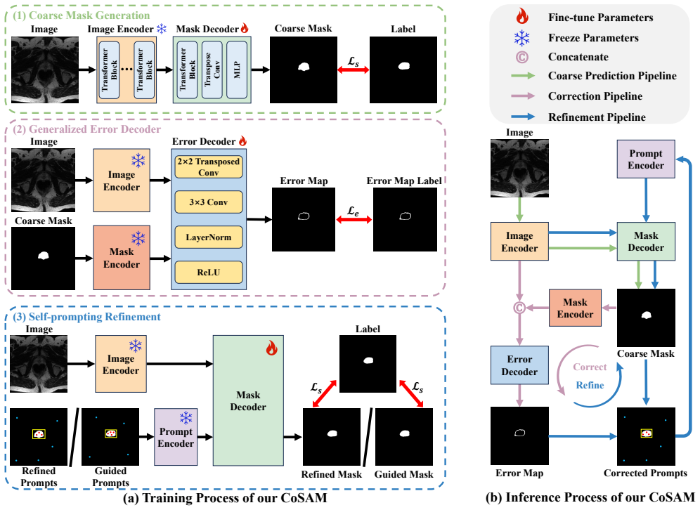
The load-bearing component is the generalized error decoder, a U-Net decoder that takes the concatenation of SAM's image embedding and mask embeddings from SAM's dense prompt encoder, which is reused as a mask encoder, and predicts an error map in which correct pixels are 0 and error pixels are 1. It is trained with random Bernoulli flips of the binary coarse mask and a class-balanced weighted loss, which is meant to simulate distribution shift and clinician correction. The self-correcting loop, driven by corrected masks and diverse point, box, and mask prompts, is what turns the predicted error map into improved segmentation.
What would settle it
Take a target-domain image whose scanner or protocol never appears in training, compute CoSAM's predicted error map, and compare it with the true error map defined as the XOR of the coarse mask and the clinician label; if the self-correction loop's improvement over the coarse mask disappears whenever the predicted error map misses a large fraction of true error pixels, the core claim fails.
Extended reading notes
Core claim
CoSAM claims that coarse masks produced by a prompt-free SAM are a sufficient starting point for prompting, provided the model can learn to correct them. During training, random Bernoulli perturbations of the coarse mask make the error decoder more robust to distribution shifts in the error-prediction task. At inference, corrected masks are converted into three prompt types—top-K point prompts from error locations, a box prompt from the largest foreground-connected region, and a mask prompt from the corrected mask—and fed back into SAM. The loop repeats for up to T iterations and stops early when the predicted number of error points stops falling. CoSAM reports that this loop outperforms SAM and five SAM-based baselines on both benchmark tasks.
Load-bearing premise
The load-bearing premise is that an error decoder trained on source images, with random flips applied only to the binary mask and never to image appearance, will predict which pixels of the coarse mask are wrong accurately enough on an unseen target domain to drive useful corrections.
Editorial extensions
If this is right
- A test-time pipeline can avoid manual prompts and separate trainable prompt generators, using SAM's own coarse mask as the seed.
- Predicted error maps can serve as a quality monitor, since the refinement loop stops when the estimated number of error points would increase.
- Combining point, box, and mask prompts gives more consistent gains than any single prompt type in the reported ablations.
- On the two benchmarks, the improvement is concentrated in average performance across leave-one-domain-out settings rather than in every individual source domain.
Reading between the lines
- Nothing in the method is organ-specific, so the same correct-then-reprompt cycle should transfer to other promptable segmentation backbones and other 2D medical targets, a claim the paper leaves untested.
- Because the perturbation used to train the error decoder only flips mask pixels, real scanner-level appearance shifts remain the likeliest failure mode; adding image-level augmentations on top of the mask perturbation would be a direct test of that boundary.
- The early-stop rule could be reinterpreted as a confidence signal and used to reject unreliable corrections rather than simply halting refinement, an extension the paper does not explore.
Editorial analysis
A structured set of objections, weighed in public.
Referee Report
Summary. The paper proposes CoSAM, a SAM-based method for domain generalization in 2D medical image segmentation. CoSAM first produces a coarse mask with SAM in a prompt-free manner, then uses a learned error decoder to predict an error map between the coarse mask and the ground truth, and corrects the mask accordingly. The corrected mask and a set of diverse prompts (point, box, mask) are fed back into SAM's mask decoder, and this refinement is repeated for T iterations with an early-stop mechanism. The method is evaluated with leave-one-domain-out experiments on prostate MRI segmentation (six domains) and joint optic disc/cup segmentation (four domains), reporting average DSC improvements over five SAM-based baselines.
Significance. If the reported results are reproducible, CoSAM contributes a conceptually simple way to leverage SAM's prompt-based design without requiring a prompt generator or manual prompts, addressing a real need in clinical deployment under distribution shifts. The use of a dedicated error decoder to guide iterative refinement is a sensible idea, and the reported improvements over multiple baselines on two benchmarks are encouraging. However, the verification is weakened by the absence of statistical significance tests and by an internal inconsistency in the error-decoder training objective described below.
major comments (3)
- [Section 4.3, Eq. (4)-(5), Algorithm 1 lines 7-9] The error decoder receives the perturbed mask ˆyp as input but is supervised with the error map e = XOR(ˆy, y) of the unperturbed coarse mask. At inference (Algorithm 2, line 7) the decoder receives the unperturbed current mask and is expected to predict its error map. As written, the training objective teaches the decoder to produce error maps for a mask it does not see, so the decoder cannot learn the conditional mapping required for self-correction. This internal inconsistency undermines the attribution of the Le contribution in Table 3 and makes the method unreproducible. The authors likely meant to compute e = XOR(ˆyp, y) or to feed the unperturbed mask in Eq. (4); either way the manuscript must be corrected and the experiments rerun.
- [Tables 1-5] No standard deviations, confidence intervals, or significance tests are reported for any DSC value, and the reported improvements over the best baselines are modest (average DSC gains of 1.64 and 1.72 points on the two tasks). Since the main claim is superiority over existing methods, the absence of repeated-run statistics makes it difficult to judge whether the improvements are stable. Please report mean and standard deviation over at least three runs, and ideally paired significance tests.
- [Section 4.3, Eq. (3)] The perturbation used to train the error decoder acts only on the binary mask, not on the image appearance. Because the decoder also receives image embeddings E(x) as input, the domain shift in image statistics (scanner/protocol variations) is not simulated, so the claim that the decoder is 'generalized' across image domains is not directly supported. The authors should provide evidence that the error decoder maintains accuracy on target domains, for example by reporting error-map quality or by ablating the perturbation under image-level augmentation.
minor comments (6)
- [Eq. (5)] The weighted BCE is written as Le(˜e, e) = ωe log(˜e) + (1− e) log(1− ˜e), which is ambiguous about whether ω multiplies e and where the minus sign is. Please write the standard form Le = −[ω e log(˜e) + (1−e) log(1−˜e)].
- [Section 5.3] The baseline 'SAM' is described as 'fine-tuning SAM with the full image size as box prompts'; this conflicts with the earlier description of prompt-free methods. Please clarify the exact prompting configuration of the SAM baseline.
- [Table 5] The rows for Random-K and Top-K contain formatting artifacts (font-size shift) in the submitted PDF; please ensure the table renders uniformly.
- [References] References [5] and [6] are the same paper (Chen et al., 'Treasure in distribution...'); duplicate bibliography entries should be merged.
- [Section 4.3, text after Eq. (6)] The definition of nw as 'the number of correct and error points in ˜e' is contradictory; from the equation nw should refer only to error points and nr to correct points. Please fix the wording.
- [Section 4.5 and Algorithm 2] The early-stop logic in Algorithm 2 is described only in the caption of Figure 2; consider stating it explicitly in the main text of Section 4.5 for clarity.
Circularity Check
No significant circularity: CoSAM's reported gains come from held-out target domains; the only self-referential element is reuse of the same group's earlier error-map loss, which is not load-bearing.
full rationale
The central empirical claims are evaluated under a leave-one-domain-out protocol on held-out target domains, with results reported in Tables 1 and 2. The reported DSC values therefore cannot reduce to fitted training values or to target-domain labels reused as predictions. The error decoder is trained on source-domain images with supervision e = XOR(y_hat, y), an externally defined target derived from source labels, and at inference it is applied to unseen target images; the iterative refinement loop uses only the model's own outputs and an early-stop criterion, not target annotations. The self-citations, including reference [44] for the class-balance weight in Eq. (6), point to independently published prior work and are not used to forbid alternatives or to force the benchmark outcomes. There is a genuine internal-consistency concern in Section 4.3: Eq. (4) feeds the perturbed mask y_hat_p to the error decoder, while the error label in Eq. (5) and Algorithm 1 line 9 is computed from the unperturbed binarized mask y_hat, so the decoder is not trained on the exact input it receives at inference. That is a reproducibility and correctness issue, not a circularity: the error map is still supervised by source labels, and the final predictions are not defined in terms of the target outputs they claim to predict.
Assumptions & free parameters
free parameters (6)
- perturbation probability alpha =
0.2 (prostate), 0.1/0.2 (OD/OC)
- number of point prompts K =
64 (prostate), 8/16 (OD/OC)
- number of refinement iterations T =
4 (prostate), 1 (OC), 4 (OD)
- refined-loss weight lambda_r =
1.0
- guided-loss weight lambda_g =
0.1 (prostate), 0.1/0.25 (OD/OC)
- binarization threshold =
0.5
assumptions (3)
- domain assumption SAM's frozen image encoder and mask decoder provide strong representations across domains due to billion-mask pretraining.
- domain assumption The error-map loss from [44] is a valid objective for learning error prediction with class imbalance.
- domain assumption Leave-one-domain-out evaluation on two benchmarks is representative of domain generalization performance in medical imaging.
Cite this review
Pith. "Pith review of CoSAM: Self-Correcting SAM for Domain Generalization in 2D Medical Image Segmentation." pith.science (2026). https://pith.science/paper/EM4N2GYT
@misc{pith2026241110136,
  author       = {Pith},
  title        = {Pith review of: CoSAM: Self-Correcting SAM for Domain Generalization in 2D Medical Image Segmentation},
  year         = {2026},
  howpublished = {\url{https://pith.science/paper/EM4N2GYT}},
  note         = {Machine review of arXiv:2411.10136}
}
read the original abstract
Medical images often exhibit distribution shifts due to variations in imaging protocols and scanners across different medical centers. Domain Generalization (DG) methods aim to train models on source domains that can generalize to unseen target domains. Recently, the segment anything model (SAM) has demonstrated strong generalization capabilities due to its prompt-based design, and has gained significant attention in image segmentation tasks. Existing SAM-based approaches attempt to address the need for manual prompts by introducing prompt generators that automatically generate these prompts. However, we argue that auto-generated prompts may not be sufficiently accurate under distribution shifts, potentially leading to incorrect predictions that still require manual verification and correction by clinicians. To address this challenge, we propose a method for 2D medical image segmentation called Self-Correcting SAM (CoSAM). Our approach begins by generating coarse masks using SAM in a prompt-free manner, providing prior prompts for the subsequent stages, and eliminating the need for prompt generators. To automatically refine these coarse masks, we introduce a generalized error decoder that simulates the correction process typically performed by clinicians. Furthermore, we generate diverse prompts as feedback based on the corrected masks, which are used to iteratively refine the predictions within a self-correcting loop, enhancing the generalization performance of our model. Extensive experiments on two medical image segmentation benchmarks across multiple scenarios demonstrate the superiority of CoSAM over state-of-the-art SAM-based methods.
Figures


Reference graph
Works this paper leans on
-
[1]
Mi-segnet: Mu- tual information-based us segmentation for unseen domain generalization
Yuan Bi, Zhongliang Jiang, Ricarda Clarenbach, Reza Ghotbi, Angelos Karlas, and Nassir Navab. Mi-segnet: Mu- tual information-based us segmentation for unseen domain generalization. In Int. Conf. Med. Image Comput. Comput.- Assist. Intervent., pages 130–140. Springer, 2023. 2
work page 2023
-
[2]
Biosam: Generating sam prompts from superpixel graph for biological instance segmentation
Miaomiao Cai, Xiaoyu Liu, Zhiwei Xiong, and Xuejin Chen. Biosam: Generating sam prompts from superpixel graph for biological instance segmentation. IEEE Jour. Bio. and Heal. Info, 2024. 2
work page 2024
-
[3]
Ma-sam: Modality-agnostic sam adap- tation for 3d medical image segmentation.Med
Cheng Chen, Juzheng Miao, Dufan Wu, Aoxiao Zhong, Zhiling Yan, Sekeun Kim, Jiang Hu, Zhengliang Liu, Lichao Sun, Xiang Li, et al. Ma-sam: Modality-agnostic sam adap- tation for 3d medical image segmentation.Med. Image Anal., 98:103310, 2024. 2
work page 2024
-
[4]
Sam-adapter: Adapting segment any- thing in underperformed scenes
Tianrun Chen, Lanyun Zhu, Chaotao Deng, Runlong Cao, Yan Wang, Shangzhan Zhang, Zejian Li, Lingyun Sun, Ying Zang, and Papa Mao. Sam-adapter: Adapting segment any- thing in underperformed scenes. In ICCVW, pages 3367– 3375, 2023. 2, 3, 7
work page 2023
-
[6]
Treasure in distribution: a domain randomiza- tion based multi-source domain generalization for 2d medi- cal image segmentation
Ziyang Chen, Yongsheng Pan, Yiwen Ye, Hengfei Cui, and Yong Xia. Treasure in distribution: a domain randomiza- tion based multi-source domain generalization for 2d medi- cal image segmentation. In Int. Conf. Med. Image Comput. Comput.-Assist. Intervent., pages 89–99. Springer, 2023. 2
2023
-
[7]
PAM: A Propagation-Based Model for Segmenting Any 3D Objects across Multi-Modal Medical Images
Zifan Chen, Xinyu Nan, Jiazheng Li, Jie Zhao, Haifeng Li, Zilin Lin, Haoshen Li, Heyun Chen, Yiting Liu, Bin Dong, et al. Propsam: A propagation-based model for segment- ing any 3d objects in multi-modal medical images. arXiv preprint arXiv:2408.13836, 2024. 3
work page Pith review arXiv 2024
-
[8]
Un-sam: Universal prompt-free segmentation for generalized nuclei images
Zhen Chen, Qing Xu, Xinyu Liu, and Yixuan Yuan. Un-sam: Universal prompt-free segmentation for generalized nuclei images. arXiv preprint arXiv:2402.16663, 2024. 2, 3
arXiv 2024
-
[9]
Unleashing the potential of sam for medical adaptation via hierarchical de- coding
Zhiheng Cheng, Qingyue Wei, Hongru Zhu, Yan Wang, Liangqiong Qu, Wei Shao, and Yuyin Zhou. Unleashing the potential of sam for medical adaptation via hierarchical de- coding. In CVPR, pages 3511–3522, 2024. 2, 3, 7
work page 2024
Show all 50 references
-
[10]
An image is worth 16x16 words: Transformers for image recognition at scale
Alexey Dosovitskiy. An image is worth 16x16 words: Transformers for image recognition at scale. arXiv preprint arXiv:2010.11929, 2020. 3
2010 arXiv
-
[11]
Dyno: Dynamic normalization based test-time adaptation for 2d medical image segmentation
Yihang Fu, Ziyang Chen, Yiwen Ye, and Yong Xia. Dyno: Dynamic normalization based test-time adaptation for 2d medical image segmentation. In International Workshop on Machine Learning in Medical Imaging , pages 269–279. Springer, 2024. 6
2024
-
[12]
Desam: Decoupled segment anything model for gen- eralizable medical image segmentation
Yifan Gao, Wei Xia, Dingdu Hu, Wenkui Wang, and Xin Gao. Desam: Decoupled segment anything model for gen- eralizable medical image segmentation. In Int. Conf. Med. Image Comput. Comput.-Assist. Intervent. , pages 509–519. Springer, 2024. 2, 3, 7
2024
-
[13]
Transfer learning for domain adapta- tion in mri: Application in brain lesion segmentation
Mohsen Ghafoorian, Alireza Mehrtash, Tina Kapur, Nico Karssemeijer, Elena Marchiori, Mehran Pesteie, Charles RG Guttmann, Frank-Erik de Leeuw, Clare M Tempany, Bram Van Ginneken, et al. Transfer learning for domain adapta- tion in mri: Application in brain lesion segmentation....
2017
-
[14]
Apseg: Auto-prompt network for cross-domain few-shot semantic segmentation
Weizhao He, Yang Zhang, Wei Zhuo, Linlin Shen, Jiaqi Yang, Songhe Deng, and Liang Sun. Apseg: Auto-prompt network for cross-domain few-shot semantic segmentation. In CVPR, pages 23762–23772, 2024. 2
2024
-
[15]
Lora: Low-rank adaptation of large language models
Edward J Hu, Yelong Shen, Phillip Wallis, Zeyuan Allen- Zhu, Yuanzhi Li, Shean Wang, Lu Wang, and Weizhu Chen. Lora: Low-rank adaptation of large language models. arXiv preprint arXiv:2106.09685, 2021. 3
2021 arXiv
-
[16]
Domain and content adaptive convolution based multi- source domain generalization for medical image segmenta- tion
Shishuai Hu, Zehui Liao, Jianpeng Zhang, and Yong Xia. Domain and content adaptive convolution based multi- source domain generalization for medical image segmenta- tion. IEEE Trans. Med. Imaging, 42(1):233–244, 2022. 6
2022
-
[17]
nnu-net: a self-configuring method for deep learning-based biomedical image segmen- tation
Fabian Isensee, Paul F Jaeger, Simon AA Kohl, Jens Pe- tersen, and Klaus H Maier-Hein. nnu-net: a self-configuring method for deep learning-based biomedical image segmen- tation. Nature methods, 18(2):203–211, 2021. 6
2021
-
[18]
Ultralytics yolov8
Glenn Jocher, Ayush Chaurasia, and Jing Qiu. Ultralytics yolov8. 2023. URL https://github.com/ultralytics/ultralytics,
2023
-
[19]
Adam: A method for stochastic opti- mization
Diederik P Kingma. Adam: A method for stochastic opti- mization. arXiv preprint arXiv:1412.6980, 2014. 6
2014 arXiv
-
[20]
Segment any- thing
Alexander Kirillov, Eric Mintun, Nikhila Ravi, Hanzi Mao, Chloe Rolland, Laura Gustafson, Tete Xiao, Spencer White- head, Alexander C Berg, Wan-Yen Lo, et al. Segment any- thing. In ICCV, pages 4015–4026, 2023. 2, 3, 7
2023
-
[21]
Frequency-mixed single-source domain generalization for medical image segmentation
Heng Li, Haojin Li, Wei Zhao, Huazhu Fu, Xiuyun Su, Yan Hu, and Jiang Liu. Frequency-mixed single-source domain generalization for medical image segmentation. In Int. Conf. Med. Image Comput. Comput.-Assist. Intervent., pages 127–
-
[22]
Asps: Augmented segment anything model for polyp segmentation
Huiqian Li, Dingwen Zhang, Jieru Yao, Longfei Han, Zhongyu Li, and Junwei Han. Asps: Augmented segment anything model for polyp segmentation. In Int. Conf. Med. Image Comput. Comput.-Assist. Intervent. , pages 118–128. Springer, 2024. 2, 3
2024
-
[23]
Am- sam: Automated prompting and mask calibration for seg- ment anything model
Yuchen Li, Li Zhang, Youwei Liang, and Pengtao Xie. Am- sam: Automated prompting and mask calibration for seg- ment anything model. arXiv preprint arXiv:2410.09714 ,
-
[24]
A survey on active deep learning: from model driven to data driven
Peng Liu, Lizhe Wang, Rajiv Ranjan, Guojin He, and Lei Zhao. A survey on active deep learning: from model driven to data driven. ACM Computing Surveys (CSUR) , 54(10s): 1–34, 2022. 2
2022
-
[25]
Shape-aware meta-learning for generalizing prostate mri segmentation to 9 unseen domains
Quande Liu, Qi Dou, and Pheng-Ann Heng. Shape-aware meta-learning for generalizing prostate mri segmentation to 9 unseen domains. In Int. Conf. Med. Image Comput. Comput.- Assist. Intervent., pages 475–485. Springer, 2020. 6
2020
-
[26]
Semi-supervised meta-learning with disentangle- ment for domain-generalised medical image segmentation
Xiao Liu, Spyridon Thermos, Alison O’Neil, and Sotirios A Tsaftaris. Semi-supervised meta-learning with disentangle- ment for domain-generalised medical image segmentation. In Int. Conf. Med. Image Comput. Comput.-Assist. Intervent., pages 307–317. Springer, 2021. 3
2021
-
[27]
Decoupled weight decay regularization
I Loshchilov. Decoupled weight decay regularization. arXiv preprint arXiv:1711.05101, 2017. 6
2017 arXiv
-
[28]
Segment anything in medical images
Jun Ma, Yuting He, Feifei Li, Lin Han, Chenyu You, and Bo Wang. Segment anything in medical images. Nature Communications, 15(1):654, 2024. 3
2024
-
[29]
A survey on domain generalization for medical image analysis
Ziwei Niu, Shuyi Ouyang, Shiao Xie, Yen-wei Chen, and Lanfen Lin. A survey on domain generalization for medical image analysis. arXiv preprint arXiv:2402.05035, 2024. 2
2024 arXiv
-
[30]
Bias in data-driven artificial intelligence systems—an introductory survey
Eirini Ntoutsi, Pavlos Fafalios, Ujwal Gadiraju, Vasileios Iosifidis, Wolfgang Nejdl, Maria-Esther Vidal, Salvatore Ruggieri, Franco Turini, Symeon Papadopoulos, Emmanouil Krasanakis, et al. Bias in data-driven artificial intelligence systems—an introductory survey. Wiley Inte...
2020
-
[31]
Refuge challenge: A unified framework for evaluat- ing automated methods for glaucoma assessment from fun- dus photographs
Jos ´e Ignacio Orlando, Huazhu Fu, Jo ˜ao Barbosa Breda, Karel Van Keer, Deepti R Bathula, Andr ´es Diaz-Pinto, Ruogu Fang, Pheng-Ann Heng, Jeyoung Kim, JoonHo Lee, et al. Refuge challenge: A unified framework for evaluat- ing automated methods for glaucoma assessment from fun...
2020
-
[32]
S-sam: Svd-based fine-tuning of segment anything model for medical image segmentation
Jay N Paranjape, Shameema Sikder, S Swaroop Vedula, and Vishal M Patel. S-sam: Svd-based fine-tuning of segment anything model for medical image segmentation. arXiv preprint arXiv:2408.06447, 2024. 2, 3
2024 arXiv
-
[33]
Db-sam: Delving into high quality universal medical image segmentation
Chao Qin, Jiale Cao, Huazhu Fu, Fahad Shahbaz Khan, and Rao Muhammad Anwer. Db-sam: Delving into high quality universal medical image segmentation. In Int. Conf. Med. Image Comput. Comput.-Assist. Intervent. , pages 498–508. Springer, 2024. 2
2024
-
[34]
Dataset shift in ma- chine learning
Joaquin Quinonero-Candela, Masashi Sugiyama, Anton Schwaighofer, and Neil D Lawrence. Dataset shift in ma- chine learning. Mit Press, 2008. 2
2008
-
[35]
U-net: Convolutional networks for biomedical image segmentation
Olaf Ronneberger, Philipp Fischer, and Thomas Brox. U-net: Convolutional networks for biomedical image segmentation. In Int. Conf. Med. Image Comput. Comput.-Assist. Intervent., pages 234–241. Springer, 2015. 5
2015
-
[36]
Learning from synthetic data: Addressing domain shift for semantic segmentation
Swami Sankaranarayanan, Yogesh Balaji, Arpit Jain, Ser Nam Lim, and Rama Chellappa. Learning from synthetic data: Addressing domain shift for semantic segmentation. In CVPR, pages 3752–3761, 2018. 2
2018
-
[37]
Drishti-gs: Retinal image dataset for optic nerve head (onh) segmentation
Jayanthi Sivaswamy, SR Krishnadas, Gopal Datt Joshi, Mad- hulika Jain, and A Ujjwaft Syed Tabish. Drishti-gs: Retinal image dataset for optic nerve head (onh) segmentation. In IEEE Int. Symp. on Bio. Imaging, pages 53–56. IEEE, 2014. 6
2014
-
[38]
Rethinking data augmentation for single-source domain generalization in medical image segmentation
Zixian Su, Kai Yao, Xi Yang, Kaizhu Huang, Qiufeng Wang, and Jie Sun. Rethinking data augmentation for single-source domain generalization in medical image segmentation. In AAAI, pages 2366–2374, 2023. 2
2023
-
[39]
Rethinking domain generalization for face anti- spoofing: Separability and alignment
Yiyou Sun, Yaojie Liu, Xiaoming Liu, Yixuan Li, and Wen- Sheng Chu. Rethinking domain generalization for face anti- spoofing: Separability and alignment. In CVPR, pages 24563–24574, 2023. 2
2023
-
[40]
Cross-domain face presentation attack detection via multi- domain disentangled representation learning
Guoqing Wang, Hu Han, Shiguang Shan, and Xilin Chen. Cross-domain face presentation attack detection via multi- domain disentangled representation learning. In CVPR, pages 6678–6687, 2020. 2
2020
-
[41]
Lever- aging sam for single-source domain generalization in medi- cal image segmentation
Hanhui Wang, Huaize Ye, Yi Xia, and Xueyan Zhang. Lever- aging sam for single-source domain generalization in medi- cal image segmentation. arXiv preprint arXiv:2401.02076,
-
[42]
Prompting segment anything model with domain-adaptive prototype for generalizable medical image segmentation
Zhikai Wei, Wenhui Dong, Peilin Zhou, Yuliang Gu, Zhou Zhao, and Yongchao Xu. Prompting segment anything model with domain-adaptive prototype for generalizable medical image segmentation. In Int. Conf. Med. Image Com- put. Comput.-Assist. Intervent. , pages 533–543. Springer, 2024
2024
-
[43]
Masksam: Towards auto-prompt sam with mask classifi- cation for medical image segmentation
Bin Xie, Hao Tang, Bin Duan, Dawen Cai, and Yan Yan. Masksam: Towards auto-prompt sam with mask classifi- cation for medical image segmentation. arXiv preprint arXiv:2403.14103, 2024. 2
2024 arXiv
-
[44]
Sesv: Accurate medical image segmentation by predicting and correcting errors
Yutong Xie, Jianpeng Zhang, Hao Lu, Chunhua Shen, and Yong Xia. Sesv: Accurate medical image segmentation by predicting and correcting errors. IEEE Trans. Med. Imaging, 40(1):286–296, 2020. 5
2020
-
[45]
Esp-medsam: Efficient self-prompting sam for universal domain-generalized medical image seg- mentation
Qing Xu, Jiaxuan Li, Xiangjian He, Ziyu Liu, Zhen Chen, Wenting Duan, Chenxin Li, Maggie M He, Fiseha B Tesema, Wooi P Cheah, et al. Esp-medsam: Efficient self-prompting sam for universal domain-generalized medical image seg- mentation. arXiv preprint arXiv:2407.14153, 2024. 2, 3
2024 arXiv
-
[46]
Customized segment any- thing model for medical image segmentation
Kaidong Zhang and Dong Liu. Customized segment any- thing model for medical image segmentation. arXiv preprint arXiv:2304.13785, 2023. 2, 3, 7
2023 arXiv
-
[47]
Origa-light: An online retinal fundus image database for glaucoma analysis and research
Zhuo Zhang, Feng Shou Yin, Jiang Liu, Wing Kee Wong, Ngan Meng Tan, Beng Hai Lee, Jun Cheng, and Tien Yin Wong. Origa-light: An online retinal fundus image database for glaucoma analysis and research. In Int. Conf. IEEE Eng. in Med. and Bio., pages 3065–3068. IEEE, 2010. 6
2010
-
[48]
Domain generalization with adversarial intensity attack for medical image segmentation
Zheyuan Zhang, Bin Wang, Lanhong Yao, Ugur Demir, Debesh Jha, Ismail Baris Turkbey, Boqing Gong, and Ulas Bagci. Domain generalization with adversarial intensity attack for medical image segmentation. arXiv preprint arXiv:2304.02720, 2023. 2
2023 arXiv
-
[49]
Do- main adaptive ensemble learning
Kaiyang Zhou, Yongxin Yang, Yu Qiao, and Tao Xiang. Do- main adaptive ensemble learning. IEEE TIP, 30:8008–8018,
-
[50]
Domain generalization: A survey
Kaiyang Zhou, Ziwei Liu, Yu Qiao, Tao Xiang, and Chen Change Loy. Domain generalization: A survey. IEEE TPAMI, 45(4):4396–4415, 2022. 2
2022
-
[51]
Localized adversarial domain generalization
Wei Zhu, Le Lu, Jing Xiao, Mei Han, Jiebo Luo, and Adam P Harrison. Localized adversarial domain generalization. In CVPR, pages 7108–7118, 2022. 2 10
2022
Reviewed August 12, 2026 · model on record in the stance chip above.
Discussion (0). Continue with ORCID to comment.