REVIEW 3 major objections 6 minor 1 cited by
Visualizing strongly focused 3D light fields in an atomic vapor
T0 review · 3 major / 6 minor · reviewed 2026-08-07 · deepseek-v4-flash
Pith's one-line read Strongly focused radial light develops a component of the electric field along the optical axis, and the paper shows this axial component can be observed directly as absorption of pi transitions in a rubidium vapor.
desk verdict A genuinely new atomic-vapor way to image the axial polarization of focused vector beams; the qualitative evidence is strong, but the quantitative claims and an artifact control need work. read the letter →
The pith
A machine-rendered reading of the paper's core claim, the machinery that carries it, and where it could break.
The reading
What carries the argument
The mechanism is the electric-dipole selection rule of rubidium-87 in the hyperfine Paschen-Back regime at 1.6 T: with the magnetic field (and therefore the atomic quantization axis) along the optical axis, a polarization component along the axis drives $\pi$ transitions, while transverse circular polarization components drive $\sigma^{\pm}$ transitions. Because the Zeeman splitting exceeds the Doppler width, the $\pi$ lines are spectrally isolated, so their absorption reports the axial field at the focus. A lens with NA 0.4 or 0.7 performs the conversion from radial input polarization to axial field, and the experiment compares the measured optical-density images at a $\pi$ line with the simulated axial intensity distribution.
What would settle it
Record the $\pi$ absorption depth and its spatial map while rotating the input polarization from azimuthal to radial, using a Stokes measurement of the actual input beam; if a substantial $\pi$ signal remains for the azimuthal case, and if that residual does not vanish or scale predictably when the beam-cell-magnet alignment is deliberately varied, then the absorption is not a faithful map of the axial field. The paper itself notes residual $\pi$ absorption for azimuthal beams, attributed to alignment and cell-wall refraction, so the decisive test is whether this residual can be eliminated or quantitatively accounted for by the measured misalignment.
Extended reading notes
Core claim
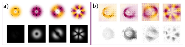
Under strong focusing, part of the radial polarization of an input beam is converted into an electric-field component along the optical axis, while an azimuthally polarized input remains purely transverse. The paper's central discovery is that this axial component can be observed as absorption of $\pi$ transitions in rubidium-87 vapor in the hyperfine Paschen-Back regime, using a single beam and a magnetic field parallel to the beam. The strength of the $\pi$ absorption grows as the input polarization is rotated from azimuthal to radial, and the spatial optical-density map recorded at a $\pi$ resonance reproduces the positions where the focused field is expected to have axial polarization. The authors take this as a direct mapping from the three-dimensional vector light field onto atomic transition strength, and as experimental validation of the Richards-Wolf vectorial diffraction predictions.
Load-bearing premise
The result assumes that the observed $\pi$ absorption is dominated by the genuine axial field component and not by systematic effects such as a small tilt between the beam axis and the magnetic field or refraction by the tilted vapor-cell walls, which can create spurious $\pi$ signals even for azimuthal input.
Editorial extensions
If this is right
- A single-beam atomic-vapor setup can measure transverse and longitudinal polarization components simultaneously in one spectrum, without moving any detector.
- The optical-density map at a $\pi$ transition provides a two-dimensional image of where the focused field has axial polarization, so the focal structure of a vector beam can be visualized directly.
- The observed linear growth of $\pi$ absorption with the input beam's radial content offers a quantitative test of vectorial diffraction theory.
- Because the $\pi$ transition is driven only by the axial field, the method can imprint the rotational symmetry of the input polarization onto the spatial pattern of atomic excitation, enabling polarization-selective control of atoms.
- The scheme works with a hot vapor, a permanent magnet, and standard photodiode or CCD detection, and the paper argues it should be orders of magnitude more efficient than earlier single-molecule or nanoparticle probes of the same quantity.
Reading between the lines
- Beyond the paper: because the $\pi$ absorption is spectrally resolved, the same setup could in principle map not only the amplitude but the frequency-dependent response of the axial field, potentially revealing phase structure through detuning lineshapes; the paper does not itself extract phase.
- Beyond the paper: a controlled experiment that tilts the magnetic field by small known angles relative to the beam would convert the residual $\pi$ absorption for azimuthal input into a quantitative calibration of alignment error, allowing the residual signal to be subtracted rather than merely attributed to imperfections.
- Beyond the paper: the linear mapping between input radiality and $\pi$ strength suggests a route to a compact atomic polarimeter that measures the local degree of radial polarization of a focused beam; this would be a new sensing mode not proposed in the paper.
- Beyond the paper: extending the measurements to other alkali species or to higher NA should make the axial-field contrast larger, and comparing the measured $\pi$ maps with full Richards-Wolf simulations including the measured input Stokes profile would test whether the technique can reconstruct the complete 3D field, not just its radial components.
Editorial analysis
A structured set of objections, weighed in public.
Referee Report
Summary. The manuscript reports an experiment in which a 780-nm laser, shaped into radial, azimuthal, and higher-order vector-vortex polarization profiles, is strongly focused (NA = 0.4–0.7) into a 1-mm 87Rb vapor cell placed in a 1.6-T magnetic field along the optical axis. In the hyperfine Paschen-Back regime, the D2 transitions split into spectrally resolved σ± and π lines, and the authors record integrated and spatially resolved absorption as the laser frequency is scanned. They find that π-line absorption is strongest for radial input polarization, nearly absent for azimuthal input, and modulated with two-fold and six-fold symmetry for the e2 and e6 vector modes, in qualitative agreement with Richards-Wolf calculations of the longitudinal electric-field component. From this they conclude that atomic π absorption directly evidences and visualizes the axial polarization component of strongly focused vector light.
Significance. The proposed detection method is of genuine interest: it uses the atom itself as a polarization-sensitive sensor for the longitudinal field component that is normally invisible to cameras and waveplates, and the hyperfine Paschen-Back splitting provides a clean spectral separation of the π and σ channels. The paper's strengths are that the predictions are generated with an independent forward model (Richards-Wolf focusing plus the ElecSus susceptibility code) without fitting a normalization constant, that a control comparison of focused versus unfocused linearly polarized light appears in the Supplement, and that the qualitative symmetry mapping across four input polarization classes is internally consistent. The main limitations are that the quantitative linear-dependence claim lacks error bars and a fitted model, and that the systematic sources of spurious π absorption admitted in the text are not bounded. These issues do not invalidate the qualitative demonstration, but they do mean the paper currently overstates the strength of the quantitative evidence.
major comments (3)
- [Sec. 4, Fig. 4(c)] The quantitative claim that the π-transition depth 'depends linearly on the radial component' is not supported by the presented data. The plot has no error bars, no fitted curve, and no defined x-axis quantity: the axis is labeled HWP angle, and for the beams of Eq. (2) the radial amplitude is cos(2θ) while the radial power fraction is cos^2(2θ). Since the measured absorption depth is a power/intensity quantity, a linear dependence on the radial amplitude would imply a quadratic dependence on cos(2θ), whereas a linear dependence on the radial power fraction would be cos^2(2θ); the text does not state which is meant. In addition, at the elevated temperature of ~125 °C the vapor may be optically thick, so the line depth is not simply proportional to the integrated longitudinal intensity. Please provide the fitted functional form, the uncertainty budget, and the definition of 'radiality' used for the horizontal axis.
- [Sec. 3 and Supplemental D] The manuscript itself identifies two sources of artificial π absorption: misalignment between the optical axis and the magnetic field (Sec. 3) and refraction at the tilted cell windows (Supplemental D), which is said to be 'in part responsible for the residual π transition observed even for azimuthal input beams.' The residual π signal for azimuthal input is therefore nonzero, but its magnitude relative to the radial-beam signal is never quantified. Because these artifact sources are fixed in the lab frame while the input polarization patterns in Figs. 5–6 are each measured at only one orientation, a lab-fixed longitudinal contribution could partly mimic the observed two-fold or six-fold angular pattern. Please add a control in which the input polarization pattern is rotated about the optical axis (e.g., the e2 beam) and show that the π-absorption map rotates with it, or otherwise provide a quantitative upper bound on the artifact contribution to the measured π depth.
- [Sec. 4 and Supplemental D] The absolute frequency axis is not calibrated: the paper states that the temperature scan is nonlinear, is not referenced to an atomic standard, and that fits used to relate frequency to simulations carry large errors. This matters for the quantitative analysis because the π-transition depth in Fig. 4(c) and the identification of the 'leftmost π transition' in Figs. 5–6 depend on knowing which spectral line is being integrated and on the width and position of that line. Please provide a relative frequency calibration (for example, an etalon signal recorded simultaneously) or quantify the resulting uncertainty in the transition depth and line assignment.
minor comments (6)
- [Data availability] The manuscript ends with 'Data underlying the results presented in this paper are available in XXX'; the placeholder must be replaced before publication.
- [Sec. 3] The quoted peak intensity '0.005 μW/m^2' cannot be correct; presumably it is μW/μm^2 or nW/μm^2. Please correct the units.
- [Fig. 4(b) caption] The caption reads 'was taking at a vapor temperature' and should read 'was taken at a vapor temperature.'
- [Supplemental Fig. 8] The caption refers to 'circularly polarized light,' whereas the surrounding section discusses linearly polarized light; please reconcile the caption with the section text.
- [Sec. 4] The term 'radiality' is used without a formal definition; please define it in terms of the input state of Eq. (2), specifying whether amplitude or power fraction is meant.
- [Sec. 5] The conclusion's phrase 'directly measured this axial component' is stronger than what the experiment provides: the measurement is a far-field absorption signal integrated along the 1-mm cell, and the spatial maps are far-field beam profiles rather than focal-plane images. Please soften the wording to 'inferred through atomic absorption.'
Circularity Check
No significant circularity: the pi-absorption maps are forward-model predictions from Richards–Wolf and ElecSus, not fits to the data they explain.
full rationale
The central claim is that the depth and azimuthal modulation of π-transition absorption under strong focusing tracks the longitudinal electric-field component, which is a forward prediction of Richards–Wolf vector diffraction theory (Ref. 27, external) applied to the measured input polarization states. The atomic transition positions and relative strengths are calculated with the ElecSus code (Ref. 53); although this code comes from Ifan Hughes's group, it is a parameter-free, independently published susceptibility solver and is not adjusted to force agreement with the present data. No fitting constant connects the plotted π-transition optical density to the simulated |E_z|^2 maps: Fig. 4c is normalized only to the radial-beam value, and Figs. 5–6 compare the azimuthal symmetry of the data with the computed symmetry of the axial field. The paper's own caveats about beam–field misalignment and tilted cell walls (Section 3 and Supplemental D) identify systematic uncertainties in attributing residual π signal to a genuine axial component, but these are validity concerns, not circular definitions: the observed signal is not used as the definition of the input polarization or as an input to the model. The derivation chain is therefore self-contained rather than circular.
Assumptions & free parameters
assumptions (3)
- domain assumption Vectorial diffraction theory (Richards-Wolf) correctly predicts focal fields for the experimental input profiles and numerical apertures.
- domain assumption At 1.6 T, the hyperfine Paschen-Back level structure and electric dipole selection rules map transverse polarization to sigma transitions and axial polarization to pi transitions.
- domain assumption Misalignment between the optical axis and the magnetic field, plus refraction from tilted cell walls, is small enough that residual pi absorption does not dominate the observed signal.
Cite this review
Pith. "Pith review of Visualizing strongly focused 3D light fields in an atomic vapor." pith.science (2026). https://pith.science/paper/FCB4HJBF
@misc{pith2026250601680,
  author       = {Pith},
  title        = {Pith review of: Visualizing strongly focused 3D light fields in an atomic vapor},
  year         = {2026},
  howpublished = {\url{https://pith.science/paper/FCB4HJBF}},
  note         = {Machine review of arXiv:2506.01680}
}
read the original abstract
Structured light, when strongly focused, generates highly confined vectorial electromagnetic field distributions which may feature a polarization component along the optical axis. Manipulating and detecting such 3D light fields is challenging, as conventional optical elements and detectors do not interact with the axial polarization component. Vector light can, however, be mapped onto atomic polarizations, making electric dipole transitions an ideal candidate to sense such 3D light configurations. Working in the hyperfine Paschen-Back regime, where the electric dipole transitions are spectrally resolved, we demonstrate direct evidence of the axial polarization component of strongly focused radial light. We investigate the influence of various input polarization states, including radial, azimuthal, and higher-order optical vortices, on atomic absorption profiles. Our results confirm a clear mapping between the 3D vector light and the atomic transition strength. This work provides new insights into vectorial light-matter interaction, and opens avenues for novel quantum sensing applications.
Figures


Figures from the paper (7 more)




Forward citations
Cited by 1 Pith paper
-
Vortex Light at the Nanoscale: Twists, Spins, and Surprises -- A Review
This review unifies non-paraxial optical vortex physics, showing that tight focusing generates transverse spin, orbit-induced spin, and polarization-independent optical chirality.
Reference graph
Works this paper leans on
-
[1]
H. Rubinsztein-Dunlop, A. Forbes, M. V. Berry,et al., “Roadmap on structured light,” J. Opt.19, 013001 (2016)
work page 2016
-
[2]
Special Section Guest Editorial: Spatial Light Modulators: Devices and Applications,
I. Moreno, M. del Mar Sánchez-López, A. Forbes, and Y. Hayasaki, “Special Section Guest Editorial: Spatial Light Modulators: Devices and Applications,” Opt. Eng.59, 041201 (2020)
work page 2020
-
[3]
Subwavelength gratings for polarization conversion and focusing of laser light,
S. S. Stafeev, V. V. Kotlyar, A. G. Nalimov,et al., “Subwavelength gratings for polarization conversion and focusing of laser light,” Photonics Nanostructures - Fundam. Appl.27, 32–41 (2017)
work page 2017
-
[4]
K. A. Forbes and D. Green, “Customized optical chirality of vortex structured light through state and degree-of- polarization control,” Phys. Rev. A107 (2023)
work page 2023
-
[5]
Superresolution structured illumination microscopy reconstruction algorithms: A review,
X. Chen, S. Zhong, Y. Hou,et al., “Superresolution structured illumination microscopy reconstruction algorithms: A review,” Light. Sci. & Appl.12(2023)
work page 2023
-
[6]
Dynamic holographic optical tweezers,
J. E. Curtis, B. A. Koss, and D. G. Grier, “Dynamic holographic optical tweezers,” Opt. Commun.207, 169–175 (2002)
work page 2002
-
[7]
Storage and retrieval of vector beams of light in a multiple-degree-of- freedom quantum memory,
V. Parigi, V. D’Ambrosio, C. Arnold,et al., “Storage and retrieval of vector beams of light in a multiple-degree-of- freedom quantum memory,” Nat Commun.13, 7706 (2015)
work page 2015
-
[8]
Optical networking for quantum key distribution and quantum communica- tions,
T. Chapuran, P. Toliver, N. Peters,et al., “Optical networking for quantum key distribution and quantum communica- tions,” New J. Phys.11 (2009)
work page 2009
Show all 76 references
-
[9]
From Maxwell to paraxial wave optics,
M. Lax, W. H. Louisell, and W. B. McKnight, “From Maxwell to paraxial wave optics,” Phys. Rev. A11, 1365–1370 (1975)
1975
-
[10]
Novotny and B
L. Novotny and B. Hecht,Principles of nano-optics(Cambridge university press, 2012)
2012
-
[11]
Zhan,Vectorial optical fields: Fundamentals and applications(World scientific, 2013)
Q. Zhan,Vectorial optical fields: Fundamentals and applications(World scientific, 2013)
2013
-
[12]
C. S. Adams and I. Hughes,Optics f2f: from Fourier to Fresnel(Oxford University Press, 2019)
2019
-
[13]
Vector-vortex Bessel–Gauss beams and their tightly focusing properties,
K. Huang, P. Shi, G. W. Cao,et al., “Vector-vortex Bessel–Gauss beams and their tightly focusing properties,” Opt. Lett. 36, 888 (2011)
2011
-
[14]
Observation of optical polarization Möbius strips,
T. Bauer, P. Banzer, E. Karimi,et al., “Observation of optical polarization Möbius strips,” Science347, 964–966 (2015)
2015
-
[15]
Creating Complex Optical Longitudinal Polarization Structures,
F. Maucher, S. Skupin, S. Gardiner, and I. Hughes, “Creating Complex Optical Longitudinal Polarization Structures,” Phys. Rev. Lett.120 (2018)
2018
-
[16]
Sculpting complex polarization singularity networks,
E. Otte and C. Denz, “Sculpting complex polarization singularity networks,” Opt. Lett.43, 5821 (2018)
2018
-
[17]
Singular knot bundle in light,
D. Sugic and M. R. Dennis, “Singular knot bundle in light,” JOSA A35, 1987–1999 (2018)
2018
-
[18]
Generating and converting special focal field patterns in a 4𝜋 focusing system illuminated by azimuthally polarized vortex beam,
Y. Yang, X. Yan, Y. He,et al., “Generating and converting special focal field patterns in a 4𝜋 focusing system illuminated by azimuthally polarized vortex beam,” Opt. Rev.29, 320–326 (2022)
2022
-
[19]
Focusing of high numerical aperture cylindrical-vector beams,
K. S. Youngworth and T. G. Brown, “Focusing of high numerical aperture cylindrical-vector beams,” Opt. Express7, 77–87 (2000)
2000
-
[20]
Sharper focus for a radially polarized light beam,
R. Dorn, S. Quabis, and G. Leuchs, “Sharper focus for a radially polarized light beam,” Phys. Rev. Lett.91, 233901 (2003)
2003
-
[21]
Superoscillation: from physics to optical applications,
G. Chen, Z.-Q. Wen, and C.-W. Qiu, “Superoscillation: from physics to optical applications,” Light. Sci. & Appl.8 (2019)
2019
-
[22]
Kozawa and S
Y. Kozawa and S. Sato,Small focal spot formation by vector beams(Elsevier, 2021), p. 35–90
2021
-
[23]
Creation of a needle of longitudinally polarized light in vacuum using binary optics,
H. Wang, L. Shi, B. Lukyanchuk,et al., “Creation of a needle of longitudinally polarized light in vacuum using binary optics,” Nat. Photonics2, 501–505 (2008)
2008
-
[24]
Optimization-free customization of optical tightly focused fields: uniform needles and hotspot chains,
J. He, J. Zhuang, L. Ding, and K. Huang, “Optimization-free customization of optical tightly focused fields: uniform needles and hotspot chains,” Appl. Opt.60, 3081 (2021)
2021
-
[25]
Plasmonic Nanolenses Produced by Cylindrical Vector Beam Printing for Sensing Applications,
S. A. Syubaev, A. Y. Zhizhchenko, D. V. Pavlov,et al., “Plasmonic Nanolenses Produced by Cylindrical Vector Beam Printing for Sensing Applications,” Sci. Reports9(2019)
2019
-
[26]
Selective Excitation of Dark Plasmon Modes Using Cylindrical Vector Beams Studied by Microscopic Imaging of Nonlinear Photoluminescence,
S. Hasegawa, H. Ichikawa, and K. Imura, “Selective Excitation of Dark Plasmon Modes Using Cylindrical Vector Beams Studied by Microscopic Imaging of Nonlinear Photoluminescence,” The J. Phys. Chem. C128, 2536–2542 (2024)
2024
-
[27]
Electromagnetic diffraction in optical systems, II. Structure of the image field in an aplanatic system,
B. Richards and E. Wolf, “Electromagnetic diffraction in optical systems, II. Structure of the image field in an aplanatic system,” Royal Soc. Publ.253 (1959)
1959
-
[28]
Dynamical characteristics of tightly focused vortex beams with different states of polarization,
Z. Cui, J. Sun, N. M. Litchinitser, and Y. Han, “Dynamical characteristics of tightly focused vortex beams with different states of polarization,” J. Opt.21, 015401 (2018)
2018
-
[29]
3D Stokes parameters for vector focal fields,
A. V. Andreev, O. A. Shoutova, S. M. Trushin, and S. Y. Stremoukhov, “3D Stokes parameters for vector focal fields,” J. Opt. Soc. Am. B39, 1775 (2022)
2022
-
[30]
Local characterization of the polarization state of 3D electromagnetic fields: an alternative approach,
R. Martínez-Herrero, D. Maluenda, M. Aviñoá,et al., “Local characterization of the polarization state of 3D electromagnetic fields: an alternative approach,” Photonics Res.11, 1326 (2023)
2023
-
[31]
Transverse Angular Momentum and Geometric Spin Hall Effect of Light,
A. Aiello, N. Lindlein, C. Marquardt, and G. Leuchs, “Transverse Angular Momentum and Geometric Spin Hall Effect of Light,” Phys. Rev. Lett.103 (2009)
2009
-
[32]
Measuring the Transverse Spin Density of Light,
M. Neugebauer, T. Bauer, A. Aiello, and P. Banzer, “Measuring the Transverse Spin Density of Light,” Phys. Rev. Lett. 114 (2015)
2015
-
[33]
Dual Coaxial Longitudinal Polarization Vortex Structures,
Z. Man, Z. Xi, X. Yuan,et al., “Dual Coaxial Longitudinal Polarization Vortex Structures,” Phys. Rev. Lett.124 (2020)
2020
-
[34]
Longitudinal field modes probed by single molecules,
L. Novotny, M. R. Beversluis, K. S. Youngworth, and T. G. Brown, “Longitudinal field modes probed by single molecules,” Phys. Rev. Lett.86, 5251–5254 (2001)
2001
-
[35]
Single-molecule orientations determined by direct emission pattern imaging,
M. A. Lieb, J. M. Zavislan, and L. Novotny, “Single-molecule orientations determined by direct emission pattern imaging,” J. Opt. Soc. Am. B300, 2530 (2004)
2004
-
[36]
Nanointerferometric amplitude and phase reconstruction of tightly focused vector beams,
T. Bauer, S. Orlov, U. Peschel,et al., “Nanointerferometric amplitude and phase reconstruction of tightly focused vector beams,” Nat. Photonics8, 23–27 (2013)
2013
-
[37]
An indirect method of imaging the Stokes parameters of a submicron particle with sub-diffraction scattering,
K. Ullah, B. Garcia-Camara, M. Habib,et al., “An indirect method of imaging the Stokes parameters of a submicron particle with sub-diffraction scattering,” J. Quant. Spectrosc. Radiat. Transf.213, 35–40 (2018)
2018
-
[38]
Polarization nano-tomography of tightly focused light landscapes by self-assembled monolayers,
E. Otte, K. Tekce, S. Lamping,et al., “Polarization nano-tomography of tightly focused light landscapes by self-assembled monolayers,” Nat. Commun.10(2019)
2019
-
[39]
Generalization of the optical theorem: experimental proof for radially polarized beams,
A. V. Krasavin, P. Segovia, R. Dubrovka,et al., “Generalization of the optical theorem: experimental proof for radially polarized beams,” Light. Sci. &; Appl.7 (2018)
2018
-
[40]
Theoreticalinvestigationonasymmetricalspinningandorbitingmotionsofparticles in a tightly focused power-exponent azimuthal-variant vector field,
Y.Zhang,Y.Xue,Z.Zhu, etal.,“Theoreticalinvestigationonasymmetricalspinningandorbitingmotionsofparticles in a tightly focused power-exponent azimuthal-variant vector field,” Opt. Express26, 4318 (2018)
2018
-
[41]
Stokes-basedanalysisfortheestimationof3Ddipolaremission,
I.Herrera,L.Alemán-Castañeda,S.Brasselet, etal.,“Stokes-basedanalysisfortheestimationof3Ddipolaremission,” J. Opt. Soc. Am. A Opt. p. 41 (2024)
2024
-
[42]
Atoms in complex twisted light,
M. Babiker, D. L. Andrews, and V. E. Lembessis, “Atoms in complex twisted light,” J. Opt.21, 013001 (2018)
2018
-
[43]
Vectorial light–matter interaction: Exploring spatially structured complex light fields,
J. Wang, F. Castellucci, and S. Franke-Arnold, “Vectorial light–matter interaction: Exploring spatially structured complex light fields,” AVS Quantum Sci.2 (2020)
2020
-
[44]
MeasuringtheOpticalConcurrenceofVectorBeamswithanAtomic-State Interferometer,
J.Wang,S.J.Svensson,T.W.Clark, etal.,“MeasuringtheOpticalConcurrenceofVectorBeamswithanAtomic-State Interferometer,” Phys. Rev. Lett.132 (2024)
2024
-
[45]
I. Experimental researches in electricity.—Nineteenth series,
M. Faraday, “I. Experimental researches in electricity.—Nineteenth series,” Phil. Trans. R. Soc. L. pp. 1–20 (1846)
-
[46]
Zur Theorie der magneto-optischen Erscheinungen,
W. Voigt, “Zur Theorie der magneto-optischen Erscheinungen,” Ann. der Physik303, 345–365 (1899)
-
[47]
How to build an optical filter with an atomic vapor cell,
D. Uhland, H. Dillmann, Y. Wang, and I. Gerhardt, “How to build an optical filter with an atomic vapor cell,” New J. Phys. 25, 125001 (2023)
2023
-
[48]
Na-Faraday rotation filtering: The optimal point,
W. Kiefer, R. Löw, J. Wrachtrup, and I. Gerhardt, “Na-Faraday rotation filtering: The optimal point,” Sci. Reports4, 6552 (2014)
2014
-
[49]
Atomic Compass: Detecting 3D Magnetic Field Alignment with Vector Vortex Light,
F. Castellucci, T. W. Clark, A. Selyem,et al., “Atomic Compass: Detecting 3D Magnetic Field Alignment with Vector Vortex Light,” Phys. Rev. Lett.127 (2021)
2021
-
[50]
Interaction of vector light beams with atoms exposed to a time-dependent magnetic field,
S. Ramakrishna, R. P. Schmidt, A. A. Peshkov,et al., “Interaction of vector light beams with atoms exposed to a time-dependent magnetic field,” Phys. Rev. A110 (2024)
2024
-
[51]
Polarization singularities: Progress, fundamental physics, and prospects,
Q. Wang, C.-H. Tu, Y.-N. Li, and H.-T. Wang, “Polarization singularities: Progress, fundamental physics, and prospects,” APL Photonics6(2021)
2021
-
[52]
Permanent magnets for Faraday rotators inspired by the design of the magic sphere,
G. Trénec, W. Volondat, O. Cugat, and J. Vigué, “Permanent magnets for Faraday rotators inspired by the design of the magic sphere,” Appl. optics50, 4788–4797 (2011)
2011
-
[53]
𝐸𝑙𝑒𝑐𝑆𝑢𝑠 : A program to calculate the electric susceptibility of an atomic ensemble,
M. A. Zentile, J. Keaveney, L. Weller,et al., “𝐸𝑙𝑒𝑐𝑆𝑢𝑠 : A program to calculate the electric susceptibility of an atomic ensemble,” Comput. Phys. Commun.189, 162–174 (2015)
2015
-
[54]
Laser spectroscopy of hot atomic vapours: from ‘scope to theoretical fit,
D. Pizzey, J. D. Briscoe, F. D. Logue,et al., “Laser spectroscopy of hot atomic vapours: from ‘scope to theoretical fit,” New J. Phys.24, 125001 (2022)
2022
-
[55]
Saturated absorption technique used in potassium microcells for magnetic field sensing,
A. Sargsyan, R. Momier, C. Leroy, and D. Sarkisyan, “Saturated absorption technique used in potassium microcells for magnetic field sensing,” Laser Phys.32, 105701 (2022)
2022
-
[56]
Zeeman-and Paschen-Back-effect of the hyperfine structure of the sodium D 1-line,
L. Windholz, “Zeeman-and Paschen-Back-effect of the hyperfine structure of the sodium D 1-line,” Zeitschrift für Physik A Atoms Nucl.322, 203–206 (1985)
1985
-
[57]
Zeeman-and Paschen-Back-effect of the hyperfine structure of the sodium D 2-line,
L. Windholz and M. Musso, “Zeeman-and Paschen-Back-effect of the hyperfine structure of the sodium D 2-line,” Zeitschrift für Physik D Atoms, Mol. Clust.8, 239–249 (1988)
1988
-
[58]
Absorption profiles of alkali-metal D lines in the presence of a static magnetic field,
P. Tremblay, A. Michaud, M. Levesque,et al., “Absorption profiles of alkali-metal D lines in the presence of a static magnetic field,” Phys. Rev. A42, 2766 (1990)
1990
-
[59]
Investigations of the sodium and lithium D-lines in strong magnetic fields,
C. Umfer, L. Windholz, and M. Musso, “Investigations of the sodium and lithium D-lines in strong magnetic fields,” Zeitschrift für Physik D Atoms, Mol. Clust.25, 23–29 (1992)
1992
-
[60]
Optical pumping and spectroscopy of Cs vapor at high magnetic field,
B. A. Olsen, B. Patton, Y.-Y. Jau, and W. Happer, “Optical pumping and spectroscopy of Cs vapor at high magnetic field,” Phys. Rev. A84, 063410 (2011)
2011
-
[61]
Decoupling of hyperfine structure of Cs D 1 line in strong magnetic field studied by selective reflection from a nanocell,
A. Sargsyan, E. Klinger, G. Hakhumyan,et al., “Decoupling of hyperfine structure of Cs D 1 line in strong magnetic field studied by selective reflection from a nanocell,” JOSA B34, 776–784 (2017)
2017
-
[62]
Simultaneous two-photon resonant optical laser locking (STROLLing) in the hyperfine Paschen–Back regime,
R. S. Mathew, F. Ponciano-Ojeda, J. Keaveney,et al., “Simultaneous two-photon resonant optical laser locking (STROLLing) in the hyperfine Paschen–Back regime,” Opt. Lett.43, 4204–4207 (2018)
2018
-
[63]
Quantitative optical spectroscopy of 87Rb vapour in the Voigt geometry in DC magnetic fields up to 0.4 T,
J. Keaveney, F. S. Ponciano-Ojeda, S. M. Rieche,et al., “Quantitative optical spectroscopy of 87Rb vapour in the Voigt geometry in DC magnetic fields up to 0.4 T,” J. Phys. B: At. Mol. Opt. Phys.52, 055003 (2019)
2019
-
[64]
Precisionmeasurementoftheexcitedstatelandég-factoranddiamagnetic shift of the cesium D 2 line,
H.Stærkind,K.Jensen,J.H.Müller, etal.,“Precisionmeasurementoftheexcitedstatelandég-factoranddiamagnetic shift of the cesium D 2 line,” Phys. Rev. X13, 021036 (2023)
2023
-
[65]
Electromagnetically induced transparency and optical pumping in the hyperfine Paschen-Back regime,
R. Mottola, G. Buser, and P. Treutlein, “Electromagnetically induced transparency and optical pumping in the hyperfine Paschen-Back regime,” Phys. Rev. A108, 062820 (2023)
2023
-
[66]
Optical Memory in a Microfabricated Rubidium Vapor Cell,
R. Mottola, G. Buser, and P. Treutlein, “Optical Memory in a Microfabricated Rubidium Vapor Cell,” Phys. Rev. Lett. 131, 260801 (2023)
2023
-
[67]
Indirect measurement of atomic magneto-optical rotation via Hilbert transform,
J. D. Briscoe, D. Pizzey, S. A. Wrathmall, and I. G. Hughes, “Indirect measurement of atomic magneto-optical rotation via Hilbert transform,” J. Phys. B: At. Mol. Opt. Phys.57, 175401 (2024)
2024
-
[68]
High-field optical cesium magnetometer for magnetic resonance imaging,
H. Stærkind, K. Jensen, J. H. Müller,et al., “High-field optical cesium magnetometer for magnetic resonance imaging,” PRX Quantum5, 020320 (2024)
2024
-
[69]
Fine-structure changing collisions in 87Rb upon D2 excitation in the hyperfine Paschen-Back regime,
C. R. Higgins, D. Pizzey, and I. G. Hughes, “Fine-structure changing collisions in 87Rb upon D2 excitation in the hyperfine Paschen-Back regime,” J. Phys. B: At. Mol. Opt. Phys.57, 235002 (2024)
2024
-
[70]
Measurement of nuclear spin,
G. Breit and I. I. Rabi, “Measurement of nuclear spin,” Phys. Rev.38, 2082 (1931)
1931
-
[71]
G. K. Woodgate,Elementary Atomic Structure, Oxford science publications (Clarendon Press, 1980)
1980
-
[72]
Electromagneticallyinducedabsorptioninanondegeneratethree-level ladder system,
D.J.Whiting,E.Bimbard,J.Keaveney, etal.,“Electromagneticallyinducedabsorptioninanondegeneratethree-level ladder system,” Opt. Lett.40, 4289–4292 (2015)
2015
-
[73]
Direct measurement of excited-state dipole matrix elements using electromagnetically induced transparency in the hyperfine Paschen-Back regime,
D. J. Whiting, J. Keaveney, C. S. Adams, and I. G. Hughes, “Direct measurement of excited-state dipole matrix elements using electromagnetically induced transparency in the hyperfine Paschen-Back regime,” Phys. Rev. A93, 043854 (2016)
2016
-
[74]
Single-photon interference due to motion in an atomic collective excitation,
D. J. Whiting, N. Šibalić, J. Keaveney,et al., “Single-photon interference due to motion in an atomic collective excitation,” Phys. review letters118, 253601 (2017)
2017
-
[75]
Four-wave mixing in a non-degenerate four-level diamond configuration in the hyperfine Paschen–Back regime,
D. J. Whiting, R. S. Mathew, J. Keaveney,et al., “Four-wave mixing in a non-degenerate four-level diamond configuration in the hyperfine Paschen–Back regime,” J. Mod. Opt.65, 713–722 (2018)
2018
-
[76]
Electromagnetically induced transparency in a V-system with 87Rb vapour in the hyperfine Paschen-Back regime,
C. R. Higgins and I. G. Hughes, “Electromagnetically induced transparency in a V-system with 87Rb vapour in the hyperfine Paschen-Back regime,” J. Phys. B: At. Mol. Opt. Phys.54, 165403 (2021)
2021
Reviewed August 7, 2026 · model on record in the stance chip above.
Discussion (0). Sign in to comment.