REVIEW 4 major objections 4 minor 18 references
Predicting Genetic Mutations from Single-Cell Bone Marrow Images in Acute Myeloid Leukemia Using Noise-Robust Deep Learning Models
T0 review · 4 major / 4 minor · reviewed 2026-08-07 · deepseek-v4-flash
Pith's one-line read The paper's central claim is that a two-stage deep learning pipeline can detect leukemic blasts at about 90% accuracy and classify four acute myeloid leukemia mutation subtypes at 85% accuracy from single-cell bone marrow images, using…
desk verdict The 85% mutation accuracy is unverifiable from the text: the split may leak slide identity, and the noise rate is estimated from the model's own predictions. read the letter →
The pith
A machine-rendered reading of the paper's core claim, the machinery that carries it, and where it could break.
The reading
What carries the argument
The load-bearing object is the two-stage convolutional pipeline built on a ResNet-34 feature extractor pretrained on ImageNet, with a binary blast/no-blast stage and a four-class mutation stage. At the mutation stage, smooth cross-entropy loss replaces plain cross-entropy; it mixes the one-hot target with a uniform distribution so the model is not forced to overfit the possibly wrong label, and the paper sets it to tolerate an assumed 20% label noise. Two aggregation rules complete the machinery: a 5% patient-level threshold in the blast detector, and majority voting over single-cell predictions in the mutation classifier.
What would settle it
Hold out whole patients rather than individual cells from Dataset 2 and measure slide-level majority-vote accuracy; if accuracy falls to near chance, the 85% figure was learning slide or staining-batch identity rather than mutation-specific morphology. A sharper test would image FACS-sorted blasts from one genetically confirmed NPM1-mutant patient, split them into two sets, and check whether per-cell predictions match the known mutation in both sets.
Extended reading notes
Core claim
On its own terms, the paper's discovery is that a per-cell AML mutation classifier can reach 85% accuracy from Giemsa-stained bone marrow images even though its ground truth is a bag-level label shared by every cell from the same slide. The authors first validated their blast detector on a separate large unlabeled dataset with two hematopathologists, estimating that roughly 20% of cell labels are wrong, then used that estimate to set the smoothing strength of the cross-entropy loss. A majority vote over the cell predictions converts the noisy per-cell scores into a slide-level call that matches expert-annotated benchmarks. The four mutation classes are the PML:RARA fusion, NPM1 mutation, CBFB:MYH11 fusion, and RUNX1:RUNX1T1 fusion.
Load-bearing premise
The whole argument rests on the assumption that a slide-level mutation label applies correctly to every blast cell image cut from that slide and that the mutation is expressed in single-cell morphology; if either fails, the per-cell classifier has no valid target.
Editorial extensions
If this is right
- If this holds, AML mutation screening could start from the same stained smear used for diagnosis, giving a same-day result where molecular tests take days to weeks.
- The noise-tolerant loss means that large archives of slides with known genetics but no per-cell labels can be repurposed for training, removing the main annotation bottleneck.
- The 5% threshold and majority voting give explicit rules for converting noisy per-cell outputs into patient-level decisions, which could be ported to other blood cancers.
- The pipeline's 85% accuracy across four WHO-defined subtypes positions it as a morphology-based complement to genetic confirmation, especially where molecular profiling is unavailable.
Reading between the lines
- The paper leaves implicit that the single-cell accuracy is probably lower than the 85% slide-level figure; majority voting over many cells from the same slide inflates the patient-level number, and the per-cell number should be reported.
- Because the 20% noise estimate came from validating blast labels, not mutation labels, the same smoothing strength may be miscalibrated for the mutation task; a testable fix is to re-estimate noise directly on mutation labels via expert review or contradictory stains.
- The same two-stage design could be applied to FLT3-ITD, CEBPA, and other prognostically important AML mutations, with the added prediction that mutations with stronger morphological correlates will show higher per-cell accuracy.
- A reader should be cautious that slide-level labels can leak through staining batches or scanning conditions; the held-out-patient test described in the falsifier would separate genetic morphology from batch artifacts.
Editorial analysis
A structured set of objections, weighed in public.
Referee Report
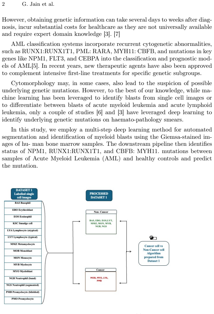
Summary. The manuscript proposes a two-stage deep learning pipeline for acute myeloid leukemia (AML) assessment from single-cell bone marrow images. Stage one trains a ResNet-18 binary classifier to distinguish leukemic blasts from non-leukemic cells on the AML-Cytomorphology MLL Helmholtz dataset (Dataset 1, over 170,000 cells from 945 patients), reporting roughly 90% accuracy. Stage two applies a ResNet-34 four-class classifier to Dataset 2 (189 slides with slide-level genetic mutation labels) to predict mutation categories, using smooth cross-entropy with an assumed 20% label-noise rate and majority voting over cells; the authors report 85% accuracy across the four mutation classes. The paper argues that this demonstrates noise-robust, clinically useful mutation prediction from morphological images alone.
Significance. If the reported results were supported by rigorous slide-disjoint evaluation, the work would be a useful contribution to morphology-based AML subtyping, potentially enabling faster and cheaper screening for clinically relevant mutations. The paper has notable strengths: it uses a large public dataset, explicitly addresses the noisy-label problem, and introduces a practical majority-voting mechanism. However, the current evaluation contains load-bearing methodological gaps—most importantly the unclear split granularity for Dataset 2 and the circular estimation of the label-noise parameter—so the central claim of 85% mutation-classification accuracy is not currently established. The clinical relevance and novelty of the question justify further work, but the evidence as presented does not meet the standard for publication.
major comments (4)
- [Section 3] The 72/18/10 split for Dataset 2 is not stated to be slide-disjoint. Since Dataset 2 consists of 189 slides with 99–500 cells per slide and each slide carries a single bag-level mutation label (Section 2.1), a cell-level split will place cells from the same slide into both training and test partitions. This permits the model to memorize slide-specific covariates such as staining, scanner, and patient background, which can inflate the reported 85% accuracy. The authors must evaluate at slide granularity, e.g., by reporting per-slide majority-vote accuracy under slide-level cross-validation, and they should state explicitly whether any cells from a slide in the training partition appear in the test partition.
- [Section 2.2 and Abstract] The 20% label-noise parameter in smooth cross-entropy is estimated by applying the first-stage blast classifier to unlabeled Dataset 2 and having pathologists review the model's predictions, as described in the Abstract. This estimate concerns blast versus non-blast labeling errors, not mutation-label noise; mutation labels come from genetic tests and follow a different error-generating process. Moreover, using the model's own predictions to estimate the noise rate and then fixing that rate in the loss is circular and does not justify the noise model applied to mutation classification. The authors should either independently estimate mutation-label noise (e.g., by expert re-review of a labeled subset) or report sensitivity of the results across a range of noise parameters.
- [Section 3] The cancer detection model is evaluated only on an 80/20 training/validation split with no independent test set. The reported 'approximately 90% accuracy' is the validation accuracy after early stopping; the manuscript does not report confidence intervals, patient-disjoint grouping, or a separate test cohort. Because the 5% threshold is applied to patient-level aggregated predictions, the paper should report patient-level sensitivity and specificity on a held-out set of patients, not just cell-level validation accuracy.
- [Sections 2.1 and 2.2] Dataset 2 is described as including four AML subtypes (PML:RARA, NPM1, CBFB:MYH11, RUNX1:RUNX1T1) plus a control group of healthy stem cell donors, but the mutation classifier is described as a four-class model. It is unclear whether the healthy controls are included in the 189 slides, whether they form a fifth class, or whether they are excluded from training and testing. If they are included but not modeled, the reported accuracy could be misleading; the authors should clarify the class label set and report per-class accuracy, precision, and recall for each mutation type.
minor comments (4)
- [Section 3] Table 1 is referenced in Section 3 but is not included in the manuscript; the accuracy, precision, recall, and F1 values that the text claims to report should be shown in the table.
- [Introduction] The Introduction lists NPM1, RUNX1:RUNX1T1, and CBFB:MYH11 mutations but not PML:RARA, while Section 2.1 includes PML:RARA among the four subtypes; please make the mutation list consistent throughout.
- [References] The reference numbering appears inconsistent: two distinct entries are both numbered [10] (Döhner et al. and Kockwelp et al.), and the in-text citations in Section 4 do not reliably match the reference list.
- [Abstract and Ethics Statement] The phrase 'tumor label noise' in the Abstract is ambiguous because the noise is in cell-level morphological labels rather than tumor-level labels, and the Ethics Statement says the study 'does not deal with human/animal data' despite using human bone marrow smears; please rephrase both to accurately describe the use of de-identified public human data.
Circularity Check
No significant circularity: the central mutation-classification result is an empirical outcome, not a reduction to fitted inputs or self-citations.
full rationale
The paper's derivation chain does not contain a step that is equivalent to its inputs by construction. The binary blast classifier is trained on expert-annotated Dataset 1 and evaluated on a validation split, so its 90% figure is an independent empirical result. The 20% label-noise estimate is obtained by applying that model to unlabeled Dataset 2 and having pathologists review the predictions; it is an external human-review measurement, not a value derived from the mutation labels or from the mutation classifier itself. The mutation classifier is then trained on Dataset 2 with slide-level mutation labels and smooth cross-entropy using a 20% noise hyperparameter, and its reported 85% accuracy is an empirical outcome, not an algebraic consequence of that hyperparameter. The paper's self-citations are background or methodological references and do not carry the central claim. The main concerns—that the Dataset 2 train/validation/test split is not explicitly stated to be slide-disjoint, and that a blast/non-blast disagreement rate is assumed as the mutation-label noise rate—are validity, leakage, and label-modeling risks, not circularity. They would be appropriate material for a correctness critique, but they do not make the prediction reduce to its own inputs. Therefore no circular step is established.
Assumptions & free parameters
free parameters (2)
- noisy_label_percentage =
20%
- blast_presence_threshold =
5%
assumptions (4)
- domain assumption Each slide-level mutation label in Dataset 2 applies to all cells from that slide, so every blast image can be trained with the slide's label.
- domain assumption Mutations NPM1, RUNX1:RUNX1T1, CBFB:MYH11, and PML:RARA produce morphology detectable in single Giemsa-stained cells at 40x.
- domain assumption A model trained on bone marrow smear images (Dataset 1) transfers to peripheral blood smear images (Dataset 2).
- ad hoc to paper The 20% label-noise estimate from pathologists reviewing the model's own predictions is unbiased.
Cite this review
Pith. "Pith review of Predicting Genetic Mutations from Single-Cell Bone Marrow Images in Acute Myeloid Leukemia Using Noise-Robust Deep Learning Models." pith.science (2026). https://pith.science/paper/FR4YUHDH
@misc{pith2026250612798,
  author       = {Pith},
  title        = {Pith review of: Predicting Genetic Mutations from Single-Cell Bone Marrow Images in Acute Myeloid Leukemia Using Noise-Robust Deep Learning Models},
  year         = {2026},
  howpublished = {\url{https://pith.science/paper/FR4YUHDH}},
  note         = {Machine review of arXiv:2506.12798}
}
read the original abstract
In this study, we propose a robust methodology for identification of myeloid blasts followed by prediction of genetic mutation in single-cell images of blasts, tackling challenges associated with label accuracy and data noise. We trained an initial binary classifier to distinguish between leukemic (blasts) and non-leukemic cells images, achieving 90 percent accuracy. To evaluate the models generalization, we applied this model to a separate large unlabeled dataset and validated the predictions with two haemato-pathologists, finding an approximate error rate of 20 percent in the leukemic and non-leukemic labels. Assuming this level of label noise, we further trained a four-class model on images predicted as blasts to classify specific mutations. The mutation labels were known for only a bag of cell images extracted from a single slide. Despite the tumor label noise, our mutation classification model achieved 85 percent accuracy across four mutation classes, demonstrating resilience to label inconsistencies. This study highlights the capability of machine learning models to work with noisy labels effectively while providing accurate, clinically relevant mutation predictions, which is promising for diagnostic applications in areas such as haemato-pathology.
Figures

Reference graph
Works this paper leans on
-
[1]
Computer Methods and Programs in Biomedicine202, 105999 (2021)
Boldú, L., Merino, A., Acevedo, A., Molina, A., Rodellar, J.: A deep learning model (alnet) for the diagnosis of acute leukaemia lineage using peripheral blood cell images. Computer Methods and Programs in Biomedicine202, 105999 (2021)
work page 2021
-
[2]
In: IEEE Conference on Computer Vision and Pattern Recognition
Deng, J., Dong, W., Socher, R., Li, L.J., Li, K., Fei-Fei, L.: Imagenet: A large-scale hierarchical image database. In: IEEE Conference on Computer Vision and Pattern Recognition. pp. 248–255 (2009). https://doi.org/10.1109/CVPR.2009.5206848
arXiv 2009
-
[3]
Blood, The Journal of the American Society of Hematology140(12), 1345–1377 (2022)
Döhner, H., Wei, A.H., Appelbaum, F.R., Craddock, C., DiNardo, C.D., Dombret, H., Ebert, B.L., Fenaux, P., Godley, L.A., Hasserjian, R.P., et al.: Diagnosis and management of aml in adults: 2022 recommendations from an international ex- pert panel on behalf of the eln. Blood, The Journal of the American Society of Hematology140(12), 1345–1377 (2022)
work page 2022
-
[4]
Gajendra, S., Gupta, R., Thakral, D., Gupta, S.K., Jain, G., Bakhshi, S., Sharma, A., Sahoo, R.K., Kumar, L., Rai, S., et al.: Cd34 negative hla-dr negative acute myeloid leukaemia: A higher association with npm1 and flt3-itd mutations. Inter- national Journal of Laboratory Hematology45(2), 221–228 (2023) Title Suppressed Due to Excessive Length 9
work page 2023
-
[5]
American Journal of Blood Research10(5), 179 (2020)
Haider, I., Kumar, C., Jain, G., Bakhshi, S., Kumar, L., Chopra, A., Ranjan, A., Tanwar, P.: Hotspots mutational analysis of wilms tumor 1 gene in acute myeloid leukaemia; prevalence and clinical correlation in north indian population. American Journal of Blood Research10(5), 179 (2020)
work page 2020
-
[6]
PLOS Digital Health2(3), e0000187 (2023)
Hehr, M., Sadafi, A., Matek, C., Lienemann, P., Pohlkamp, C., Haferlach, T., Spiekermann, K., Marr, C.: Explainable ai identifies diagnostic cells of genetic aml subtypes. PLOS Digital Health2(3), e0000187 (2023)
2023
-
[7]
American Journal of Blood Research10(5), 257 (2020)
Jain, G., Kumar, C., Malhotra, A., Mallick, S.R., Bakhshi, S., Chopra, A.: Periph- eral blood involvement in angioimmunoblastic t-cell lymphoma: A case report and review of the literature. American Journal of Blood Research10(5), 257 (2020)
work page 2020
-
[8]
American Journal of Blood Research10(4), 134 (2020)
Jain,G.,Thakral,D.,Sahoo,R.K.,Kumar,I.,Vashishtha,S.,Verma,P.,Gupta,R.: Next generation sequencing guided treatment modulation and prognosis in acute myeloid leukemia: Case vignettes. American Journal of Blood Research10(4), 134 (2020)
work page 2020
Show all 18 references
-
[9]
In: IEEE Conference on Computer Vision & Pattern Recognition
Jian, S., Kaiming, H., Shaoqing, R., Xiangyu, Z.: Deep residual learning for image recognition. In: IEEE Conference on Computer Vision & Pattern Recognition. pp. 770–778 (2016)
2016
-
[10]
Blood advances8(1), 70–79 (2024)
Kockwelp, J., Thiele, S., Bartsch, J., Haalck, L., Gromoll, J., Schlatt, S., Exeler, R., Bleckmann, A., Lenz, G., Wolf, S., et al.: Deep learning predicts therapy-relevant genetics in acute myeloid leukemia from pappenheim-stained bone marrow smears. Blood advances8(1), 70–79 (2024)
2024
-
[11]
In: Seminars in Diagnostic Pathology
Kurzer, J.H., Weinberg, O.K.: Updates in molecular genetics of acute myeloid leukemia. In: Seminars in Diagnostic Pathology. vol. 40, pp. 140–151. Elsevier (2023)
2023
-
[12]
In: Proceedings of the IEEE/CVF Interna- tional Conference on Computer Vision (ICCV)
Liu, Z., Lin, Y., Qi, Y., et al.: Swin transformer: Hierarchical vision trans- former using shifted windows. In: Proceedings of the IEEE/CVF Interna- tional Conference on Computer Vision (ICCV). pp. 10012–10022 (2021), https://arxiv.org/abs/2103.14030
2021 arXiv
-
[13]
arXiv preprint arXiv:2201.03545 (2022), https://arxiv.org/abs/2201.03545
Liu, Z., Wang, J., Si, X., et al.: Convnext: Revisiting convolutional neu- ral networks for visual recognition. arXiv preprint arXiv:2201.03545 (2022), https://arxiv.org/abs/2201.03545
2022 arXiv
-
[14]
Scientific Reports13(1), 13364 (2023)
Lv, Z., Cao, X., Jin, X., Xu, S., Deng, H.: High-accuracy morphological identifica- tion of bone marrow cells using deep learning-based morphogo system. Scientific Reports13(1), 13364 (2023)
2023
-
[15]
In: 2024 IEEE 24th International Conference on Bioinformatics and Bioengineering (BIBE)
Sekhar, A., Goel, V., Jain, G., Patil, A., Gupta, R.K., Bameta, T., Rane, S., Sethi, A.: Her2 and fish status prediction in breast biopsy h&e-stained images using deep learning. In: 2024 IEEE 24th International Conference on Bioinformatics and Bioengineering (BIBE). pp. 1–8. I...
2024
-
[16]
In: International Conference on Learning Representations (2015), https://arxiv.org/abs/1409.1556
Simonyan, K., Zisserman, A.: Very deep convolutional networks for large-scale im- age recognition. In: International Conference on Learning Representations (2015), https://arxiv.org/abs/1409.1556
2015 arXiv
-
[17]
In: Proceedings of the IEEE conference on computer vision and pattern recognition
Szegedy, C., Vanhoucke, V., Ioffe, S., Shlens, J., Wojna, Z.: Rethinking the incep- tion architecture for computer vision. In: Proceedings of the IEEE conference on computer vision and pattern recognition. pp. 2818–2826 (2016)
2016
-
[18]
In: International conference on machine learning
Tan, M., Le, Q.: Efficientnet: Rethinking model scaling for convolutional neural networks. In: International conference on machine learning. pp. 6105–6114. PMLR (2019)
2019
Reviewed August 7, 2026 · model on record in the stance chip above.
Discussion (0). Continue with ORCID to comment.