REVIEW 3 major objections 5 minor 37 references
MRExtrap: Longitudinal Aging of Brain MRIs using Linear Modeling in Latent Space
T0 review · 3 major / 5 minor · reviewed 2026-08-15 · deepseek-v4-flash
Pith's one-line read A straight-line latent model beats a GAN at predicting brain aging.
desk verdict A simple, credible linear-extrapolation baseline for brain aging MRI, with a strong global-prior result but an evaluation that needs a few gaps closed before it can be published as-is. read the letter →
The pith
A machine-rendered reading of the paper's core claim, the machinery that carries it, and where it could break.
The reading
What carries the argument
The central object is the latent progression rate $\beta$, a tensor in the same shape as the latent code, together with the linear identity $z_i - z_j = \beta(a_i - a_j)$ in the autoencoder's latent space. Because decoded regional volumes are approximately linear in the latent interpolation factor, and age-to-volume relationships in the literature are approximately linear, the paper hypothesizes age-to-latent linearity; prediction then anchors at the latest scan and adds $\beta$ times the age gap. The Global Prior sets $\beta = \mu_{\text{global}}$, the population mean of per-subject fitted rates; amortized UNet and diffusion priors predict subject-specific $\beta$ from a baseline scan; and additional scans are incorporated through closed-form Gaussian posterior updates.
What would settle it
Look at subjects with long follow-ups, fit $\beta$ from the first two years of scans, and check whether predictions for years five to ten beat the population prior in regional-volume MAE; if per-subject early-rate extrapolation is no better than the group average, or if the error grows with the absolute change in the regional atrophy rate between the conditioning and prediction windows, the constant-$\beta$ linear model is wrong.
Extended reading notes
Core claim
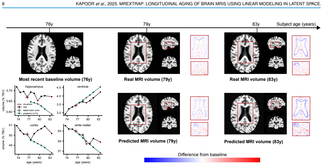
MRExtrap claims that aging trajectories in the latent space of 3D convolutional autoencoders are approximately linear, so future brain MRIs can be generated by linear extrapolation of the latent code. The paper supports this with the identity $z_i - z_j = \beta(a_i - a_j)$, where $\beta$ is a subject-specific progression rate; using the population average $\mu_{\text{global}}$ as $\beta$, it reports hippocampus MAE of 0.020 versus 0.060 for the GAN baseline, with comparable or better performance in grey and white matter and a mixed result for ventricles. It further shows that $\beta$, measured by its $\ell^1$ norm, rises from healthy to MCI to dementia, matching known volumetric atrophy patterns across diagnostic groups and age bins.
Load-bearing premise
The load-bearing premise is that each person's brain code travels along a straight line with age, so the slope measured today keeps holding into the future.
Editorial extensions
If this is right
- A single scan plus the population-average progression rate yields future brain volumes whose hippocampus, grey matter, and white matter errors are lower than the compared GAN baseline; ventricle errors are comparable or mixed.
- The framework accepts any number of past scans at arbitrary ages, using closed-form Bayesian updates to refine the progression rate whenever more scans arrive.
- When a subject's regional atrophy rate changes between the conditioning period and the prediction window, posterior updating does not help on average; the population prior can be the better forecast.
- The estimated latent progression rate carries disease and age information: its $\ell^1$ norm increases from healthy to MCI to dementia and separates groups when binned by age at first scan.
Reading between the lines
- A natural test outside the paper: train the same autoencoder and linear extrapolation on longitudinal MRI of other organs or on other brain modalities, where atrophy rates are also slow; if the Global Prior stays competitive, the result is a generic aging baseline rather than a brain-specific model.
- Because posterior updating fails when early and late progression rates differ, the framework suggests a modification the paper does not pursue: model $\beta$ as a slowly varying latent process, for example piecewise linear in age, which would keep interpretation while allowing acceleration in dementia.
- The correlation between the norm of $\beta$ and diagnosis status implies $\beta$ could serve as a scalar biomarker; one could test whether $\beta$ measured from a single scan and the population prior separates future converters from stable MCI better than standard volumetric measures.
Signed reviews
Editorial analysis
A structured set of objections, weighed in public.
Referee Report
Summary. The paper proposes MRExtrap, a method for predicting future 3D brain MRIs from one or more observed scans by modeling brain aging as a linear progression in the latent space of a convolutional autoencoder. The latent code at a future age is obtained by linear extrapolation from the most recent latent using a progression rate beta, which is estimated either as a global population-averaged prior, an amortized subject-specific prior (Gaussian or diffusion), or a Bayesian posterior updated from additional longitudinal observations. On ADNI, the authors report that the simple Global Prior achieves mean absolute errors of 0.020% TBV for hippocampus, 0.322% for ventricles, 0.509% for grey matter, and 0.399% for white matter for single-scan 2-4 year prediction, beating the reported DANINet baseline in hippocampus, grey matter, and white matter. The paper also analyzes multi-scan posterior updating, finding that it does not consistently improve over the Global Prior, and shows that the inferred progression rates correlate with disease status and age in a manner consistent with volumetric atrophy literature. The manuscript is transparent about its central linearity assumption and includes a discussion of its limitations.
Significance. If the single-scan results are robust, MRExtrap would be a valuable contribution: it provides an extremely simple, interpretable, and computationally inexpensive baseline for brain-aging MRI generation, and the released code and closed-form Bayesian update make the method easy to reproduce and extend. The observation that a global average progression rate in latent space can compete with a purpose-built GAN has practical value as a benchmark. The paper also honestly reports that the linear constancy assumption breaks down over longer horizons and for some MCI/dementia subjects, which is a measured assessment. However, the headline single-scan claim currently lacks a decisive control: the no-change baseline beta=0 is not reported, and the DANINet comparison relies on previously published numbers under a different pipeline. These gaps must be addressed before the claimed predictive accuracy can be accepted.
major comments (3)
- [Sec. VI-A, Table I] The single-scan evaluation never reports a no-progression control (beta=0, i.e., decoding the latest latent z_N unchanged). Since the evaluation horizon is only 2-4 years and the metric is volume MAE as a percentage of total brain volume, a predictor that outputs no aging could achieve small MAE if the average absolute volume change over the horizon is comparable to the reported values. In particular, the hippocampus MAE of 0.020% TBV is the same order of magnitude as typical annual hippocampal volume loss expressed in % TBV, so the reported error does not by itself show that the age-dependent beta term contributes. Please report the beta=0 MAE for all four regions, together with the mean absolute volume change from baseline over the test horizon, and state whether the full extrapolation is significantly better than this trivial baseline. This control is decisive for the central claim that MRExtrap accurately predicts aging rather than merely reproducing the latest scan.
- [Sec. VI-A and Sec. V (Baselines)] The comparison with DANINet uses numbers reported directly by Ravi et al. rather than re-running the baseline under the preprocessing, resolution, and evaluation pipeline used in this paper. The authors preprocess with ROI clipping, bias-field correction, affine registration to MNI152, and skull stripping at 160x192x160 resolution, while the reported DANINet results are for 2D slices at 128x128x100 per volume. Because the z-test in Sec. VI-A is computed from the reported DANINet mean and standard error, any protocol difference in registration, segmentation, or subject-level pairing could affect the comparison. Please either re-run DANINet under the same pipeline or clearly qualify the comparison as cross-protocol and soften the claim that MRExtrap 'outperforms' DANINet.
- [Sec. VI-B and Sec. VIII] The paper's own posterior-updating experiments show that the constant-beta assumption breaks down for longer trajectories, with clear shifts in regional progression rates between the conditioning and prediction periods, especially for MCI/dementia subjects. This is acknowledged in Sec. VIII, but the single-scan conclusion in Sec. VI-A is stated without an explicit horizon limitation. Since the linearity assumption is central to the method, the authors should state more precisely the range of age gaps over which the single-scan Global Prior remains accurate relative to beta=0, and should present the single-scan and multi-scan claims as separate regimes rather than as one general 'accurate aging prediction' claim.
minor comments (5)
- [Sec. III-B1] There is a duplicated phrase: 'corresponding to corresponding to scans separated by approximately 2-4 years' should be corrected.
- [Sec. VI-A] The word 'inhertently' should be 'inherently' in the sentence about adapting a population-level estimate from a single scan.
- [Sec. VI-D title] The section title 'Behavior of of beta across disease labels' contains a duplicated 'of'.
- [Sec. V, Evaluation Metrics] The evaluation is based solely on four regional volume MAEs. Since the method generates full 3D MRI volumes, reporting additional image-level fidelity metrics such as SSIM or LPIPS on the predicted scans, or at least the reconstruction-quality numbers for the decoder outputs used in the prediction, would help the reader assess voxel-level prediction quality beyond the qualitative examples.
- [Sec. III-B2] The derivation of z(a) = beta*a + z0 as motivation is understandable, but the subject-specific mapping C is never estimated, so this should be framed purely as a motivation rather than as a verified derivation. The paper already says the linear relationship is hypothesized, which is appropriate; making this framing explicit in the final version would avoid confusion.
Circularity Check
No significant circularity: the progression rate is estimated on training subjects and applied to held-out test scans, and the linearity assumption is an empirically motivated hypothesis rather than an input redefined as the output.
full rationale
The central prediction rule z* = z_N + beta*(a* - a_N) is an extrapolation formula whose slope beta is estimated from training data (Sec. IV-A: beta_i = LinearRegression(Delta a, Delta z)) and then averaged into the Global Prior mu_global (Sec. IV-B). Applying mu_global to held-out test latents is standard supervised generalization, not a tautology: the test volumes are never used to set beta, and the decoded predictions are compared against real unseen scans. The linearity assumption is explicitly proposed as a hypothesis (Sec. III-B2: 'we hypothesize a direct linear relationship between age and latent representations') and is supported by empirical interpolation plots and PCA, not derived from the target predictions. The algebra with v = Cz + c and v = Ba + b is motivational and does not define the reported errors. The paper itself flags the linearity assumption as approximate in Sec. VIII, which is a limitation rather than a circular step. The only self-citations ([2], [15]) appear in the related-work survey and are not load-bearing for any derivation or evaluation. The absence of a beta = 0 no-progression control is an evaluation-strength concern, not a circularity concern: even if the learned slope contributed little, the prediction would not be equivalent to its input by construction in the sense required by the circularity criteria.
Assumptions & free parameters
free parameters (4)
- Global prior mean mu_global (population-averaged progression rate) =
not reported numerically
- Observation noise variance sigma^2_obs =
estimated from training residuals, not reported
- Autoencoder loss weights gamma1, gamma2 =
gamma1 = 0.005, gamma2 = 1e-5
- Latent dimension =
4 x 20 x 24 x 20
assumptions (5)
- domain assumption Regional brain volumes v(a) are approximately linear in age a: v(a) = B a + b.
- domain assumption Within a subject, regional volumes v are approximately linear in latent interpolation factor alpha: v_interp ~ alpha v1 + (1-alpha) v2.
- ad hoc to paper Latent representations z(a) are linear in age with constant slope beta: z_i - z_j = beta(a_i - a_j).
- domain assumption The autoencoder's reconstructions are accurate enough that latent-space linear operations transfer to voxel space.
- standard math The Bayesian linear regression update (Eq. 4) correctly models observation noise as i.i.d. Gaussian with fixed variance sigma^2_obs.
Cite this review
Pith. "Pith review of MRExtrap: Longitudinal Aging of Brain MRIs using Linear Modeling in Latent Space." pith.science (2026). https://pith.science/paper/FTTPSK2W
@misc{pith2026250819482,
  author       = {Pith},
  title        = {Pith review of: MRExtrap: Longitudinal Aging of Brain MRIs using Linear Modeling in Latent Space},
  year         = {2026},
  howpublished = {\url{https://pith.science/paper/FTTPSK2W}},
  note         = {Machine review of arXiv:2508.19482}
}
abstract
Simulating aging in 3D brain MRI scans can reveal disease progression patterns in neurological disorders such as Alzheimer's disease. Current deep learning-based generative models typically approach this problem by predicting future scans from a single observed scan. We investigate modeling brain aging via linear models in the latent space of convolutional autoencoders (MRExtrap). Our approach, MRExtrap, is based on our observation that autoencoders trained on brain MRIs create latent spaces where aging trajectories appear approximately linear. We train autoencoders on brain MRIs to create latent spaces, and investigate how these latent spaces allow predicting future MRIs through linear extrapolation based on age, using an estimated latent progression rate $\boldsymbol{\beta}$. For single-scan prediction, we propose using population-averaged and subject-specific priors on linear progression rates. We also demonstrate that predictions in the presence of additional scans can be flexibly updated using Bayesian posterior sampling, providing a mechanism for subject-specific refinement. On the ADNI dataset, MRExtrap predicts aging patterns accurately and beats a GAN-based baseline for single-volume prediction of brain aging. We also demonstrate and analyze multi-scan conditioning to incorporate subject-specific progression rates. Finally, we show that the latent progression rates in MRExtrap's linear framework correlate with disease and age-based aging patterns from previously studied structural atrophy rates. MRExtrap offers a simple and robust method for the age-based generation of 3D brain MRIs, particularly valuable in scenarios with multiple longitudinal observations.
Figures


Figures from the paper (2 more)

Reference graph
Works this paper leans on
-
[1]
C. Bass, M. da Silva, C. H. Sudre, et al., “ICAM-reg: Interpretable classification and regression with feature attribution for mapping neurological phenotypes in individual scans,” in Medical Imaging with Deep Learning, 2021
work page 2021
-
[2]
Visual feature attribution using wasserstein gans,
C. F. Baumgartner, L. M. Koch, K. C. Tezcan, J. X. Ang, and E. Konukoglu, “Visual feature attribution using wasserstein gans,” in 2018 IEEE Conference on Computer Vision and Pattern Recognition, CVPR 2018, Salt Lake City, UT, USA, June 18-22, 2018 , 2018
work page 2018
-
[3]
B. Billot, C. Magdamo, Y . Cheng, S. E. Arnold, S. Das, and J. E. Iglesias, “Robust machine learning segmentation for large-scale analysis of heterogeneous clinical brain mri datasets,” Proceedings of the National Academy of Sciences , 2023
work page 2023
-
[4]
Prediction of the progression of subcortical brain structures in alzheimer’s disease from baseline,
A. Bône, M. Louis, A. Routier, J. Samper, M. Bacci, B. Charlier, O. Colliot, and S. Durrleman, “Prediction of the progression of subcortical brain structures in alzheimer’s disease from baseline,” in Graphs in Biomedical Image Analysis, Computational Anatomy and Imaging Genetics, 2017
work page 2017
-
[5]
The use of neuroimaging techniques in the early and differential diagnosis of dementia,
L. Chouliaras and J. T. O’Brien, “The use of neuroimaging techniques in the early and differential diagnosis of dementia,” Molecular Psychiatry, vol. 28, no. 10, pp. 4084–4097, 2023
work page 2023
-
[6]
Diffusion models beat gans on image synthesis,
P. Dhariwal and A. Q. Nichol, “Diffusion models beat gans on image synthesis,” in Advances in Neural Information Processing Systems , M. Ranzato, A. Beygelzimer, Y . N. Dauphin, P. Liang, and J. W. Vaughan, Eds., 2021
work page 2021
-
[7]
An image is worth 16x16 words: Transformers for image recognition at scale,
A. Dosovitskiy, L. Beyer, A. Kolesnikov, et al., “An image is worth 16x16 words: Transformers for image recognition at scale,”International Conference On Learning Representations , 2020
work page 2020
-
[8]
Patterns of progressive atrophy vary with age in alzheimer’s disease patients,
C. M. Fiford, G. R. Ridgway, D. M. Cash, et al., “Patterns of progressive atrophy vary with age in alzheimer’s disease patients,” Neurobiology of aging, vol. 63, pp. 22–32, 2018
work page 2018
Show all 37 references
-
[9]
High consistency of regional cortical thinning in aging across multiple samples,
A. M. Fjell, L. T. Westlye, I. Amlien, et al. , “High consistency of regional cortical thinning in aging across multiple samples,” Cereb Cortex, vol. 19, no. 9, pp. 2001–2012, 2009
2001
-
[10]
Characterization of Brain V olume Changes in Aging Individuals With Normal Cognition Using Serial Magnetic Resonance Imaging,
S. Fujita, S. Mori, K. Onda, et al., “Characterization of Brain V olume Changes in Aging Individuals With Normal Cognition Using Serial Magnetic Resonance Imaging,” JAMA Network Open , 2023
2023
-
[11]
Denoising diffusion probabilistic models,
J. Ho, A. Jain, and P. Abbeel, “Denoising diffusion probabilistic models,” in NeurIPS, H. Larochelle, M. Ranzato, R. Hadsell, M. Balcan, and H. Lin, Eds., 2020
2020
-
[12]
Huszar, Gaussian distributions are soap bubbles , https : / / www
F. Huszar, Gaussian distributions are soap bubbles , https : / / www . inference . vc / high - dimensional - gaussian - distributions-are-soap-bubble/ , 2017
2017
-
[13]
Image-to-image translation with conditional adversarial networks,
P. Isola, J. -Y . Zhu, T. Zhou, and A. A. Efros, “Image-to-image translation with conditional adversarial networks,” arXiv preprint arXiv: 1611.07004, 2016
2016 arXiv
-
[14]
Conditional gan with 3d discriminator for mri generation of alzheimer’s disease progression,
E. Jung, M. Luna, and S. H. Park, “Conditional gan with 3d discriminator for mri generation of alzheimer’s disease progression,” Pattern Recognition, vol. 133, p. 109 061, 2023
2023
-
[15]
Multiscale metamor- phic vae for 3d brain mri synthesis,
J. Kapoor, J. H. Macke, and C. F. Baumgartner, “Multiscale metamor- phic vae for 3d brain mri synthesis,” NeurIPS Workshop on Medical Imaging, 2022
2022
-
[16]
Ad course map charts alzheimer’s disease progression,
I. Koval, A. Bône, M. Louis, et al., “Ad course map charts alzheimer’s disease progression,” Scientific Reports, vol. 11, no. 1, p. 8020, 2021
2021
-
[17]
Generation of 3d brain MRI using auto-encoding generative adversarial networks,
G. Kwon, C. Han, and D. Kim, “Generation of 3d brain MRI using auto-encoding generative adversarial networks,” in MICCAI 2019
2019
-
[18]
Cerebral atrophy in mild cognitive impairment and alzheimer disease: Rates and acceleration,
K. K. Leung, J. W. Bartlett, J. Barnes, E. N. Manning, S. Ourselin, N. C. Fox, and A. D. N. Initiative, “Cerebral atrophy in mild cognitive impairment and alzheimer disease: Rates and acceleration,” Neurology, vol. 80, 2013
2013
-
[19]
Decoupled weight decay regularization,
I. Loshchilov and F. Hutter, “Decoupled weight decay regularization,” International Conference on Learning Representations , 2017
2017
-
[20]
Diffusion probabilistic models beat gans on medical images,
G. Müller-Franzes, J. M. Niehues, F. Khader, et al. , “Diffusion probabilistic models beat gans on medical images,” arXiv preprint arXiv: Arxiv-2212.07501, 2022
2022 arXiv
-
[21]
Autoencoder image interpolation by shaping the latent space,
A. Oring, Z. Yakhini, and Y . Hel-Or, “Autoencoder image interpolation by shaping the latent space,” in Proceedings of the 38th International Conference on Machine Learning , M. Meila and T. Zhang, Eds., ser. Proceedings of Machine Learning Research, vol. 139, PMLR, 18-24 Jul ...
2021
-
[22]
Alzheimer’s disease neuroimaging initiative (adni) clinical characterization,
R. C. Petersen, P. S. Aisen, L. A. Beckett, et al., “Alzheimer’s disease neuroimaging initiative (adni) clinical characterization,” Neurology, vol. 74, no. 3, pp. 201–209, 2010
2010
-
[23]
Brain imaging generation with latent diffusion models,
W. H. Pinaya, P.-D. Tudosiu, J. Dafflon, P. F. D. Costa, V . Fernandez, P. Nachev, S. Ourselin, and M. Cardoso, “Brain imaging generation with latent diffusion models,” DGM4MICCAI Workshop, MICCAI, 2022
2022
-
[24]
Equitable modelling of brain imaging by counterfactual augmentation with morphologically constrained 3d deep generative models,
G. Pombo, R. Gray, M. Cardoso, S. Ourselin, G. Rees, J. Ashburner, and P. Nachev, “Equitable modelling of brain imaging by counterfactual augmentation with morphologically constrained 3d deep generative models,” Medical Image Anal. , 2021
2021
-
[25]
Enhancing spatiotemporal disease progression models via latent diffusion and prior knowledge,
L. Puglisi, D. C. Alexander, and D. Ravì, “Enhancing spatiotemporal disease progression models via latent diffusion and prior knowledge,” MICCAI, 2024
2024
-
[26]
Degenerative adversarial neuroimage nets for brain scan simulations: Application in ageing and dementia,
D. Ravi, S. B. Blumberg, S. Ingala, F. Barkhof, D. C. Alexander, N. Oxtoby, et al., “Degenerative adversarial neuroimage nets for brain scan simulations: Application in ageing and dementia,” Medical Image Analysis, vol. 75, p. 102 257, 2022
2022
-
[27]
High- resolution image synthesis with latent diffusion models,
R. Rombach, A. Blattmann, D. Lorenz, P. Esser, and B. Ommer, “High- resolution image synthesis with latent diffusion models,” CVPR, 2022
2022
-
[28]
Thinning of the Cerebral Cortex in Aging,
D. H. Salat, R. L. Buckner, A. Z. Snyder, D. N. Greve, R. S. Desikan, E. Busa, J. C. Morris, A. M. Dale, and B. Fischl, “Thinning of the Cerebral Cortex in Aging,” Cerebral Cortex, vol. 14, no. 7, pp. 721–730, 2004
2004
-
[29]
Predicting brain atrophy from tau pathology: A summary of clinical findings and their translation into personalized models,
A. Schäfer, P. Chaggar, T. B. Thompson, A. Goriely, and E. Kuhl, “Predicting brain atrophy from tau pathology: A summary of clinical findings and their translation into personalized models,” Brain Multi- physics, vol. 2, 2021
2021
-
[30]
Generalised dice overlap as a deep learning loss function for highly unbalanced segmentations,
C. Sudre, W. Li, T. K. M. Vercauteren, S. Ourselin, and M. J. Cardoso, “Generalised dice overlap as a deep learning loss function for highly unbalanced segmentations,” DLMIA/ML-CDS@MICCAI, 2017
2017
-
[31]
Morphology- preserving autoregressive 3d generative modelling of the brain,
P.-D. Tudosiu, W. H. L. Pinaya, M. S. Graham, et al., “Morphology- preserving autoregressive 3d generative modelling of the brain,” in SASHIMI Worksop, MICCAI, 2022
2022
-
[32]
Neural discrete representation learning,
A. Van Den Oord, O. Vinyals, et al., “Neural discrete representation learning,” NeurIPS, 2017
2017
-
[33]
Sampling generative networks,
T. White, “Sampling generative networks,” arXiv preprint arXiv: 1609.04468, 2016
2016 arXiv
-
[34]
Sadm: Sequence- aware diffusion model for longitudinal medical image generation,
J. S. Yoon, C. Zhang, H. -I. Suk, J. Guo, and X. Li, “Sadm: Sequence- aware diffusion model for longitudinal medical image generation,” in Information Processing in Medical Imaging , 2023
2023
-
[35]
Adding conditional control to text-to-image diffusion models,
L. Zhang, A. Rao, and M. Agrawala, “Adding conditional control to text-to-image diffusion models,” IEEE International Conference on Computer Vision, 2023. DOI: 10.1109/ICCV51070.2023.00355
2023
-
[36]
The unreasonable effectiveness of deep features as a perceptual metric,
R. Zhang, P. Isola, A. A. Efros, E. Shechtman, and O. Wang, “The unreasonable effectiveness of deep features as a perceptual metric,” arXiv preprint arXiv: 1801.03924 , 2018
2018 arXiv
-
[37]
Variational autoencoder for regression: Application to brain aging analysis,
Q. Zhao, E. Adeli, N. Honnorat, T. Leng, and K. M. Pohl, “Variational autoencoder for regression: Application to brain aging analysis,” in MICCAI 2019
2019
Reviewed August 15, 2026 · model on record in the stance chip above.
Discussion (0). Continue with ORCID to comment.