REVIEW 3 major objections 6 minor 55 references
A Gaussian process approach for rapid evaluation of skin tension
T0 review · 3 major / 6 minor · reviewed 2026-08-07 · deepseek-v4-flash
Pith's one-line read A Gaussian process trained on 10,000 finite-element skin simulations maps Rayleigh and supersonic wave speeds to stress and pre-stretch, with cross-validated $R^2 = 0.9570$.
desk verdict A solid simulation-based proof of concept for GP inversion of skin wave speeds, but the central in vivo claim is untested because the supersonic wave was never detected experimentally. read the letter →
The pith
A machine-rendered reading of the paper's core claim, the machinery that carries it, and where it could break.
The reading
What carries the argument
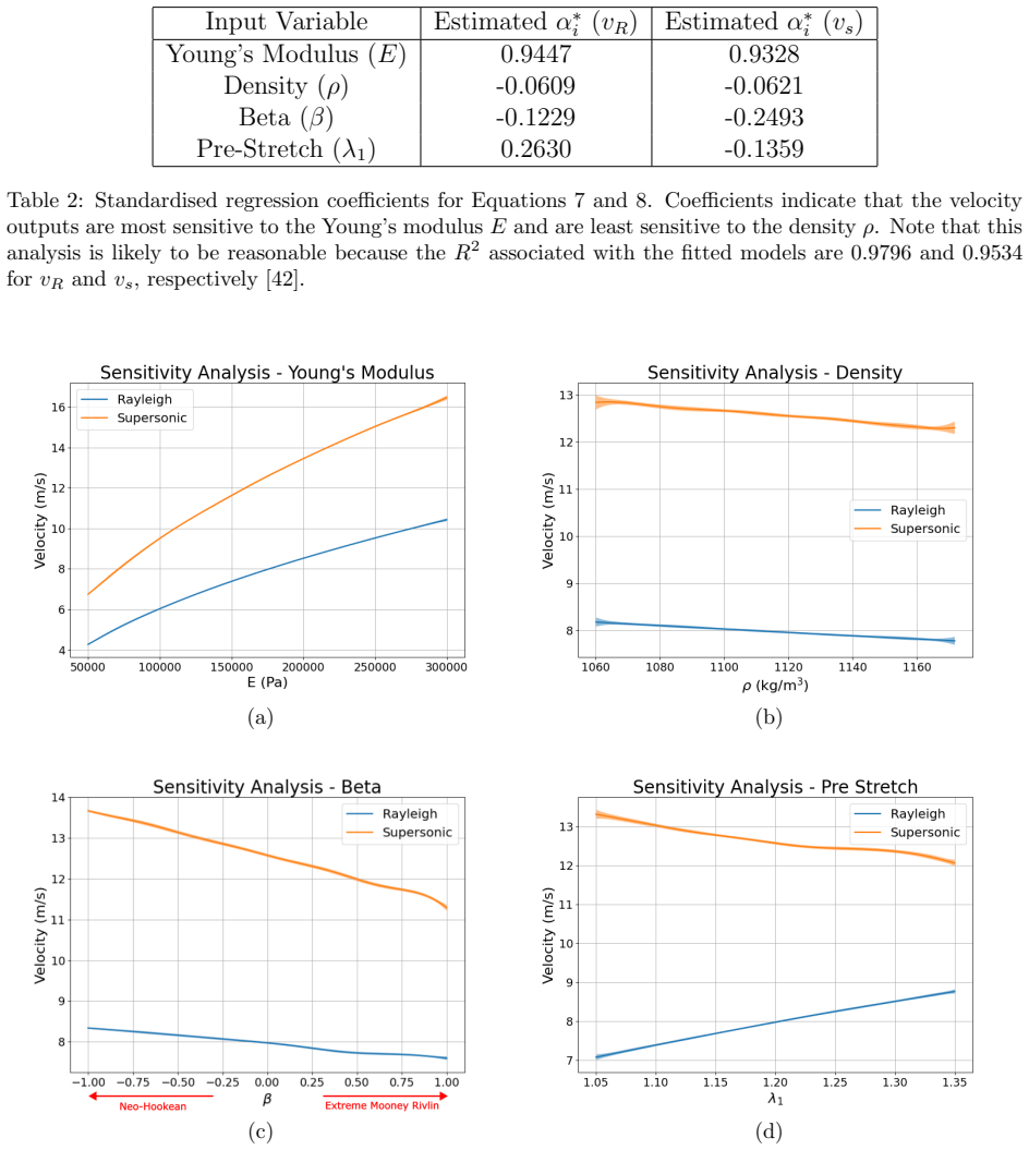
The machinery is a two-dimensional finite-element model of a pre-stretched skin block that simulates a surface wave propagation experiment, generating 10,000 training cases across two hyperelastic material descriptions, coupled with a multi-output Gaussian process regression using a radial-basis-function kernel. A Gaussian process is a statistical model that predicts an output value along with an uncertainty from previously seen training examples. The GP serves two roles: as an emulator it reproduces the finite-element wave speeds with $R^2 = 0.9993$ at about four orders of magnitude lower computational cost; as an inverse solver it maps $(v_R, v_s)$ to $(S_{11}, \lambda_1)$. The inversion works because increasing pre-stretch raises the Rayleigh speed while lowering the supersonic shear speed, so the speed pair carries far more information about stretch than either speed alone.
What would settle it
Run an in vivo study on human skin with a sensor that can resolve both the Rayleigh and supersonic arrivals, then compare the GP-predicted $S_{11}$ and $\lambda_1$ against an independent measurement such as excision retraction; if the supersonic wave is undetectable or the predictions disagree beyond the training range, the central claim fails.
Extended reading notes
Core claim
The central claim is that the ill-posed inverse problem of determining in vivo skin stress and pre-stretch from surface wave speeds can be solved in real time with a statistical surrogate. Using the pair of measurable speeds, $v_R$ and $v_s$, a Gaussian process regression trained on finite-element outputs predicts the steady-state stress $S_{11}$ and natural pre-stretch $\lambda_1$ with cross-validated $R^2 = 0.9570$. The supersonic wave is load-bearing: dropping it leaves pre-stretch almost unidentifiable ($R^2 = 0.1405$), while including it makes pre-stretch recoverable. Experimental Rayleigh wave speeds from uniaxially stretched synthetic skin agree with the FE predictions up to moderate stretch, and a model trained only on simulations predicts the synthetic skin's Young's modulus near the 146 kPa measured by independent destructive characterisation. The paper concludes that elastic wave measurements combined with machine learning are a viable non-invasive route to patient-specific skin tension.
Load-bearing premise
The load-bearing premise is that the supersonic shear wave speed can be measured reliably in living human skin; the paper's own experimental device did not detect it, and without it pre-stretch predictions degrade to $R^2 = 0.1405$.
Editorial extensions
If this is right
- With two measured wave speeds, the inverse GP returns $S_{11}$ and $\lambda_1$ in near real time, replacing iterative inverse finite-element fitting for this problem.
- A Rayleigh-only device can still estimate stiffness, as demonstrated on synthetic skin, but cannot recover pre-stretch ($R^2 = 0.1405$), so device design should target reliable detection of the supersonic shear wave.
- The simplified fitted equations for $S_{11}$ and $\lambda_1$ provide closed-form approximations that portable clinical devices could evaluate without running the GP.
- The training pipeline extends by adding new simulator runs, so additional material models or three-dimensional geometries can be incorporated as future training data.
Reading between the lines
- Beyond the paper: if clinical sensors cannot detect the supersonic shear wave, the two-speed inversion presented here cannot be deployed as-is, and the Rayleigh-only fallback gives stiffness but not pre-stretch.
- Beyond the paper: a decisive engineering test would move receivers closer to the impact site and increase excitation amplitude to see whether the supersonic wave survives attenuation in living human skin.
- Beyond the paper: the same emulator-plus-inverse-GP recipe transfers to other pre-stressed soft tissues with expensive forward simulations and cheap wave measurements, such as tendon or arterial wall.
- Beyond the paper: because the training data are deterministic and noise-free, adding realistic sensor noise or retraining on real waveforms is the natural next step toward clinical robustness, as the paper itself notes but does not test.
Editorial analysis
A structured set of objections, weighed in public.
Referee Report
Summary. This paper proposes a machine-learning pipeline for non-invasive estimation of in vivo skin stress and pre-stretch from surface wave speed measurements. The authors simulate 10,000 uniaxial pre-stretch and wave-propagation finite element experiments over a Latin hypercube of material parameters, train a Gaussian process emulator mapping (E, β, ρ, λ1) to Rayleigh and supersonic shear wave speeds, and then train a second Gaussian process to invert (v_R, v_s) to steady-state stress S11 and natural pre-stretch λ1. The inverse model achieves 10-fold cross-validation R²=0.9570. An experimental device with piezoelectric sensors is used on synthetic skin at four pre-stretch levels; Rayleigh wave speeds are measured and agree reasonably with the FE/analytical predictions, but the supersonic wave is not detected. A separate GP trained on FE data with pre-stretch and v_R as inputs predicts the synthetic skin's Young's modulus within one standard deviation of an independent measurement. The paper concludes that elastic wave measurements combined with machine learning provide a viable non-invasive method for determining in vivo skin tension.
Significance. If the two-input inverse mapping were validated on physical skin, the approach would be a meaningful advance: a cheap, fast, non-invasive measurement of skin pre-stretch and stress could inform surgical planning. The paper's strengths are its large simulator-based dataset, the GP emulator's agreement with the analytical Rayleigh solution (R²=0.9951, Section 3.1), stable 10-fold cross-validation with a small standard deviation, and a successful experimental prediction of Young's modulus for synthetic skin using a FE-trained GP. However, the headline inverse model (v_R, v_s) → (S11, λ1) is not tested on experimental data because v_s could not be measured; the experimental validation exercises a different model. The paper is therefore best read as a simulation-based proof of concept with a partial experimental feasibility demonstration.
major comments (3)
- [Section 2.5, Section 3.4, Conclusions] The central inverse model (Section 2.4) has not been validated experimentally. Section 2.5 states that "the supersonic wave was not visible" with the piezoelectric sensors, and Section 3.4 consequently trains a new GP that uses pre-stretch and v_R to predict Young's modulus, explicitly noting that the Section 2.4 model could not be validated. Because Section 3.3 shows that λ1 prediction collapses to R²=0.1405 when only v_R is used, the usefulness of the method depends critically on v_s detectability. The abstract's statement that the method provides "real time non-invasive access to in vivo stretch and stress" and the corresponding concluding claim are therefore not supported by the experimental results; either demonstrate v_s detection with a more sensitive receiver or explicitly scope the claims to the simulated setting.
- [Section 3.3 and Section 4] The 10-fold cross-validation R²=0.9570 is computed on noiseless, deterministic simulator outputs. The authors acknowledge in Section 4 that no noise is present in the training data. Real wave-speed measurements on synthetic skin show 1–3 m/s scatter for a fixed Young's modulus (Figure 12a), and the experimental device could not resolve the weaker v_s signal. Since the reported R² does not include measurement noise, it likely overstates the real-world performance of the inverse model. A noise-robustness study (e.g., adding realistic jitter to the simulated inputs during training, or reporting prediction intervals under measurement error) would substantially strengthen the generalization claim.
- [Section 2.4 and Section 3.3] The inverse problem is described as "ill-posed", but the identifiability of the target pair (S11, λ1) from the two wave speeds is not examined. Because the forward simulator maps four inputs (E, β, ρ, λ1) to two outputs, different parameter combinations can in principle produce identical (v_R, v_s); in that case the GP would regress to a conditional mean and the R² value would hide the non-uniqueness. A check of the Jacobian of the forward map over the input hypercube, or an analysis of how many training points with similar (v_R, v_s) have different targets, would clarify the actual degree of ill-posedness.
minor comments (6)
- [Section 2.2, Eq. (6)] The third formula should read D1 = (9−18ν)/(E(1+ν)); as written, "ν=" is a typo that obscures the intended relationship.
- [Section 2.5] The distance between the two sensors (17.13 mm) appears only in the Figure 5 caption; please state it in the text, along with the distance from the impact site to the first sensor.
- [Section 3.2 and Section 4] "transistion" should be "transition" and "feasability" should be "feasibility".
- [Equations 9–12] Specify the units of all fitted coefficients (e.g., E in kPa, wave speeds in m/s, S11 in Pa) and note that Equations 9 and 10 include a quadratic term in E, so they are not strictly linear regression models as described in the text.
- [Figure 12b] Clarify that the GP used for Young's modulus prediction is the newly trained model from Section 3.4 (inputs: pre-stretch and v_R), not the two-input inverse model of Section 2.4.
- [Section 3.3] When comparing the two-input and one-input GP models, specify whether the same kernel and hyperparameter settings were used for both, so that the comparison between R²=0.9570 and R²=0.1405 is meaningful.
Circularity Check
No significant circularity: the GP emulator and inverse model are held-out cross-validated on the same FE simulator, which is standard emulator practice, and the experimental benchmark uses an independently measured Young's modulus; the central gap is missing experimental validation of the inverse model, not circular reasoning.
full rationale
The paper's derivation chain is not circular. The forward GP emulator (Section 3.2) maps (E, beta, rho, lambda1) to (vR, vs) and is tested on held-out FE folds (R2 = 0.9993); this is a standard emulator fit, and the FE wave speeds are independently checked against the analytical Rayleigh solution of Eq. 1 (R2 = 0.9951). The inverse GP (Section 3.3) maps (vR, vs) to (S11, lambda1) on the same simulator dataset; although both training and test data come from the same simulator, the held-out cross-validation genuinely tests the learnability of the simulated mapping rather than fitting the test outputs. Equations 9-12 are explicitly described as data-driven interpolations of the emulator, not physics-derived predictions, so no target quantity is assumed into the inputs. There are minor self-citations: [11] (same first author) motivates the wave-speed premise, and [49] (overlapping co-author) supplies E = 146 kPa as the experimental benchmark. Neither is load-bearing: [11] is background motivation, and the Kho et al. modulus was not used to train the GP that predicted it. The paper itself flags the important limitation (Sections 2.5 and 3.4) that the supersonic wave was not visible experimentally and that the Section 2.4 inverse model could not be experimentally validated; with vR alone, lambda1 prediction degrades to R2 = 0.1405. Section 4 further cautions that the models are a proof of concept. Those are external-validity and transferability concerns, not circular reductions.
Assumptions & free parameters
free parameters (3)
- GP kernel hyperparameters (RBF length scale and alpha)
- Linear regression coefficients in Eqs. 9-12 =
Coefficient values printed in the equations
- Poisson ratio nu =
0.495
assumptions (6)
- domain assumption Skin is modeled as a homogeneous, hyperelastic, nearly incompressible material using either neo-Hookean or Mooney-Rivlin models; anisotropy, viscoelasticity, and layered structure are ignored.
- domain assumption A 2D pre-stretched block with plane behavior adequately represents in vivo skin wave propagation.
- domain assumption The sampled input ranges (E 50-300 kPa, beta -1 to 1, rho +/-5%, lambda1 1.05-1.35) cover the clinically relevant population.
- standard math The analytical solution in Eq. 1 (Flavin) correctly describes Rayleigh wave speed in a pre-stressed Mooney-Rivlin half-space, used as a reference for FE validation.
- domain assumption A Gaussian process with RBF kernel is sufficient to represent the FE simulator output and the inverse mapping.
- domain assumption The synthetic skin sample is uniform with density 1116 kg/m3 and Young's modulus 146 kPa, per Kho et al.
Cite this review
Pith. "Pith review of A Gaussian process approach for rapid evaluation of skin tension." pith.science (2026). https://pith.science/paper/FTVRQ3AW
@misc{pith2026250605118,
  author       = {Pith},
  title        = {Pith review of: A Gaussian process approach for rapid evaluation of skin tension},
  year         = {2026},
  howpublished = {\url{https://pith.science/paper/FTVRQ3AW}},
  note         = {Machine review of arXiv:2506.05118}
}
read the original abstract
Skin tension plays a pivotal role in clinical settings, it affects scarring, wound healing and skin necrosis. Despite its importance, there is no widely accepted method for assessing in vivo skin tension or its natural pre-stretch. This study aims to utilise modern machine learning (ML) methods to develop a model that uses non-invasive measurements of surface wave speed to predict clinically useful skin properties such as stress and natural pre-stretch. A large dataset consisting of simulated wave propagation experiments was created using a simplified two-dimensional finite element (FE) model. Using this dataset, a sensitivity analysis was performed, highlighting the effect of the material parameters and material model on the Rayleigh and supersonic shear wave speeds. Then, a Gaussian process regression model was trained to solve the ill-posed inverse problem of predicting stress and pre-stretch of skin using measurements of surface wave speed. This model had good predictive performance (R2 = 0.9570) and it was possible to interpolate simplified parametric equations to calculate the stress and pre-stretch. To demonstrate that wave speed measurements could be obtained cheaply and easily, a simple experiment was devised to obtain wave speed measurements from synthetic skin at different values of pre-stretch. These experimental wave speeds agree well with the FE simulations and a model trained solely on the FE data provided accurate predictions of synthetic skin stiffness. Both the simulated and experimental results provide further evidence that elastic wave measurements coupled with ML models are a viable non-invasive method to determine in vivo skin tension.
Figures


Figures from the paper (10 more)




Reference graph
Works this paper leans on
-
[1]
E. Proksch, J. M. Brandner, J.-M. Jensen, The skin: an indispensable barrier, Experimental Dermatol- ogy 17 (12) (2008) 1063–1072, publisher: John Wiley & Sons, Ltd.doi:10.1111/j.1600-0625.2008. 00786.x. URLhttps://doi.org/10.1111/j.1600-0625.2008.00786.x
-
[2]
H. I.-C. Harn, R. Ogawa, C.-K. Hsu, M. W. Hughes, M.-J. Tang, C.-M. Chuong, The tension biology of wound healing, Experimental Dermatology 28 (4) (2019) 464–471, publisher: John Wiley & Sons, Ltd.doi:10.1111/exd.13460. URLhttps://doi.org/10.1111/exd.13460
-
[3]
S. Paul, Biodynamic excisional skin tension lines for surgical excisions: untangling the science, The Annals of The Royal College of Surgeons of England 100 (4) (2018) 330–337, eprint: https://doi.org/10.1308/rcsann.2018.0038.doi:10.1308/rcsann.2018.0038. URLhttps://doi.org/10.1308/rcsann.2018.0038
arXiv 2018
-
[4]
D. Son, A. Harijan, Overview of Surgical Scar Prevention and Management, J Korean Med Sci 29 (6) (2014) 751–757. URLhttps://doi.org/10.3346/jkms.2014.29.6.751
-
[5]
URLhttp://bmjopen.bmj.com/content/9/6/e021289.abstract
Natalia Ziolkowski, Simon C Kitto, Dahn Jeong, Jennifer Zuccaro, Thomasin Adams-Webber, Anna Miroshnychenko, Joel S Fish, Psychosocial and quality of life impact of scars in the surgical, traumatic and burn populations: a scoping review protocol, BMJ Open 9 (6) (2019) e021289.doi:10.1136/ bmjopen-2017-021289. URLhttp://bmjopen.bmj.com/content/9/6/e021289.abstract
work page 2019
-
[6]
J. L. Azzi, C. Thabet, A. J. Azzi, M. S. Gilardino, Complications of tissue expansion in the head and neck, Head & Neck 42 (4) (2020) 747–762, publisher: John Wiley & Sons, Ltd.doi:10.1002/hed. 26017. URLhttps://doi.org/10.1002/hed.26017
work page doi:10.1002/hed 2020
-
[7]
G. C. Yalanis, S. Nag, J. R. Georgek, C. M. Cooney, M. A. Manahan, G. D. Rosson, J. M. Sacks, Mastectomy Weight and Tissue Expander Volume Predict Necrosis and Increased Costs Associ- ated with Breast Reconstruction, Plastic and Reconstructive Surgery – Global Open 3 (7) (2015). doi:10.1097/GOX.0000000000000408. URLhttps://journals.lww.com/prsgo/fulltext/...
-
[8]
Langer, On the anatomy and physiology of the skin
K. Langer, On the anatomy and physiology of the skin. I. The cleavability of the cutis. (Translated from Langer, K. (1861). Zur Anatomie und Physiologie der Haut. I. Uber die Spaltbarkeit der Cutis. Sitzungsbericht der Mathematisch-naturwissenschaftlichen Classe der Kaiserlichen Academie der Wis- senschaften, 44, 19.), Br J Plast Surg. (1978)
work page 1978
Show all 55 references
-
[9]
A. F. Borges, Relaxed Skin Tension Lines (RSTL) versus Other Skin Lines, Plastic and Reconstructive Surgery 73 (1) (1984).doi:10.1097/00006534-198401000-00036. URLhttps://journals.lww.com/plasreconsurg/fulltext/1984/01000/relaxed_skin_tension_ lines__rstl__versus_other.36.aspx
1984 doi
-
[10]
Deroy, M
C. Deroy, M. Destrade, A. Aidan, A. N ´ ı Annaidh, Non-invasive evaluation of skin tension lines with elastic waves, Skin Research and Technology 23 (2017) 326–335.doi:10.1111/srt.12339
2017 doi
-
[11]
Nagle, S
M. Nagle, S. Price, A. Trotta, M. Destrade, M. Fop, A. N ´ ı Annaidh, Analysis of In Vivo Skin Anisotropy Using Elastic Wave Measurements and Bayesian Modelling, Annals of Biomedical Engineering 51 (8) (2023) 1781–1794.doi:10.1007/s10439-023-03185-2. URLhttps://doi.org/10.1007...
2023 doi
-
[12]
N ´ ı Annaidh, M
A. N ´ ı Annaidh, M. Destrade, Tension Lines of the Skin, in: G. Limbert (Ed.), Skin Biophysics: From Experimental Characterisation to Advanced Modelling, Springer International Publishing, Cham, 2019, pp. 265–280.doi:10.1007/978-3-030-13279-8_9. URLhttps://doi.org/10.1007/978...
2019 doi
-
[13]
Seo, S.-j
H. Seo, S.-j. Kim, F. Cordier, J. Choi, K. Hong, Estimating dynamic skin tension lines in vivo using 3D scans, Computer-Aided Design 45 (2) (2013) 551–555.doi:10.1016/j.cad.2012.10.044. 25
2013 doi
-
[14]
Laiacona, J
D. Laiacona, J. M. Cohen, K. Coulon, Z. W. Lipsky, C. Maiorana, R. Boltyanskiy, E. R. Dufresne, G. K. German, Non-invasive in vivo quantification of human skin tension lines, Acta Biomaterialia 88 (2019) 141–148.doi:https://doi.org/10.1016/j.actbio.2019.02.003. URLhttps://www....
2019 doi
-
[15]
Boyer, L
G. Boyer, L. Laqui` eze, A. Le Bot, S. Laqui` eze, H. Zahouani, Dynamic indentation on human skin in vivo: ageing effects, Skin Research and Technology 15 (1) (2009) 55–67, publisher: John Wiley & Sons, Ltd.doi:10.1111/j.1600-0846.2008.00324.x. URLhttps://doi.org/10.1111/j.160...
2009
-
[16]
S. P. Paul, Biodynamic Excisional Skin Tension Lines for Excisional Surgery of the Lower Limb and the Technique of Using Parallel Relaxing Incisions to Further Reduce Wound Tension, Plastic and Reconstructive Surgery – Global Open 5 (12) (2017).doi:10.1097/GOX.0000000000001614...
2017 doi
-
[17]
E. C. J. Ruvolo, G. N. Stamatas, N. Kollias, Skin viscoelasticity displays site- and age-dependent angular anisotropy., Skin Pharmacol Physiol. (2007).doi:10.1159/000108147
2007 doi
-
[18]
Dauendorffer, S
J. Dauendorffer, S. Bastuji-Garin, S. Gu´ ero, N. Brousse, S. Fraitag, Shrinkage of skin excision spec- imens: formalin fixation is not the culprit, British Journal of Dermatology 160 (4) (2009) 810–814. doi:10.1111/j.1365-2133.2008.08994.x. URLhttps://doi.org/10.1111/j.1365-2...
2009
-
[19]
J. W. Y. Jor, M. P. Nash, P. M. F. Nielsen, P. J. Hunter, Estimating material parameters of a struc- turally based constitutive relation for skin mechanics, Biomechanics and Modeling in Mechanobiology 10 (5) (2011) 767–778.doi:10.1007/s10237-010-0272-0. URLhttps://doi.org/10.1...
2011 doi
-
[20]
S. P. Paul, J. Matulich, N. Charlton, A New Skin Tensiometer Device: Computational Analyses To Understand Biodynamic Excisional Skin Tension Lines, Scientific Reports 6 (1), publisher: Springer Science and Business Media LLC (Jul. 2016).doi:10.1038/srep30117. URLhttps://doi.or...
2016 doi
-
[21]
J. N. FLA VIN, SURF ACE W A VES IN PRE-STRESSED MOONEY MATERIAL, The Quarterly Jour- nal of Mechanics and Applied Mathematics 16 (4) (1963) 441–449.doi:10.1093/qjmam/16.4.441. URLhttps://doi.org/10.1093/qjmam/16.4.441
1963 doi
-
[22]
G.-Y. Li, X. Feng, A. Ramier, S.-H. Yun, Supershear surface waves reveal prestress and anisotropy of soft materials, Journal of the Mechanics and Physics of Solids 169 (2022) 105085, publisher: Elsevier BV.doi:10.1016/j.jmps.2022.105085. URLhttps://doi.org/10.1016%2Fj.jmps.2022.105085
2022
-
[23]
Feng, G.-Y
X. Feng, G.-Y. Li, A. Ramier, A. M. Eltony, S.-H. Yun, In vivo stiffness measurement of epidermis, dermis, and hypodermis using broadband Rayleigh-wave optical coherence elastography, Acta Bioma- terialia 146 (2022) 295–305.doi:https://doi.org/10.1016/j.actbio.2022.04.030. URL...
2022 doi
-
[24]
Virtanen, R
P. Virtanen, R. Gommers, T. E. Oliphant, M. Haberland, T. Reddy, D. Cournapeau, E. Burovski, P. Peterson, W. Weckesser, J. Bright, S. J. van der Walt, M. Brett, J. Wilson, K. J. Millman, N. May- orov, A. R. J. Nelson, E. Jones, R. Kern, E. Larson, C. J. Carey, I. Polat, Y. Fen...
2020
-
[25]
M. D. McKay, R. J. Beckman, W. J. Conover, A Comparison of Three Methods for Selecting Values of Input Variables in the Analysis of Output from a Computer Code, Technometrics 21 (2) (1979) 239–245.doi:10.2307/1268522. URLhttp://www.jstor.org/stable/1268522
1979
-
[26]
Behera, P
D. Behera, P. Roy, E. Madenci, Peridynamic correspondence model for finite elastic deformation and rupture in Neo-Hookean materials, International Journal of Non-Linear Mechanics 126 (2020) 103564. doi:https://doi.org/10.1016/j.ijnonlinmec.2020.103564. URLhttps://www.sciencedi...
2020
-
[27]
Smith, ABAQUS/Standard User’s Manual, Version 6.9, Dassault Syst` emes Simulia Corp, United States, 2009
M. Smith, ABAQUS/Standard User’s Manual, Version 6.9, Dassault Syst` emes Simulia Corp, United States, 2009
2009
-
[28]
C. Li, G. Guan, R. Reif, Z. Huang, R. K. Wang, Determining elastic properties of skin by measuring surface waves from an impulse mechanical stimulus using phase-sensitive optical coherence tomography, Journal of The Royal Society Interface 9 (70) (2011) 831–841, publisher: Roy...
2011
-
[29]
N ´ ı Annaidh, K
A. N ´ ı Annaidh, K. Bruy` ere, M. Destrade, M. D. Gilchrist, M. Ott´ enio, Characterization of the anisotropic mechanical properties of excised human skin, Journal of the Mechanical Behavior of Biomed- ical Materials 5 (1) (2012) 139–148.doi:https://doi.org/10.1016/j.jmbbm.20...
2012 doi
-
[30]
Liang, S
X. Liang, S. A. Boppart, Biomechanical Properties of In Vivo Human Skin From Dynamic Optical Coherence Elastography, IEEE Transactions on Biomedical Engineering 57 (4) (2010) 953–959.doi: 10.1109/TBME.2009.2033464
2010
-
[31]
Diridollou, V
S. Diridollou, V. Vabre, M. Berson, L. Vaillant, D. Black, J. M. Lagarde, J. M. Gr´ egoire, Y. Gall, F. Patat, Skin ageing: changes of physical properties of human skin in vivo, International Journal of Cosmetic Science 23 (6) (2001) 353–362, publisher: John Wiley & Sons, Ltd....
2001
-
[32]
Joodaki, M
H. Joodaki, M. B. Panzer, Skin mechanical properties and modeling: A review, Proceedings of the Institution of Mechanical Engineers, Part H: Journal of Engineering in Medicine 232 (4) (2018) 323– 343.doi:10.1177/0954411918759801
2018 doi
-
[33]
D. L. Bader, P. Bowker, Mechanical characteristics of skin and underlying tissues in vivo, Biomaterials 4 (4) (1983) 305–308.doi:https://doi.org/10.1016/0142-9612(83)90033-9. URLhttps://www.sciencedirect.com/science/article/pii/0142961283900339
1983
-
[34]
Grahame, P
R. Grahame, P. Holt, The Influence of Ageing on the in vivo Elasticity of Human Skin, Gerontologia 15 (2-3) (2009) 121–139.doi:10.1159/000211681. URLhttps://doi.org/10.1159/000211681
2009 doi
-
[35]
Sanders, Torsional elasticity of human skin in vivo, Pfl¨ ugers Archiv 342 (3) (1973) 255–260.doi: 10.1007/BF00591373
R. Sanders, Torsional elasticity of human skin in vivo, Pfl¨ ugers Archiv 342 (3) (1973) 255–260.doi: 10.1007/BF00591373. URLhttps://doi.org/10.1007/BF00591373
1973 doi
-
[36]
F. M. Hendriks, D. Brokken, J. T. W. M. Van Eemeren, C. W. J. Oomens, F. P. T. Baaijens, J. B. A. M. Horsten, A numerical-experimental method to characterize the non-linear mechanical behaviour of human skin, Skin Research and Technology 9 (3) (2003) 274–283, publisher: John W...
2003
-
[37]
Reihsner, B
R. Reihsner, B. Balogh, E. J. Menzel, Two-dimensional elastic properties of human skin in terms of an incremental model at the in vivo configuration, Medical Engineering & Physics 17 (4) (1995) 304–313. 27 doi:https://doi.org/10.1016/1350-4533(95)90856-7. URLhttps://www.scienc...
1995
-
[38]
A. M. Teixeira, P. Martins, A review of bioengineering techniques applied to breast tissue: Mechanical properties, tissue engineering and finite element analysis, Frontiers in Bioengineering and Biotechnology 11 (2023) 1161815.doi:10.3389/fbioe.2023.1161815. URLhttps://www.fro...
2023
-
[39]
L. S. Bastos, A. O’Hagan, Diagnostics for Gaussian Process Emulators, Technometrics 51 (4) (2009) 425–438, publisher: Taylor & Francis eprint: https://doi.org/10.1198/TECH.2009.08019.doi:10. 1198/TECH.2009.08019. URLhttps://doi.org/10.1198/TECH.2009.08019
2009 arXiv
-
[40]
Sacks, W
J. Sacks, W. J. Welch, T. J. Mitchell, H. P. Wynn, Design and Analysis of Computer Experiments, Statistical Science 4 (4) (1989) 409 – 423, publisher: Institute of Mathematical Statistics.doi:10. 1214/ss/1177012413. URLhttps://doi.org/10.1214/ss/1177012413
1989
-
[41]
R. B. Gramacy, Surrogates: Gaussian Process Modeling, Design, and Optimization for the Applied Sciences, Chapman and Hall/CRC, 2020.doi:10.1201/9780367815493. URLhttp://dx.doi.org/10.1201/9780367815493
2020 doi
-
[42]
T. J. Santner, B. J. Williams, W. I. Notz, The Design and Analysis of Computer Experiments, Springer New York, 2018.doi:10.1007/978-1-4939-8847-1. URLhttps://doi.org/10.1007/978-1-4939-8847-1
2018 doi
-
[43]
Razavi, A
S. Razavi, A. Jakeman, A. Saltelli, C. Prieur, B. Iooss, E. Borgonovo, E. Plischke, S. L. Piano, T. Iwanaga, W. Becker, S. Tarantola, J. H. A. Guillaume, J. Jakeman, H. Gupta, N. Melillo, G. Ra- bitti, V. Chabridon, Q. Duan, X. Sun, S. Smith, R. Sheikholeslami, N. Hosseini, M....
2021
-
[44]
Villa-Vialaneix, M
N. Villa-Vialaneix, M. Follador, M. Ratto, A. Leip, A comparison of eight metamodeling techniques for the simulation of N2O fluxes and N leaching from corn crops, Environmental Modelling & Software 34 (2012) 51–66.doi:https://doi.org/10.1016/j.envsoft.2011.05.003. URLhttps://w...
2012 doi
-
[45]
Zhang, C
R. Zhang, C. Wu, A. T. C. Goh, T. B¨ ohlke, W. Zhang, Estimation of diaphragm wall deflections for deep braced excavation in anisotropic clays using ensemble learning, Geoscience Frontiers 12 (1) (2021) 365–373.doi:https://doi.org/10.1016/j.gsf.2020.03.003. URLhttps://www.scie...
2021 doi
-
[46]
Bhosekar, M
A. Bhosekar, M. Ierapetritou, Advances in surrogate based modeling, feasibility analysis, and optimiza- tion: A review, Computers & Chemical Engineering 108 (2018) 250–267.doi:https://doi.org/10. 1016/j.compchemeng.2017.09.017. URLhttps://www.sciencedirect.com/science/article/...
2018
-
[47]
C. E. Rasmussen, Gaussian Processes in Machine Learning, in: O. Bousquet, U. von Luxburg, G. R¨ atsch (Eds.), Advanced Lectures on Machine Learning: ML Summer Schools 2003, Canberra, Australia, February 2 - 14, 2003, T¨ ubingen, Germany, August 4 - 16, 2003, Revised Lectures, ...
2003 doi
-
[48]
Pedregosa, G
F. Pedregosa, G. Varoquaux, A. Gramfort, V. Michel, B. Thirion, O. Grisel, M. Blondel, P. Prettenhofer, R. Weiss, V. Dubourg, J. Vanderplas, A. Passos, D. Cournapeau, M. Brucher, M. Perrot, E. Duchesnay, Scikit-learn: Machine Learning in Python, Journal of Machine Learning Res...
2011
-
[49]
A. S. K. Kho, S. B´ eguin, E. D. O’Cearbhaill, A. N. Annaidh, Mechanical characterisation of commercial artificial skin models, Journal of the Mechanical Behavior of Biomedical Materials 147 (2023) 106090. 28 doi:https://doi.org/10.1016/j.jmbbm.2023.106090. URLhttps://www.scie...
2023
-
[50]
Hastie, R
T. Hastie, R. Tibshirani, J. Friedman, The Elements of Statistical Learning, Springer New York, 2009. doi:10.1007/978-0-387-84858-7. URLhttps://doi.org/10.1007/978-0-387-84858-7
2009 doi
-
[51]
Lewis-Beck, M
C. Lewis-Beck, M. Lewis-Beck, Applied Regression: An Introduction, Quantitative Applications in the Social Sciences, SAGE Publications, 2015. URLhttps://books.google.ie/books?id=13tZDwAAQBAJ
2015
-
[52]
H. A. Nieuwstadt, S. Fekkes, H. H. G. Hansen, C. L. d. Korte, A. v. d. Lugt, J. J. Wentzel, A. F. W. v. d. Steen, F. J. H. Gijsen, Carotid plaque elasticity estimation using ultrasound elastography, MRI, and inverse FEA – A numerical feasibility study, Medical Engineering & Ph...
2015 doi
-
[53]
R. A. Baldewsing, M. G. Danilouchkine, F. Mastik, J. A. Schaar, P. W. Serruys, A. F. W. van der Steen, An Inverse Method for Imaging the Local Elasticity of Atherosclerotic Coronary Plaques, IEEE Transactions on Information Technology in Biomedicine 12 (3) (2008) 277–289.doi:1...
2008
-
[54]
A. D. Pant, L. Kagemann, J. S. Schuman, I. A. Sigal, R. Amini, An imaged-based inverse finite element method to determine in-vivo mechanical properties of the human trabecular meshwork, J. Model. Ophthalmol. 1 (3) (2017) 100–111
2017
-
[55]
Narayanan, M
B. Narayanan, M. L. Olender, D. Marlevi, E. R. Edelman, F. R. Nezami, An inverse method for mechanical characterization of heterogeneous diseased arteries using intravascular imaging, Scientific Reports 11 (1) (2021) 22540.doi:10.1038/s41598-021-01874-3. URLhttps://doi.org/10....
2021 doi
Reviewed August 7, 2026 · model on record in the stance chip above.
Discussion (0). Sign in to comment.