Magnification effects in scanning tunneling microscopy: the role of surface radicals
Pith reviewed 2026-05-24 18:57 UTC · model grok-4.3
The pith
Polarized surface radicals angled to the normal produce magnified spurious images in STM.
A machine-rendered reading of the paper's core claim, the machinery that carries it, and where it could break.
Core claim
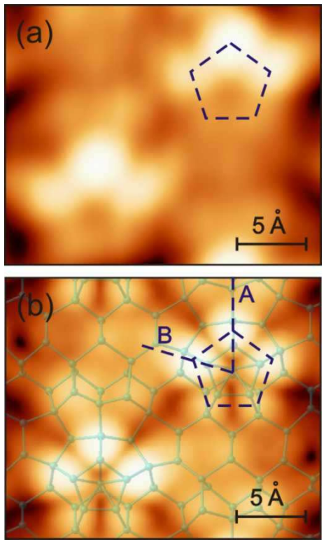
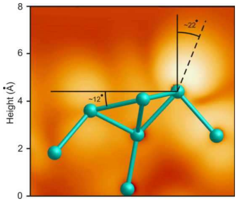
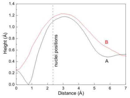
Spurious images are formed in the presence of polarized surface radicals showing a pronounced angle with respect to the surface normal; this magnification effect on pentamer-like structures has been overlooked and must be accounted for when interpreting high-resolution STM images on surfaces with directional bonds.
What carries the argument
Polarized surface radicals tilted away from the surface normal, which distort the STM tunneling current to suggest incorrect atomic positions.
If this is right
- High-resolution STM data on surfaces with directional bonds require correction for radical-induced magnification.
- Pentamer-like features on high-index Si and Ge surfaces reflect apparent rather than true atomic positions.
- Surface structure models derived solely from STM without accounting for radical angle can misidentify atomic arrangements.
- The effect applies generally to any surface where radicals form at an angle to the normal.
Where Pith is reading between the lines
- This mechanism could explain similar image distortions reported on other semiconductor or oxide surfaces with tilted bonds.
- Combining STM with non-contact AFM might separate the polarization contribution from true topography.
- Density functional theory simulations of radical tilt could quantify the magnification factor for different angles.
Load-bearing premise
The observed magnification arises primarily from the geometric angle and polarization of surface radicals rather than from other unmodeled aspects of tip-sample electronic coupling or tip geometry.
What would settle it
STM images of the same pentamer structures on Si or Ge surfaces taken with a tip or bias that eliminates interaction with radical polarization, showing no magnification.
Figures
read the original abstract
Scanning tunneling microscopy (STM) is a fundamental tool for determination of the surface atomic structure. However, the interpretation of high resolution microscopy images is not straightforward. In this paper we provide a physical insight on how STM images can suggest atomic locations which are distinctively different from the real ones. This effect should be taken into account when interpreting high-resolution STM images obtained on surfaces with directional bonds. It is shown that spurious images are formed in the presence of polarized surface radicals showing a pronounced angle with respect to the surface normal. This issue has been overlooked within the surface science community and often disregarded by experimentalists working with STM. Without loss of generality, we illustrate this effect by the magnification observed for pentamer-like structures on (110), (113) and (331) surfaces of silicon and germanium.
Editorial analysis
A structured set of objections, weighed in public.
Referee Report
Summary. The manuscript argues that STM images on surfaces with directional bonds can produce spurious, magnified atomic positions due to polarized surface radicals oriented at a pronounced angle to the surface normal. This effect is illustrated qualitatively using pentamer-like structures observed on the (110), (113), and (331) surfaces of Si and Ge, with the claim that the geometric/polarization contribution from the radicals has been overlooked in the community.
Significance. If the proposed mechanism can be shown to dominate other contributions, the result would supply a useful interpretive caution for high-resolution STM work on reconstructed surfaces. The manuscript does not, however, supply machine-checked proofs, reproducible code, or falsifiable quantitative predictions that would strengthen its standing.
major comments (2)
- [Abstract and mechanism description] The central attribution—that the observed magnification is driven primarily by the radicals' angle and polarization rather than tip geometry or orbital-overlap details—is presented without systematic variation of tip parameters or full DFT+STM simulations. This leaves the claim under-constrained, as alternative convolution mechanisms could reproduce the same apparent magnification (see skeptic note on the core assertion).
- [Illustration on Si/Ge surfaces] No quantitative metric (e.g., apparent vs. true interatomic distances, bias-dependent LDOS maps, or comparison of simulated images with and without the radical tilt) is supplied to demonstrate that the radical-angle effect is load-bearing; the illustrations remain qualitative.
minor comments (1)
- Notation for the radical polarization vector and its angle with respect to the surface normal should be defined explicitly in the first figure or equation.
Simulated Author's Rebuttal
We thank the referee for the constructive comments on our manuscript. The work is intended to highlight a qualitative geometric and polarization-based mechanism for spurious magnification in STM images that has been overlooked. We address the major comments point by point below, indicating planned revisions where appropriate.
read point-by-point responses
-
Referee: [Abstract and mechanism description] The central attribution—that the observed magnification is driven primarily by the radicals' angle and polarization rather than tip geometry or orbital-overlap details—is presented without systematic variation of tip parameters or full DFT+STM simulations. This leaves the claim under-constrained, as alternative convolution mechanisms could reproduce the same apparent magnification (see skeptic note on the core assertion).
Authors: We agree that the manuscript does not include systematic tip-parameter sweeps or full DFT+STM image simulations, which would provide stronger constraints on the relative importance of the radical-angle effect versus other convolution mechanisms. The paper's scope is limited to a conceptual geometric argument based on the known tilt and polarization of surface radicals on these reconstructed surfaces. We will revise the text to explicitly state the assumptions, discuss possible competing contributions (such as tip geometry), and note that quantitative dominance would require additional simulations beyond the present qualitative illustration. revision: partial
-
Referee: [Illustration on Si/Ge surfaces] No quantitative metric (e.g., apparent vs. true interatomic distances, bias-dependent LDOS maps, or comparison of simulated images with and without the radical tilt) is supplied to demonstrate that the radical-angle effect is load-bearing; the illustrations remain qualitative.
Authors: The illustrations are qualitative, as the manuscript aims to draw attention to the effect rather than to deliver quantitative predictions or simulated images. We will add a short supplementary note providing simple geometric projections of apparent versus true interatomic distances based on the reported radical tilt angles for the pentamer structures. Bias-dependent LDOS maps and full image simulations with/without tilt are not available in the current study and would require new computational work; we will acknowledge this limitation explicitly in the revised manuscript. revision: partial
Circularity Check
No circularity: mechanism presented as physical insight without self-referential reduction
full rationale
The paper's central claim is that polarized surface radicals at an angle to the normal produce magnified spurious STM images, illustrated on Si/Ge high-index surfaces. The provided abstract and description contain no equations, fitted parameters, self-citations used as load-bearing premises, or derivations that reduce to their own inputs by construction. The argument is framed as overlooked physical insight rather than a prediction derived from prior results of the same authors or a renaming of known patterns. No load-bearing step matches any of the enumerated circularity patterns.
Axiom & Free-Parameter Ledger
Reference graph
Works this paper leans on
-
[1]
Scanning tunneling mi- croscopy contrast in lateral Ge-Si nanostructures on Si(11 1)- √ 3 × √ 3-Bi
(1) Mysliveček, J.; Dvořák, F.; Stróżecka, A.; Voigtländer , B. Scanning tunneling mi- croscopy contrast in lateral Ge-Si nanostructures on Si(11 1)- √ 3 × √ 3-Bi. Physical Re- view B 2010, 81, 245427. (2) Zhachuk, R.; Coutinho, J. Ab initio study of height contr ast in scanning tunneling microscopy of Ge/Si surface layers grown on Si(111) in prese nce of...
work page 2010
-
[2]
(4) Hofer, W. A. Challenges and errors: interpreting high re solution images in scanning tunneling microscopy. Progress in Surface Science 2003, 71,
work page 2003
-
[3]
Physical Review B 2017, 95, 041412
surface reconstruction. Physical Review B 2017, 95, 041412. (6) Dąbrowski, J.; Müssig, H.-J.; Wolff, G. A novel surface re construction: subsurface interstitials stabilize Si(113)- 3 ×
work page 2017
-
[4]
Surface Science 1995, 331-333,
work page 1995
-
[5]
Elemental struct ure in Si(110)-" 16 × 2" revealed by scanning tunneling microscopy
(7) An, T.; Yoshimura, M.; Ono, I.; Ueda, K. Elemental struct ure in Si(110)-" 16 × 2" revealed by scanning tunneling microscopy. Physical Review B 2000, 61,
work page 2000
-
[6]
A.; Furthmüller, J.; Bechstedt, F
(8) Stekolnikov, A. A.; Furthmüller, J.; Bechstedt, F. Stru ctural elements on reconstructed Si and Ge(110) surfaces. Physical Review B 2004, 70, 045305. (9) Sakamoto, K.; Setvin, M.; Mawatari, K.; Eriksson, P. E. J .; Miki, K.; Uhrberg, R. I. G. Electronic structure of the Si(110)-( 16 ×
work page 2004
-
[7]
Physical Review B 2009, 79, 045304
surface: high-resolution ARPES and STM investigation. Physical Review B 2009, 79, 045304. (10) Teys, S. A. Different STM images of the superstructure on a clean Si(133)- 6 ×2 surface. JETP Letters 2017, 105,
work page 2009
-
[8]
Different STM imag es of the superstructure on a clean Si(133)- 6 × 2 surface
(11) Zhachuk, R.; Coutinho, J. Comment on "Different STM imag es of the superstructure on a clean Si(133)- 6 × 2 surface" (JETP Letters 105, 477 (2017)). JETP Letters 2017, 106,
work page 2017
-
[9]
D.; Haefke, H.; Reimann, P.; Güntherodt, H
(12) Schwarz, U. D.; Haefke, H.; Reimann, P.; Güntherodt, H. -J. Tip artefacts in scanning force microscopy. Journal of Microscopy 1994, 173,
work page 1994
-
[10]
N.; Fauster, T .; Lin, J.-L.; Petrovykh, D
(13) Kirakosian, A.; Bennewitz, R.; Crain, J. N.; Fauster, T .; Lin, J.-L.; Petrovykh, D. Y.; Himpsel, F. J. Atomically accurate Si grating with 5.73 nm pe riod. Applied Physics Letters 2001, 79,
work page 2001
-
[11]
(14) Teys, S. A.; Romanyuk, K. N.; Zhachuk, R. A.; Olshanetsk y, B. Z. Orientation and structure of triple step staircase on vicinal Si(111) surfa ces. Surface Science 2006, 600,
work page 2006
-
[12]
9 (15) Zhachuk, R.; Pereira, S. Comment on "Atomic structure m odel of the reconstructed Si(557) surface with a triple step structure: adatom-paral lel dimer model". Physical Review B 2009, 79, 077401. (16) Zhachuk, R.; Teys, S.; Coutinho, J.; Rayson, M. J.; Brid don, P. R. Static and dynamic buckling of reconstructions at triple steps on Si(111) surf ace...
work page 2009
-
[13]
(18) León, C. P.; Drees, H.; Wippermann, S. M.; Marz, M.; Hoffm ann-Vogel, R. Atomic- scale imaging of the surface dipole distribution of stepped surfaces. Journal of Physical Chemistry Letters 2016, 7,
work page 2016
-
[14]
(19) León, C. P.; Drees, H.; Wippermann, S. M.; Marz, M.; Hoffm ann-Vogel, R. Atomically resolved scanning force studies of vicinal Si(111). Physical Review B 2017, 95, 245412. (20) Kresse, G.; Hafner, J. Ab initio molecular dynamics for liquid metals. Physical Review B 1993, 47,
work page 2017
-
[15]
(21) Kresse, G.; Hafner, J. Ab initio molecular-dynamics si mulation of the liquid-metal- amorphous-semiconductor transition in germanium. Physical Review B 1994, 49, 14251. (22) Kresse, G.; Furthmüller, J. Efficient iterative schemes for ab initio total-energy calcu- lations using a plane-wave basis set. Physical Review B 1996, 54, 11169. (23) Kresse, G.; ...
work page 1994
-
[16]
10 (24) Perdew, J. P.; Burke, K.; Ernzerhof, M. Generalized Gra dient Approximation made simple. Physical Review Letters 1996, 77,
work page 1996
-
[17]
(25) Blöchl, P. E. Projector augmented-wave method. Physical Review B 1994, 50, 17953. (26) Kresse, G.; Joubert, D. From ultrasoft pseudopotentia ls to the projector augmented- wave method. Physical Review B 1999, 59,
work page 1994
-
[18]
(27) Monkhorst, H. J.; Pack, J. D. Special points for Brillou in-zone integrations. Physical Review B 1976, 13,
work page 1976
-
[19]
M.; Col chero, J.; Gómez-Herrero, J.; Baro, A
(28) Horcas, I.; Fernández, R.; Gómez-Rodríguez, J. M.; Col chero, J.; Gómez-Herrero, J.; Baro, A. M. WSXM: A software for scanning probe microscopy an d a tool for nan- otechnology. Review of Scientific Instruments 2007, 78, 013705. 11
work page 2007
discussion (0)
Sign in with ORCID, Apple, or X to comment. Anyone can read and Pith papers without signing in.