REVIEW 5 major objections 7 minor 122 references
The Role of AI in Early Detection of Life-Threatening Diseases: A Retinal Imaging Perspective
T0 review · 5 major / 7 minor · reviewed 2026-08-07 · deepseek-v4-flash
Pith's one-line read This review argues that AI-enhanced retinal imaging can be developed into a unified, non-invasive screening platform for life-threatening systemic diseases, and positions itself as the first cross-domain synthesis of retinal oculomics…
desk verdict A broad but carelessly assembled review whose central novelty claim is contradicted by its own references and whose citation errors undermine its evidentiary value. read the letter →
The pith
A machine-rendered reading of the paper's core claim, the machinery that carries it, and where it could break.
The reading
What carries the argument
The central object is oculomics: the treatment of the retina as a surrogate tissue whose microvasculature and neural layers mirror systemic and cerebral pathology. The engine is AI/deep learning, which learns to map fundus photographs, OCT, and OCTA scans to disease labels and risk factors without relying only on predefined features. The imaging modalities—fundus photography, optical coherence tomography, OCT angiography, and adaptive optics—supply the high-resolution structural and vascular measurements that the AI models analyze.
What would settle it
Check whether reference [18], cited for hemoglobin and red-blood-cell prediction, is actually a study of central serous chorioretinopathy, and whether reference [81], cited for five-year atherosclerosis risk prediction, is a coronary artery disease genetics paper; if either mismatch is confirmed, the corresponding claims in Sections 6.1 and 5.3 lack the stated evidentiary support.
Extended reading notes
Core claim
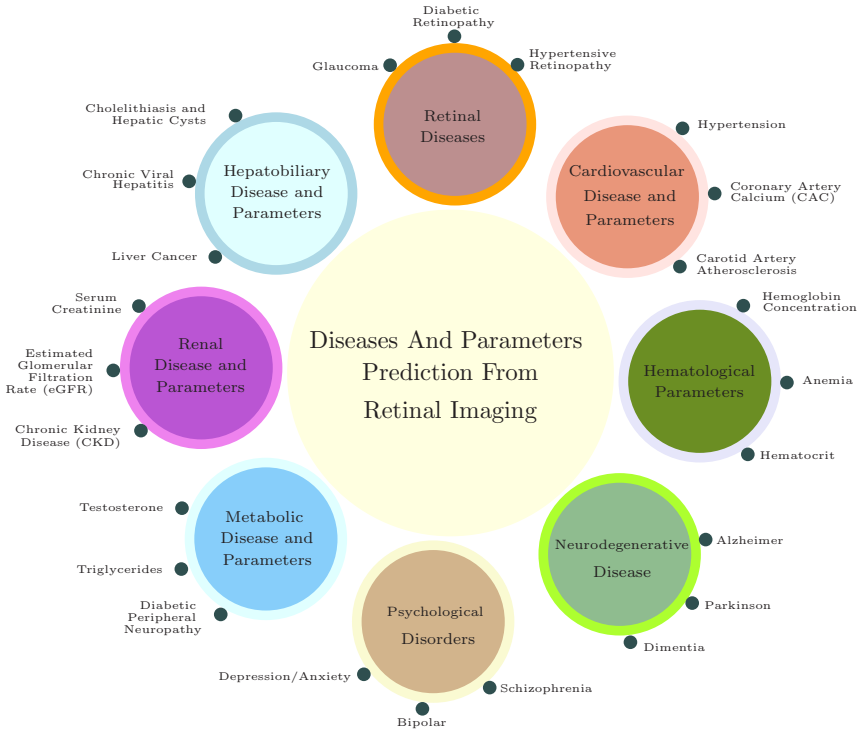
On its own terms, the paper's central discovery is that retinal biomarkers—arteriolar narrowing, venular dilation, retinal nerve fiber layer thinning, microvascular density loss, and even amyloid deposits—are consistently linked to systemic disease across multiple organ systems, and that modern imaging plus deep learning can extract these signals at scale. The authors assert that no prior review has systematically unified AI-enhanced retinal oculomics across neurological and cardiovascular disease domains, and they offer this review as the integrated framework that maps modality strengths, quantifies diagnostic performance, and outlines a roadmap for multicenter standardization and prospective validation. Their conclusion is that retinal imaging, powered by AI, is poised to become a cornerstone of precision medicine for early detection and risk stratification.
Load-bearing premise
The review's load-bearing premise is that each cited study actually supports the specific statement attached to it, so the performance figures, disease associations, and summary tables rest on accurate sourcing.
Editorial extensions
If this is right
- Retinal fundus photography could be added to primary-care screening for cardiovascular risk, since deep learning models already predict risk factors like age, blood pressure, and major adverse cardiovascular events from such images.
- OCT-based measures of retinal nerve fiber layer and macular thickness could become non-invasive biomarkers for early Alzheimer's disease and for tracking multiple sclerosis progression.
- AI analysis of retinal images could support non-invasive screening for anemia, chronic kidney disease, and liver disease, though the review notes predictive accuracy for continuous lab values such as hemoglobin remains modest.
- Standardized imaging protocols and external validation in diverse populations would be required before any of these retinal biomarkers enter routine clinical workflows.
- A unified cross-domain framework could accelerate translation by aligning regulatory, interoperability, and data-sharing standards across cardiology, neurology, nephrology, and primary care.
Reading between the lines
- The claim that this is the first unified review is a literature-positioning statement; its truth depends on the search scope and inclusion criteria, which the paper does not independently verify against a systematic protocol.
- Because the review aggregates performance numbers from heterogeneous, mostly retrospective studies without meta-analysis, the pooled figures should be treated as indicative ranges rather than settled effect sizes.
- If retinal oculomics matures, the same imaging infrastructure could be reused across multiple specialties, potentially changing the cost structure of population screening for cardiovascular and neurodegenerative disease.
- A testable extension would be a prospective study asking whether adding an AI retinal risk score to conventional risk models improves reclassification of patients for statin therapy or dementia workup.
Editorial analysis
A structured set of objections, weighed in public.
Referee Report
Summary. The manuscript is a narrative review claiming to be the first systematic unification of AI-enhanced retinal oculomics across neurological and cardiovascular disease domains, with additional coverage of metabolic, renal, hepatobiliary, and hematological applications. It surveys imaging modalities (fundus photography, OCT/OCTA, adaptive optics), AI/ML approaches, and clinical-translation challenges, and proposes a roadmap for standardization, external validation, and workflow integration. The paper includes several summary tables of reported performance figures and a cross-domain challenge table.
Significance. If the review's synthesis and performance figures were reliable, a cross-domain survey of retinal oculomics could be a useful resource for researchers and clinicians. The paper explicitly identifies important challenges such as external validation, dataset bias, and protocol standardization, and it correctly emphasizes the need for prospective multicenter studies. However, the evidentiary value of the review is heavily compromised by numerous mismatches between cited references and the claims they are supposed to support, and the stated novelty is contradicted by references within the paper's own bibliography. The tables therefore cannot be trusted as accurate summaries of the literature, and the central contribution is not established.
major comments (5)
- [§1, p. 4 (end of Introduction)] The novelty claim that 'No prior review has systematically unified AI-enhanced retinal oculomics across both neurological and cardiovascular disease domains' is contradicted by the paper's own references. Reference [29] is Wagner et al., 'Insights into systemic disease through retinal imaging-based oculomics' (Translational Vision Science & Technology, 2020), a review that explicitly spans cardiovascular and neurological conditions. Reference [30] (Barriada & Masip) reviews deep-learning methods for cardiovascular risk assessment from retinal images, and reference [62] is a systematic scoping review of retinal fundus imaging for cardiovascular markers. The authors must either remove the 'no prior review' claim or restrict the novelty to a precise, reproducible scope that remains after acknowledging these works.
- [§6.1, Table 4 and surrounding text] The claim that 'Zhang et al. [18]' reported fundus-image-based models with low R2 (0.06–0.57) for hemoglobin and RBC counts is unsupported. Reference [18] is X. Zhang, C. Z. F. Lim, J. Chhablani, Y. M. Wong, 'Central serous chorioretinopathy: Updates in the pathogenesis, diagnosis and therapeutic strategies' (Eye and Vision, 2023), which contains no hemoglobin prediction results. The R2 values appear to be misattributed, and the same reference is later used in §7.2 for CKD risk classification, where it is also inappropriate. Without a correct citation, the hematological parameter discussion and Table 4 lose their evidentiary basis.
- [§5.3, paragraph on Lieb et al.] The statement that 'Lieb et al. [81]' analyzed retinal images from diabetic patients to classify atherosclerotic risk categories and predicted atherosclerosis up to five years in advance is contradicted by reference [81], which is W. Lieb and R. S. Vasan, 'Genetics of coronary artery disease' (Circulation, 2013). This is a genetics review, not a retinal imaging study. This misattribution undermines the credibility of the AI-for-cardiovascular-risk section and indicates that the reference list does not reliably correspond to the claims made.
- [§7.5, 'Importance of Vessels Segmentaion'] This subsection is largely a list of the authors' own segmentation networks (references [105]–[117]), with more than ten self-citations and no comparative assessment against other published methods or benchmarks. The text describes general benefits of vessel segmentation but does not critically evaluate these specific methods or justify their selection over alternative approaches. At minimum, the authors should replace this self-citation-heavy passage with a balanced discussion that includes non-self references and quantitative comparisons, or remove it from a review whose scope is systemic disease detection.
- [Overall, 'systematic' synthesis and methods] The abstract and introduction describe the paper as a systematic synthesis, but no search strategy, inclusion criteria, or PRISMA-style methodology is provided. Given that the paper claims to 'systematically synthesize' a broad literature and quantify diagnostic performance, the absence of a reproducible search method makes the selection of references and the reported performance figures unverifiable. This is a load-bearing methodological gap for a review that asserts systematic coverage.
minor comments (7)
- [§3.2.1] The sentence 'A theoretical model proposed by Sabanayagam [35] suggests that nearly all individuals with PD exhibit telangiectasia in the mega-reflex' is unclear and appears to misattribute a retinal imaging finding; the cited reference [35] is a chronic kidney disease deep learning paper, not a Parkinson's disease study.
- [§3.3] 'Retinal Omics' is used to mean multimodal imaging and AI analysis, which is a nonstandard use of the term 'omics'; this should be defined or replaced with clearer terminology.
- [§5.2.1] The text cites 'Wang et al. [29]' for a mechanistic connection between oxidative-stress-driven nephron dysfunction and CAD-related stenosis, but reference [29] is Wagner et al., an oculomics review; the citation does not match the stated claim.
- [§6.2] The text attributes both 'Almonte et al. [89]' observations to the same reference, but the second finding (microvascular density loss in generalized anxiety disorder) is not supported by the title/scope of the cited Almonte and Capellà review; please verify and correct the citation.
- [Table 2 (Cheung et al. row)] The reported AUROC of 0.93 with sensitivity 93.2% and specificity 82.0% is given without confidence intervals or external-validation context; consider adding these details or tempering the summary statement.
- [Throughout] Several reference numbers are used inconsistently: for example, [78] is cited for a meta-analysis by McGeechan et al., but the reference list at [78] is McGeechan et al., so numbering is correct, yet the same number is also used in §5.2.1 for 'Wang et al. (2018)'. Please recheck all citation numbering and ensure each claim is supported by the cited source.
- [§9 Conclusions] The concluding section introduces a sentence about pregnant women with pro-atherosclerotic changes that is not connected to any prior analysis in the review; either elaborate or remove this tangential statement.
Circularity Check
No circular derivation of the review's predictions; the main self-citation burden is localized in Section 7.5, while the 'no prior review' novelty claim is contradicted by the paper's own references rather than established circularly.
-
self citation load bearing
[Section 7.5, 'Importance of Vessels Segmentaion']
"Accurate segmentation of the retinal vasculature enables precise quantification of vessel caliber, tortuosity, and branching patterns, providing early biomarkers for systemic hypertension and cardiovascular risk [105, 106, 107]. ... Hybrid deep architectures like TBConvL-Net integrate convolutional and transformer modules to enhance segmentation accuracy across heterogeneous imaging modalities [116, 117]. The maturation of segmentation algorithms is essential for integrating AI-enhanced oculomics into precision medicine, allowing personalized disease prediction and monitoring [107, 110]."
This subsection asserts that retinal vessel segmentation is an essential enabling technology for AI-based oculomics, and nearly all supporting citations are the authors' own prior segmentation papers (refs 105-117). The claim is therefore supported by the authors' own body of work rather than by independent or comparative evidence, giving the section a self-promotional character. However, this is not the paper's central thesis: the review's synthesis of externally reported biomarker studies does not depend on these self-citations, so the circularity is partial and localized.
full rationale
The paper is a narrative review with no fitted parameters, no derivation equations, and no prediction that reduces to its inputs by construction; the performance figures in Tables 1-7 are attributed to externally authored studies, albeit sometimes inaccurately. The only circularity-adjacent element is Section 7.5, where the importance of vessel segmentation is supported almost entirely by the authors' own prior segmentation networks, which raises the self-citation burden without invalidating the main external-literature synthesis. The paper's strongest novelty assertion, 'No prior review has systematically unified AI-enhanced retinal oculomics across both neurological and cardiovascular disease domains', is contradicted by its own bibliography (e.g., refs [29], [30], [62]), but that is a factual novelty failure rather than a circular derivation. Score 4 reflects the substantial self-citation usage, while acknowledging that the central review content retains independent evidentiary content.
Assumptions & free parameters
assumptions (3)
- domain assumption The cited studies accurately report their results
- domain assumption Retinal microvascular features are valid surrogates for systemic vascular health
- domain assumption AI models can predict systemic parameters from retinal images beyond predefined clinical features
Cite this review
Pith. "Pith review of The Role of AI in Early Detection of Life-Threatening Diseases: A Retinal Imaging Perspective." pith.science (2026). https://pith.science/paper/GDQWBOQD
@misc{pith2026250520810,
  author       = {Pith},
  title        = {Pith review of: The Role of AI in Early Detection of Life-Threatening Diseases: A Retinal Imaging Perspective},
  year         = {2026},
  howpublished = {\url{https://pith.science/paper/GDQWBOQD}},
  note         = {Machine review of arXiv:2505.20810}
}
read the original abstract
Retinal imaging has emerged as a powerful, non-invasive modality for detecting and quantifying biomarkers of systemic diseases-ranging from diabetes and hypertension to Alzheimer's disease and cardiovascular disorders but current insights remain dispersed across platforms and specialties. Recent technological advances in optical coherence tomography (OCT/OCTA) and adaptive optics (AO) now deliver ultra-high-resolution scans (down to 5 {\mu}m ) with superior contrast and spatial integration, allowing early identification of microvascular abnormalities and neurodegenerative changes. At the same time, AI-driven and machine learning (ML) algorithms have revolutionized the analysis of large-scale retinal datasets, increasing sensitivity and specificity; for example, deep learning models achieve > 90 \% sensitivity for diabetic retinopathy and AUC = 0.89 for the prediction of cardiovascular risk from fundus photographs. The proliferation of mobile health technologies and telemedicine platforms further extends access, reduces costs, and facilitates community-based screening and longitudinal monitoring. Despite these breakthroughs, translation into routine practice is hindered by heterogeneous imaging protocols, limited external validation of AI models, and integration challenges within clinical workflows. In this review, we systematically synthesize the latest OCT/OCT and AO developments, AI/ML approaches, and mHealth/Tele-ophthalmology initiatives and quantify their diagnostic performance across disease domains. Finally, we propose a roadmap for multicenter protocol standardization, prospective validation trials, and seamless incorporation of retinal screening into primary and specialty care pathways-paving the way for precision prevention, early intervention, and ongoing treatment of life-threatening systemic diseases.
Figures


Figures from the paper (6 more)


Reference graph
Works this paper leans on
-
[29]
S. K. Wagner, D. J. Fu, L. Faes, X. Liu, J. Huemer, H. Khalid, D. Ferraz, E. Ko- rot, C. Kelly, K. Balaskas, A. K. Denniston, P. A. Keane, Insights into systemic disease through retinal imaging-based oculomics, Translational Vision Science & Technology 9 (2) (2020) 6. doi:10.1167/tvst.9.2.6
-
[18]
X. Zhang, C. Z. F. Lim, J. Chhablani, Y . M. Wong, Central serous chorioretinopa- thy: Updates in the pathogenesis, diagnosis and therapeutic strategies, Eye and Vision 10 (1) (2023) 33. doi:10.1186/s40662-023-00349-y
-
[81]
W. Lieb, R. S. Vasan, Genetics of coronary artery disease, Circulation 128 (10) (2013) 1131–1138. doi:10.1161/CIRCULATIONAHA.113.005350
-
[35]
C. Sabanayagam, D. Xu, D. S. W. Ting, S. Nusinovici, R. Banu, H. Hamzah, C. Lim, Y .-C. Tham, C. Y . Cheung, E. S. Tai, Y . X. Wang, J. B. Jonas, C.-Y . Cheng, M. L. Lee, W. Hsu, T. Y . Wong, A deep learning algorithm to detect chronic kidney dis- ease from retinal photographs in community-based populations, The Lancet Digital Health 2 (6) (2020) e295–e30...
-
[30]
R. G. Barriada, D. Masip, An overview of deep-learning-based methods for car- diovascular risk assessment with retinal images, Diagnostics 13 (1) (2022) 68. doi:10.3390/diagnostics13010068
-
[62]
L. Y . Li, A. A. Isaksen, B. Lebiecka-Johansen, K. Funck, V . Thambawita, S. Byberg, T. H. Andersen, O. NAzrgaard, A. Hulman, Prediction of cardiovascular markers and diseases using retinal fundus images and deep learning: A systematic scoping review, European Heart Journal Digital Health 5 (6) (2024) 660–669. doi:10. 1093/ehjdh/ztae068
2024
-
[105]
M. A. Khan, T. M. Khan, T. A. Soomro, N. Mir, J. Gao, Boosting sensitivity of a retinal vessel segmentation algorithm, Pattern Analysis and Applications 22 (2) (2019) 583–599
work page 2019
-
[117]
T. M. Khan, M. Arsalan, S. Iqbal, I. Razzak, E. Meijering, Feature enhancer seg- mentation network (fes-net) for vessel segmentation, in: 2023 International Confer- ence on Digital Image Computing: Techniques and Applications (DICTA), IEEE, 2023, pp. 160–167. 66
work page 2023
Show all 122 references
-
[1]
C. W. Wong, T. Y . Wong, C.-Y . Cheng, C. Sabanayagam, Kidney and eye diseases: common risk factors, etiological mechanisms, and pathways, Kidney International 85 (6) (2014) 1290–1302. doi:10.1038/ki.2013.491
2014 doi
-
[2]
Flammer, K
J. Flammer, K. Konieczka, R. M. Bruno, A. Virdis, A. J. Flammer, S. Taddei, The eye and the heart, European Heart Journal 34 (17) (2013) 1270–1278. doi:10. 1093/eurheartj/eht023. 53
2013
-
[3]
Patton, T
N. Patton, T. M. Aslam, T. MacGillivray, I. J. Deary, B. Dhillon, R. H. Eikelboom, K. Yogesan, I. J. Constable, Retinal image analysis: Concepts, applications and potential, Progress in Retinal and Eye Research 25 (1) (2006) 99–127. doi:10. 1016/j.preteyeres.2005.07.001
2006
-
[4]
Imtiaz, T
R. Imtiaz, T. M. Khan, S. S. Naqvi, M. Arsalan, S. J. Nawaz, Screening of glau- coma disease from retinal vessel images using semantic segmentation, Computers & Electrical Engineering 91 (2021) 107036
2021
-
[5]
T. M. Khan, M. A. Khan, N. U. Rehman, K. Naveed, I. U. Afridi, S. S. Naqvi, I. Raazak, Width-wise vessel bifurcation for improved retinal vessel segmentation, Biomedical Signal Processing and Control 71 (2022) 103169
2022
-
[6]
T. M. Khan, A. Robles-Kelly, S. S. Naqvi, T-net: A resource-constrained tiny con- volutional neural network for medical image segmentation, in: Proceedings of the IEEE/CVF Winter Conference on Applications of Computer Vision, 2022, pp. 644– 653
2022
-
[7]
T. A. Soomro, T. M. Khan, M. A. Khan, J. Gao, M. Paul, L. Zheng, Impact of ica- based image enhancement technique on retinal blood vessels segmentation, IEEE Access 6 (2018) 3524–3538
2018
-
[8]
Z. U. Rehman, S. S. Naqvi, T. M. Khan, M. Arsalan, M. A. Khan, M. Khalil, Multi- parametric optic disc segmentation using superpixel based feature classification, Expert Systems with Applications 120 (2019) 461–473
2019
-
[9]
Tabassum, T
M. Tabassum, T. M. Khan, M. Arsalan, S. S. Naqvi, M. Ahmed, H. A. Madni, J. Mirza, Cded-net: Joint segmentation of optic disc and optic cup for glaucoma screening, IEEE access 8 (2020) 102733–102747
2020
-
[10]
T. M. Khan, M. Alhussein, K. Aurangzeb, M. Arsalan, S. S. Naqvi, S. J. Nawaz, Residual connection-based encoder decoder network (rced-net) for retinal vessel segmentation, IEEE Access 8 (2020) 131257–131272
2020
-
[11]
Iqbal, T
S. Iqbal, T. M. Khan, K. Naveed, S. S. Naqvi, S. J. Nawaz, Recent trends and advances in fundus image analysis: A review, Computers in Biology and Medicine 151 (2022) 106277
2022
-
[12]
T. Y . Wong, R. Klein, D. J. Couper, L. S. Cooper, E. Shahar, L. D. Hubbard, M. R. Wofford, A. R. Sharrett, Retinal microvascular abnormalities and incident stroke: the atherosclerosis risk in communities study, The Lancet 358 (9288) (2001) 1134– 1140. 54
2001
-
[13]
D. S. W. Ting, L. R. Pasquale, P. Peng, J. P. Campbell, A. Y . Lee, R. Raman, G. S. W. Tan, L. Schmetterer, P. A. Keane, T. Y . Wong, Artificial intelligence and deep learn- ing in ophthalmology, British Journal of Ophthalmology 103 (2) (2019) 167–175. doi:10.1136/bjophthalmol...
2019 doi
-
[14]
Mitani, A
A. Mitani, A. Huang, S. Venugopalan, G. S. Corrado, L. Peng, D. R. Webster, N. Hammel, Y . Liu, A. V . Varadarajan, Detection of anaemia from retinal fun- dus images via deep learning, Nature Biomedical Engineering 4 (1) (2020) 18–27. doi:10.1038/s41551-019-0487-z
2020 doi
-
[15]
Poplin, A
R. Poplin, A. V . Varadarajan, K. Blumer, Y . Liu, M. V . McConnell, G. S. Corrado, L. Peng, D. R. Webster, Prediction of cardiovascular risk factors from retinal fundus photographs via deep learning, Nature Biomedical Engineering 2 (3) (2018) 158–
2018
-
[16]
Y . D. Kim, K. J. Noh, S. J. Byun, S. Lee, T. Kim, L. Sunwoo, K. J. Lee, S.-H. Kang, K. H. Park, S. J. Park, Effects of hypertension, diabetes, and smoking on age and sex prediction from retinal fundus images, Scientific Reports 10 (2020) 4623. doi:10.1038/s41598-020-61519-9
2020 doi
-
[17]
Chatterjee, S
S. Chatterjee, S. Chattopadhyay, M. Hopea Ross, P. L. Lip, Hypertension and the eye: Changing perspectives, Journal of Human Hypertension 16 (10) (2002) 667–
2002
-
[19]
T. A. Soomro, J. Gao, T. Khan, A. F. M. Hani, M. A. Khan, M. Paul, Computerised approaches for the detection of diabetic retinopathy using retinal fundus images: a survey, Pattern Analysis and Applications 20 (2017) 927–961
2017
-
[20]
Khawaja, T
A. Khawaja, T. M. Khan, K. Naveed, S. S. Naqvi, N. U. Rehman, S. J. Nawaz, An improved retinal vessel segmentation framework using frangi filter coupled with the probabilistic patch based denoiser, IEEE Access 7 (2019) 164344–164361
2019
-
[21]
Khawaja, T
A. Khawaja, T. M. Khan, M. A. Khan, J. Nawaz, A multi-scale directional line detector for retinal vessel segmentation, Sensors 19 (22) (2019)
2019
-
[22]
Zamir, Optimality principles in arterial branching, Journal of Theoretical Biol- ogy 62 (1976) 227–251
M. Zamir, Optimality principles in arterial branching, Journal of Theoretical Biol- ogy 62 (1976) 227–251. 55
1976
-
[23]
D. B. Gould, F. C. Phalan, S. E. van Mil, J. P. Sundberg, K. Vahedi, P. Massin, M. Bousser, P. Heutink, J. H. Miner, A. Tourniera Lasserve, S. W. M. John, Role of COL4A1 in small vessel disease and hemorrhagic stroke, New England Journal of Medicine 354 (14) (2006) 1489–1496. ...
2006 doi
-
[24]
G. Liew, J. J. Wang, P. Mitchell, T. Y . Wong, Retinal vascular imaging: a new tool in microvascular disease research, Circulation: Cardiovascular Imaging 1 (3) (2008) 156–161. doi:10.1161/CIRCIMAGING.108.784876
2008 doi
-
[25]
Mainster, The fractal properties of retinal vessels: Embryological and clinical implications, Eye 4 (1990) 235–241
M. Mainster, The fractal properties of retinal vessels: Embryological and clinical implications, Eye 4 (1990) 235–241
1990
-
[26]
J. M. Harazny, M. Ritt, D. Baleanu, C. Ott, J. Heckmann, M. P. Schlaich, G. Michel- son, R. E. Schmieder, Increased wall:lumen ratio of retinal arterioles in male pa- tients with a history of a cerebrovascular event, Hypertension 50 (4) (2007) 623–
2007
-
[27]
Michelson, S
G. Michelson, S. Warntges, D. Baleanu, J. Welzenbach, A. Ohno-Jinno, P. Pogorelov, J. Harazny, Morphometric age-related evaluation of small retinal vessels by scanning laser doppler flowmetry: Determination of a vessel wall in- dex, Retina 27 (4) (2007) 490–498. doi:10.1097/01...
2007 doi
-
[28]
C. E. Riva, E. Logean, B. Falsini, Visually evoked hemodynamical response and as- sessment of neurovascular coupling in the optic nerve and retina, Progress in Reti- nal and Eye Research 24 (2005) 183–215. doi:10.1016/j.preteyeres. 2004.12.002
2005 doi
-
[31]
M. G. Syed, E. Trucco, M. R. K. Mookiah, C. C. Lang, R. J. McCrimmon, C. N. A. Palmer, E. R. Pearson, A. S. F. Doney, I. R. Mordi, Deep-learning pre- diction of cardiovascular outcomes from routine retinal images in individuals with 56 type 2 diabetes, Cardiovascular Diabetolo...
2025
-
[32]
C. Xing, B. E. K. Klein, R. Klein, G. Jun, K. E. Lee, S. K. Iyengar, Genome-wide linkage study of retinal vessel diameters in the Beaver Dam Eye Study, Hyperten- sion 47 (4) (2006) 797–802. doi:10.1161/01.HYP.0000208330.68355. 72
2006
-
[33]
N. Witt, T. Y . Wong, A. D. Hughes, N. Chaturvedi, B. E. K. Klein, R. Evans, M. Mc- Namara, S. A. M. Thom, R. Klein, Abnormalities of retinal microvascular structure and risk of mortality from ischemic heart disease and stroke, Hypertension 47 (5) (2006) 975–981. doi:10.1161/0...
2006
-
[34]
X. Ma, Z. Xie, H. Wang, Z. Tian, Y . Bi, Y . Li, L. Zhang, A cross-sectional study of retinal vessel changes based on optical coherence tomography angiography in Alzheimer disease and mild cognitive impairment, Frontiers in Aging Neuroscience 15 (2023) 1101950. doi:10.3389/fna...
2023
-
[36]
T. A. Soomro, M. A. Khan, J. Gao, T. M. Khan, M. Paul, N. Mir, Automatic retinal vessel extraction algorithm, in: 2016 International Conference on Digital Image Computing: Techniques and Applications (DICTA), IEEE, 2016, pp. 1–8
2016
-
[37]
M. A. Khan, T. A. Soomro, T. M. Khan, D. G. Bailey, J. Gao, N. Mir, Automatic retinal vessel extraction algorithm based on contrast-sensitive schemes, in: 2016 International conference on image and vision computing New Zealand (IVCNZ), IEEE, 2016, pp. 1–5
2016
-
[38]
T. Y . Wong, P. Mitchell, Hypertensive retinopathy, New England J. of Medicine 351 (22) (2004) 2310–2317
2004
-
[39]
T. Wong, P. Mitchell, The eye in hypertension, The Lancet 369 (9559) (2007) 425– 435
2007
-
[40]
Grosso, F
A. Grosso, F. Veglio, M. Porta, F. M. Grignolo, T. Y . Wong, Hypertensive retinopa- thy revisited: Some answers, more questions, British Journal of Ophthalmology 89 (12) (2005) 1646–1654. doi:10.1136/bjo.2005.072546. 57
2005 arXiv
-
[41]
Marcucci, F
R. Marcucci, F. Sofi, E. Grifoni, A. Sodi, D. Prisco, Retinal vein occlusions: A review for the internist, Internal and Emergency Medicine 6 (4) (2011) 307–314. doi:10.1007/s11739-010-0478-2
2011 doi
-
[42]
D. H. Lee, S. J. Lee, I. S. Yoon, Clinical progress in impending central retinal vein occlusion, Korean Journal of Ophthalmology 24 (2) (2010) 83
2010
-
[43]
W. W. Lee, I. Tajunisah, K. Sharmilla, M. Peyman, V . Subrayan, Retinal nerve fibre layer structure abnormalities in schizophrenia and its relationship to disease state: evidence from optical coherence tomography, Investigative Ophthalmology & Visual Science 54 (12) (2013) 778...
2013 doi
-
[44]
H. A. Quigley, A. T. Broman, The number of people with glaucoma worldwide in 2010 and 2020, Br J Ophthalmol 90 (3) (2006) 262–267. doi:10.1136/bjo. 2005.081224
2006 arXiv
-
[45]
Abdullah, R
F. Abdullah, R. Imtiaz, H. A. Madni, H. A. Khan, T. M. Khan, M. A. U. Khan, S. S. Naqvi, A review on glaucoma disease detection using computerized tech- niques, IEEE Access 9 (2021) 37311–37333. doi:10.1109/ACCESS.2021. 3061086
2021 doi
-
[46]
Imtiaz, T
R. Imtiaz, T. M. Khan, S. S. Naqvi, M. Arsalan, S. J. Nawaz, Screening of glaucoma disease from retinal vessel images using semantic segmentation, Computers & Electrical Engineering 91 (2021) 107036. doi:10.1016/j.compeleceng. 2021.107036
2021
-
[47]
Thomas, R
R. Thomas, R. S. Parikh, How to assess a patient for glaucoma, Community Eye Health 19 (59) (2006) 36
2006
-
[48]
Varma, S
D. Varma, S. Cugati, A. Lee, C. Chen, A review of central retinal artery occlusion: Clinical presentation and management, Eye 27 (6) (2013) 688–697. doi:10. 1038/eye.2013.43
2013
-
[49]
R. Lee, T. Y . Wong, C. Sabanayagam, Epidemiology of diabetic retinopathy, di- abetic macular edema and related vision loss, Eye and Vision 2 (1) (2015) 17. doi:10.1186/s40662-015-0021-y
2015 doi
-
[50]
E. J. Duh, J. K. Sun, A. W. Stitt, Diabetic retinopathy: current understanding, mech- anisms, and treatment strategies, JCI insight 2 (14) (2017)
2017
-
[51]
Claesson-Welsh, Vascular permeability the essentials, Upsala J
L. Claesson-Welsh, Vascular permeability the essentials, Upsala J. of Med. Sci. 120 (3) (2015) 135–143. 58
2015
-
[52]
Berisha, G
F. Berisha, G. T. Feke, C. L. Trempe, J. W. McMeel, C. L. Schepens, Retinal ab- normalities in early Alzheimer’s disease, Investigative Ophthalmology & Visual Science 48 (5) (2007) 2285–2289. doi:10.1167/iovs.06-1029
2007 doi
-
[53]
J. A. Cipolla, H. Jiang, A.-G. Simms, B. S. Baumel, T. Rundek, J. Wang, Impaired retinal capillary function in patients with Alzheimer disease, Jour- nal of Neuro-Ophthalmology 44 (1) (2024) 30–34. doi:10.1097/WNO. 0000000000001954
2024 doi
-
[54]
Koronyo, D
Y . Koronyo, D. Biggs, E. Barron, D. S. Boyer, J. A. Pearlman, W. J. Au, S. J. Kile, A. Blanco, D. T. Fuchs, A. Ashfaq, S. A. Frautschy, G. M. Cole, C. A. Miller, D. R. Hinton, S. R. Verdooner, K. L. Black, M. Koronyo-Hamaoui, Retinal amyloid pathology and proof-of-concept ima...
2017 doi
-
[55]
Koronyo-Hamaoui, Y
M. Koronyo-Hamaoui, Y . Koronyo, A. V . Ljubimov, C. A. Miller, M. K. H. Ko, K. L. Black, M. Schwartz, Identification of amyloid plaques in retinas from Alzheimer patients and noninvasive in vivo optical imaging of retinal plaques in a mouse model, Neuroimage 54 (2011) S204–S2...
2011 doi
-
[56]
N. A. Amir Hamzah, W. M. D. Wan Zaki, W. H. Wan Abdul Halim, R. Musta- far, A. H. Saad, Evaluating the potential of retinal photography in chronic kidney disease detection: a review, PeerJ 12 (2024) e17786. doi:10.7717/peerj. 17786
2024 doi
-
[57]
Guymer, P
C. Guymer, P. J. Wood, G. Chidlow, R. J. Casson, Neuroprotection in glaucoma: Recent advances and clinical translation, Clinical & Experimental Ophthalmology 47 (1) (2019) 88–105. doi:10.1111/ceo.13336
2019 doi
-
[58]
W. R. Kwapong, H. Ye, C. Peng, X. Zhuang, J. Wang, M. Shen, F. Lu, Retinal microvascular impairment in the early stages of Parkinson disease, Investigative Ophthalmology & Visual Science 59 (10) (2018) 4115–4122. doi:10.1167/ iovs.17-23230
2018
-
[59]
J. A. Pillai, R. Bermel, A. Bonner-Jackson, A. Rae-Grant, H. Fernandez, J. Bena, S. E. Jones, J. P. Ehlers, J. B. Leverenz, Retinal nerve fiber layer thinning in Alzheimer’s disease: A case-control study in comparison to normal ag- ing, Parkinson’s disease, and non-Alzheimer’s...
2016
-
[60]
Matlach, M
J. Matlach, M. Wagner, U. Malzahn, I. Schmidtmann, F. Steigerwald, T. Musacchio, J. V olkmann, F. Grehn, W. Goebel, S. Klebe, Retinal changes in Parkinson’s disease and glaucoma, Parkinsonism & Related Disorders 56 (2018) 41–46. doi:10. 1016/j.parkreldis.2018.06.016
2018
-
[61]
Murueta Goyena, R
A. Murueta Goyena, R. Del Pino, M. Galdas, J. Pagonabarraga, J. Vallsa Sol, R. Lopeza Blanco, E. Tolosa, A. Dela Valle, Retinal thickness predicts the risk of cognitive decline in Parkinson disease, Annals of Neurology 89 (1) (2021) 165–176. doi:10.1002/ana.25944
2021 doi
-
[63]
Baba, Detecting diabetic retinal neuropathy using fundus perimetry, Interna- tional Journal of Molecular Sciences 22 (19) (2021) 10726
T. Baba, Detecting diabetic retinal neuropathy using fundus perimetry, Interna- tional Journal of Molecular Sciences 22 (19) (2021) 10726. doi:10.3390/ ijms221910726
2021
-
[64]
E. Y .-C. Kang, Y .-T. Hsieh, C.-H. Li, Y .-J. Huang, C.-F. Kuo, J.-H. Kang, K.-J. Chen, C.-C. Lai, W.-C. Wu, Y .-S. Hwang, Deep learning-based detection of early renal function impairment using retinal fundus images: Model development and validation, JMIR Medical Informatics ...
2020
-
[65]
T. J. MacGillivray, E. Trucco, J. R. Cameron, B. Dhillon, J. G. Houston, E. J. V . Beek, Retinal imaging as a source of biomarkers for diagnosis, characterization and prognosis of chronic illness or long-term conditions (8 2014)
2014
-
[66]
den Haan, F
J. den Haan, F. D. Verbraak, P. J. Visser, F. H. Bouwman, Retinal thickness in Alzheimer’s disease and mild cognitive impairment: A systematic review and meta- analysis, Ageing Research Reviews 36 (2017) 1–11. doi:10.1016/j.arr. 2016.11.001
2017 doi
-
[67]
Cheung, R
Y . Cheung, R. Ran, A. S. Wang, T. T. Chan, V . K. Sham, S. Hilal, N. Venketasubra- manian, Y . Cheng, C. Sabanayagam, C. Tham, Y . L. Schmetterer, J. McKay, G. A. Williams, M. A. Wong, W. C. Au, L. Z. Lu, C. Yam, J. C. Tham, C. J. Chen, J. M. Dumitrascu, O. A. Heng, P. C. Y ....
2022
-
[68]
M. M. El-Kattan, S. M. Esmat, E. H. Esmail, H. A. Deraz, R. S. Ismail, Op- tical coherence tomography in patients with Parkinson’s disease, Egyptian Jour- nal of Neurology, Psychiatry and Neurosurgery 58 (2022) 21. doi:10.1186/ s41983-021-00421-1
2022
-
[69]
Elanwar, H
R. Elanwar, H. Al Masry, A. Ibrahim, M. Hussein, S. Ibrahim, M. M. Masoud, Retinal functional and structural changes in patients with Parkinson’s disease, BMC Neurology 23 (1) (2023) 330. doi:10.1186/s12883-023-03373-6
2023 doi
-
[70]
Doerr, K
J. Doerr, K. D. Wernecke, M. Bock, G. Gaede, J. T. Wuerfel, C. F. Pfueller, J. Bellmann-Strobl, A. Freing, A. U. Brandt, P. Friedemann, Association of reti- nal and macular damage with brain atrophy in multiple sclerosis, PLoS ONE 6 (4) (2011) e18132. doi:10.1371/journal.pone.0018132
2011 doi
-
[71]
Petzold, L
A. Petzold, L. J. Balcer, P. A. Calabresi, F. Costello, T. C. Frohman, E. M. Frohman, E. H. Martinez-Lapiscina, A. J. Green, R. Kardon, O. Outteryck, F. Paul, S. Schip- pling, P. Vermersch, P. Villoslada, L. J. Balk, E.-E. IMSVISUAL, Retinal layer segmentation in multiple scle...
2017 doi
-
[72]
J. B. Fisher, D. A. Jacobs, C. E. Markowitz, S. L. Galetta, N. J. V olpe, M. L. Nano- Schiavi, M. L. Baier, E. M. Frohman, H. Winslow, T. C. Frohman, P. A. Calabresi, M. G. Maguire, G. R. Cutter, L. J. Balcer, Relation of visual function to retinal nerve fiber layer thickness ...
2006
-
[73]
F. J. de Jong, E. M. C. Schrijvers, M. K. Ikram, P. J. Koudstaal, P. T. V . M. de Jong, A. Hofman, J. R. Vingerling, M. M. B. Breteler, Retinal vascular caliber and risk of dementia: the Rotterdam Study, Neurology 76 (9) (2011) 816–821. doi:10.1212/WNL.0b013e31820e7baa
2011 doi
-
[74]
R. Gao, H. Luo, S. Yan, L. Ba, S. Peng, B. Bu, X. Sun, M. Zhang, Retina as a potential biomarker for the early stage of Alzheimer’s disease spectrum, Annals of Clinical and Translational Neurology 11 (10) (2024) 2583–2596.doi:10.1002/ acn3.52172
2024
-
[75]
Cheung, D
N. Cheung, D. A. Bluemke, R. Klein, A. R. Sharrett, F. M. A. Islam, M. F. Cotch, B. E. K. Klein, M. H. Criqui, T. Y . Wong, Retinal arteriolar narrowing and left 61 ventricular remodeling: The Multi-Ethnic Study of Atherosclerosis, Journal of the American College of Cardiology...
2007
-
[76]
S. W. Holwerda, R. H. Kardon, R. Hashimoto, J. M. Full, J. K. Nellis, L. E. Dubose, J. G. Fiedorowicz, G. L. Pierce, Aortic stiffness is associated with changes in retinal arteriole flow pulsatility mediated by local vasodilation in healthy young/middle- age adults, J Appl Phy...
2020
-
[77]
F. N. Doubal, P. E. Hokke, J. M. Wardlaw, Retinal microvascular abnormalities and stroke: a systematic review, Journal of Neurology, Neurosurgery; Psychiatry 80 (2009) 158
2009
-
[78]
McGeechan, G
K. McGeechan, G. Liew, P. Macaskill, L. Irwig, R. Klein, B. E. K. Klein, J. J. Wang, P. Mitchell, J. R. Vingerling, P. T. V . M. de Jong, J. C. M. Witteman, M. M. B. Breteler, J. Shaw, P. Zimmet, T. Y . Wong, Meta-analysis: Retinal vessel caliber and risk for coronary heart di...
2009
-
[79]
Zhang, M
L. Zhang, M. Yuan, Z. An, X. Zhao, H. Wu, H. Li, Y . Wang, B. Sun, H. Li, S. Ding, X. Zeng, L. Chao, P. Li, W. Wu, Prediction of hypertension, hyperglycemia and dyslipidemia from retinal fundus photographs via deep learning: a cross-sectional study of chronic diseases in centr...
2020 doi
-
[80]
Chang, A
J. Chang, A. Ko, S. M. Park, S. Choi, K. Kim, S. M. Kim, J. M. Yun, U. Kang, I. H. Shin, J. Y . Shin, T. Ko, J. Lee, B. L. Oh, K. H. Park, Association of cardiovascular mortality and deep learning-funduscopic atherosclerosis score derived from retinal fundus images, American J...
-
[82]
A. Yuan, C. S. Lee, Retinal biomarkers for Alzheimer disease: the facts and the future, Asia-Pacific Journal of Ophthalmology 11 (2) (2022) 140–148. doi:10. 1097/APO.0000000000000505
2022
-
[83]
Klein, B
R. Klein, B. E. K. Klein, S. E. Moss, T. Y . Wong, Retinal vessel caliber and mi- crovascular and macrovascular disease in type 2 diabetes: XXI: The Wisconsin Epi- demiologic Study of Diabetic Retinopathy, Ophthalmology 114 (10) (2007) 1884–
2007
-
[84]
M. H. Meier, I. Shalev, T. E. Moffitt, S. Kapur, R. S. E. Keefe, T. Y . Wong, D. W. Belsky, H. Harrington, S. Hogan, R. Houts, A. Caspi, R. Poulton, Microvascu- lar abnormality in schizophrenia as shown by retinal imaging, American Journal of Psychiatry 170 (12) (2013) 1451–14...
2013 doi
-
[85]
S. B. Seidelmann, B. Claggett, P. E. Bravo, A. Gupta, H. Farhad, B. E. Klein, R. Klein, M. Di Carli, S. D. Solomon, Retinal vessel calibers in predicting long-term cardiovascular outcomes: The atherosclerosis risk in communities study, Circu- lation 134 (18) (2016) 1328–1338. ...
2016 doi
-
[86]
V . M. Matei, J. Y . Xia, C. Nguyen, Poor outcomes despite aspirin or statin use in high-risk patients with retinal vein occlusion, Graefe’s Archive for Clinical and Experimental Ophthalmology 255 (2017) 761–766
2017
-
[87]
Stettler, N
C. Stettler, N. Witt, R. J. Tapp, S. Thom, S. Allemann, T. Tillin, A. Stanton, E. O’Brien, N. Poulter, J. R. Gallimore, A. D. Hughes, N. Chaturvedi, Serum amy- loid a, c-reactive protein, and retinal microvascular changes in hypertensive diabetic and nondiabetic individuals: A...
2009
-
[88]
S. C. L. Reboucas, A. Cougnard-Gregoire, L. Arnould, M.-N. Delyfer, C. Schweitzer, J.-F. Korobelnik, A. Foubert-Samier, C. Y . Cheung, T. Y . Wong, C. Delcourt, C. Helmer, Retinal microvasculature and incident dementia over 10 years: The Three-City-ALIENOR cohort, Alzheimer’s ...
2023 doi
-
[89]
T. E. M. Almonte, P. Capelln, Retinal correlates of psychiatric disorders, Therapeu- tic Advances in Chronic Disease 11 (2020) 2040622320905215. doi:10.1177/ 2040622320905215
2020
-
[90]
Ascaso, M
F. Ascaso, M. Quintanilla, J. Buil, M. Ramirez, C. Pintado, Retinal nerve fiber layer thickness measured by optical coherence tomography in patients with schizophre- nia: a short report, European Journal of Psychiatry 24 (4) (2010) 227–235
2010
-
[91]
F. J. Ascaso, R. Rodriguez-Jimenez, L. Cabezon, R. Lopez-Anton, J. Santabar- bara, C. De la Camara, P. J. Modrego, M. A. Quintanilla, A. Bagney, L. Gutierrez, N. Cruz, J. A. Cristobal, A. Lobo, Retinal nerve fiber layer and macular thickness 63 in patients with schizophrenia: ...
2015 doi
-
[92]
Appaji, B
A. Appaji, B. Nagendra, D. Chako, A. Padmanabha, C. Hiremath, Retinal vascu- lar tortuosity in schizophrenia and bipolar disorder, Schizophrenia Research 212 (2019) 26–32. doi:10.1016/j.schres.2019.08.020
2019 doi
-
[93]
Sabanayagam, A
C. Sabanayagam, A. Shankar, D. H. Koh, K. S. Chia, S. M. Saw, S. C. Lim, E. S. Tai, T. Y . Wong, Retinal microvascular caliber and chronic kidney disease in an Asian population, American Journal of Epidemiology 181 (11) (2015) 791–799
2015
-
[94]
Benson, T
J. Benson, T. Estrada, M. Burge, P. Soliz, Diabetic peripheral neuropathy risk as- sessment using digital fundus photographs and machine learning, in: 2020 42nd Annual International Conference of the IEEE Engineering in Medicine & Biology Society (EMBC), 2020, pp. 1988–1991
2020
-
[95]
K. H. Gerrits, I. Klaassen, J. S. Schouten, E. van Duinkerken, F. D. Verbraak, C. D. Stehouwer, R. M. Henry, N. C. Schaper, T. T. Berendschot, Performance of a deep learning model versus existing methods for detecting diabetic neuropathy in retinal fundus photographs, Diabetes...
2020
-
[96]
M. R. Munk, A. H. Kashani, R. Tadayoni, J. Korobelnik, S. Wolf, F. Pichi, A. Koh, A. Ishibazawa, A. Gaudric, A. Loewenstein, K. B. Freund, et al., Recommendations for OCT angiography reporting in retinal vascular disease: A Delphi approach by international experts, Ophthalmolo...
2022 doi
-
[97]
Mehrabi, F
N. Mehrabi, F. Morstatter, N. Saxena, K. Lerman, A. Galstyan, A survey on bias and fairness in machine learning, arXiv preprint arXiv:1908.09635 (2019)
2019 arXiv
-
[98]
J. C. Mandel, D. A. Kreda, K. D. Mandl, I. S. Kohane, R. B. Ramoni, SMART on FHIR: a standards-based, interoperable apps platform for electronic health records, Journal of the American Medical Informatics Association 23 (5) (2016) 899–908. doi:10.1093/jamia/ocv189
2016 doi
-
[99]
S. Rego, M. Monteiro Soares, M. Dutra Medeiros, C. Dias, F. Nunes, Exploring the feasibility of opportunistic diabetic retinopathy screening with handheld fundus cameras in primary care: Insights from doctors and nurses, Diabetology 5 (6) (2024) 566–583. doi:10.3390/diabetolog...
2024 doi
-
[100]
D. S. Char, N. H. Shah, D. Magnus, Implementing machine learning in health care addressing ethical challenges, New England Journal of Medicine 378 (11) (2018) 981–983. doi:10.1056/NEJMp1714229
2018 doi
-
[101]
Rieke, J
N. Rieke, J. Hancox, W. Li, F. Milletari, H. R. Roth, S. Albarqouni, S. Bakas, M. N. Galtier, B. A. Landman, K. Maiera Hein, M. J. Cardoso, Z. Xu, J. Kalpathy Cramer, P. Heng, G. Kaissis, D. Rueckert, S. Ourselin, B. Kainz, The future of digital health with federated learning,...
2020 doi
-
[102]
W. Xiao, X. Guo, Serum lipid profiles and dyslipidaemia are associated with retinal microvascular changes in children and adolescents, Sci Rep 7 (2017) 44874. doi: 10.1038/srep44874
2017 doi
-
[103]
T. H. Rim, A. W. J. Teo, H. H. S. Yang, C. Y . Cheung, T. Y . Wong, Retinal vascular signs and cerebrovascular diseases, Journal of Neuro-Ophthalmology 40 (2020)
2020
-
[104]
Iwakiri, Pathophysiology of portal hypertension, Clinics in Liver Disease 18 (2) (2014) 281–291
Y . Iwakiri, Pathophysiology of portal hypertension, Clinics in Liver Disease 18 (2) (2014) 281–291. doi:10.1016/j.cld.2013.12.001
2014 doi
-
[106]
M. A. Khan, T. M. Khan, D. G. Bailey, T. A. Soomro, A generalized multi-scale line-detection method to boost retinal vessel segmentation sensitivity, Pattern Anal- ysis and Applications 22 (2019) 1177–1196
2019
-
[107]
T. M. Khan, S. S. Naqvi, E. Meijering, Esdmr-net: A lightweight network with expand-squeeze and dual multiscale residual connections for medical image seg- mentation, Engineering Applications of Artificial Intelligence 133 (2024) 107995
2024
-
[108]
M. A. Khan, T. M. Khan, S. S. Naqvi, M. Aurangzeb Khan, Ggm classifier with multi-scale line detectors for retinal vessel segmentation, Signal, Image and Video Processing 13 (2019) 1667–1675
2019
-
[109]
Iqbal, S
S. Iqbal, S. Naqvi, H. Ahmed, A. Saadat, T. M. Khan, G-net light: A lightweight modified GoogleNet for retinal vessel segmentation, in: Photonics, V ol. 9, MDPI, 2022, pp. 923–936. 65
2022
-
[110]
T. M. Khan, A. Robles-Kelly, S. S. Naqvi, A. Muhammad, Residual multiscale full convolutional network (rm-fcn) for high resolution semantic segmentation of retinal vasculature, in: Structural, Syntactic, and Statistical Pattern Recognition: Joint IAPR International Workshops, ...
2020
-
[111]
T. M. Khan, A. Robles-Kelly, S. S. Naqvi, Rc-net: A convolutional neural network for retinal vessel segmentation, in: 2021 Digital Image Computing: Techniques and Applications (DICTA), IEEE, 2021, pp. 1–7
2021
-
[112]
S. S. Naqvi, N. Fatima, T. M. Khan, Z. U. Rehman, M. A. Khan, Automatic optic disk detection and segmentation by variational active contour estimation in retinal fundus images, Signal, Image and Video Processing 13 (2019) 1191–1198
2019
-
[113]
T. M. Khan, M. Mehmood, S. S. Naqvi, M. F. U. Butt, A region growing and local adaptive thresholding-based optic disc detection, PLOS ONE 15 (1) (2020) e0227566
2020
-
[114]
T. M. Khan, F. Abdullah, S. S. Naqvi, M. Arsalan, M. A. Khan, Shallow vessel seg- mentation network for automatic retinal vessel segmentation, in: 2020 International Joint Conference on Neural Networks (IJCNN), IEEE, 2020, pp. 1–7
2020
-
[115]
T. M. Khan, S. S. Naqvi, M. Arsalan, M. A. Khan, H. A. Khan, A. Haider, Exploit- ing residual edge information in deep fully convolutional neural networks for retinal vessel segmentation, in: 2020 International Joint Conference on Neural Networks (IJCNN), IEEE, 2020, pp. 1–8
2020
-
[116]
Iqbal, T
S. Iqbal, T. M. Khan, S. S. Naqvi, A. Naveed, E. Meijering, Tbconvl-net: A hybrid deep learning architecture for robust medical image segmentation, Pattern Recog- nition 158 (2025) 111028
2025
-
[164]
doi:10.1038/s41551-018-0195-0
-
[413]
doi:10.7326/0003-4819-151-6-200909150-00005
-
[629]
doi:10.1161/HYPERTENSIONAHA.107.090779
-
[675]
doi:10.1038/sj.jhh.1001472
-
[1892]
doi:10.1016/j.ophtha.2007.02.023. 62
2007 doi
Reviewed August 7, 2026 · model on record in the stance chip above.
Discussion (0). Continue with ORCID to comment.