Extending Field Limits in Nanoscale Magnetic Imaging with Metamaterial-inspired Magnetic Flux Concentrators
Pith reviewed 2026-06-27 12:51 UTC · model grok-4.3
The pith
Sample-integrated magnetic flux concentrators locally amplify fields to extend nanoscale magnetic imaging beyond instrumental limits.
A machine-rendered reading of the paper's core claim, the machinery that carries it, and where it could break.
Core claim
Sample-integrated metamaterial-inspired magnetic flux concentrators locally amplify magnetic fields, allowing observation of magnetization reversal in a chain of magnetite nanoparticles at an applied field of 8 mT, substantially smaller than the 50 mT predicted by simulations in absence of MFCs, and extending the accessible field range by a factor of five for imaging the field-dependent magnetic domain structure evolution of an isolated giant magnetofossil.
What carries the argument
Micrometer-sized metamaterial-inspired magnetic flux concentrators fabricated directly on the sample surface, which concentrate magnetic flux lines to raise the local field at the magnetic specimen.
If this is right
- Magnetization reversal in magnetite nanoparticle chains becomes observable at applied fields as low as 8 mT.
- Field-dependent magnetic domain structure evolution in isolated giant magnetofossils can be imaged over a fivefold wider range of applied fields.
- MFC geometry and material parameters can be adjusted to optimize local amplification under given sample and experimental constraints.
Where Pith is reading between the lines
- The same sample-integrated concentrator approach may be adapted to other nanoscale magnetic imaging methods that are currently limited by probe-field interactions.
- Tuning concentrator designs for weaker or more fragile magnetic materials could open studies of low-field responses in biological or synthetic systems.
- Combining concentrators with variable-geometry designs might allow controlled access to effective fields higher than those reachable by external magnets alone.
Load-bearing premise
Fabricating the micrometer-sized magnetic flux concentrators directly on the samples does not alter the intrinsic magnetization processes or introduce artifacts in the XMCD contrast or PEEM imaging signal.
What would settle it
Direct side-by-side imaging showing that magnetization reversal in the nanoparticle chains occurs at the same 50 mT threshold both with and without the concentrators would falsify the claimed local amplification.
Figures
read the original abstract
Many nanoscale magnetic imaging techniques are constrained by the maximum magnetic field that can be applied during measurements, due to geometrical limitations or interactions with the probe or the detected signal (e.g., electrons). Here, it is demonstrated that sample-integrated metamaterial-inspired magnetic flux concentrators (MFCs) locally amplify magnetic fields, allowing observation of magnetization processes beyond instrumental limits. Micrometer-sized MFCs fabricated directly on the samples are tested in photoemission electron microscopy experiments employing X-ray magnetic circular dichroism as magnetic contrast mechanism. At low applied fields, substantial amplification factors enable observation of magnetization reversal in a chain of magnetite nanoparticles synthesized by magnetotactic bacteria at an applied field of 8 mT, substantially smaller than the 50 mT predicted by simulations in absence of MFCs. At higher fields, the field enhancement extends the accessible field range by a factor of five, enabling for the first time, imaging of the field-dependent magnetic domain structure evolution of an isolated giant magnetofossil. Finally, we show how MFC geometry and material parameters can be tuned to optimize performance considering sample and experimental constraints, providing a tunable and broadly applicable strategy for extending the accessible field range in a wide variety of nanoscale magnetic imaging techniques.
Editorial analysis
A structured set of objections, weighed in public.
Referee Report
Summary. The manuscript demonstrates that micrometer-scale metamaterial-inspired magnetic flux concentrators (MFCs) fabricated directly on samples can locally amplify applied magnetic fields in XMCD-PEEM imaging. This enables observation of magnetization reversal in a chain of biogenic magnetite nanoparticles at an external field of 8 mT (versus 50 mT predicted by simulations without MFCs) and extends the accessible field range by a factor of five for imaging the field-dependent domain evolution of an isolated giant magnetofossil. The work concludes by showing how MFC geometry and material parameters can be tuned to optimize performance under experimental constraints.
Significance. If the reported amplification is shown to arise solely from the MFCs without modifying the samples' intrinsic magnetic properties or the XMCD-PEEM contrast, the approach would provide a practical, tunable route to extend field limits in a range of nanoscale magnetic imaging techniques that are currently constrained by probe interactions or instrumental geometry. The direct integration on biological samples and the factor-of-five extension are potentially useful strengths.
major comments (2)
- [Abstract / Results] Abstract and Results (experimental demonstration): The headline claim that reversal is observed at 8 mT (versus 50 mT simulated without MFCs) rests on the assumption that MFC fabrication leaves the nanoparticles' intrinsic switching fields and the XMCD contrast unchanged. No pre- versus post-fabrication measurements on the same particles (e.g., hysteresis loops or reversal statistics) are described to separate field amplification from possible fabrication artifacts such as local strain, oxidation, or topographic effects.
- [Abstract] Abstract: The numerical claims (8 mT observation, 50 mT simulation, factor-of-five extension) are stated without reference to raw data, error bars, number of particles or fields of view sampled, or the precise method used to extract the 50 mT prediction. This prevents assessment of statistical robustness for the central experimental result.
Simulated Author's Rebuttal
We thank the referee for their detailed and constructive report. We address the two major comments point by point below, indicating where revisions will be made to the manuscript.
read point-by-point responses
-
Referee: [Abstract / Results] Abstract and Results (experimental demonstration): The headline claim that reversal is observed at 8 mT (versus 50 mT simulated without MFCs) rests on the assumption that MFC fabrication leaves the nanoparticles' intrinsic switching fields and the XMCD contrast unchanged. No pre- versus post-fabrication measurements on the same particles (e.g., hysteresis loops or reversal statistics) are described to separate field amplification from possible fabrication artifacts such as local strain, oxidation, or topographic effects.
Authors: We agree that pre- versus post-fabrication measurements on identical particles would provide the strongest direct validation. Such measurements are not reported because the MFC fabrication (lithography and deposition over the entire sample) precludes isolating the exact same nanoparticles for both measurements without compromising the sample. The manuscript instead relies on the quantitative match between the observed reversal field and the independently simulated amplification factor, together with control samples prepared without MFCs. In revision we will add an explicit discussion of possible fabrication artifacts (strain, oxidation, topography) and the evidence from controls indicating that these do not account for the observed shift in reversal field. revision: partial
-
Referee: [Abstract] Abstract: The numerical claims (8 mT observation, 50 mT simulation, factor-of-five extension) are stated without reference to raw data, error bars, number of particles or fields of view sampled, or the precise method used to extract the 50 mT prediction. This prevents assessment of statistical robustness for the central experimental result.
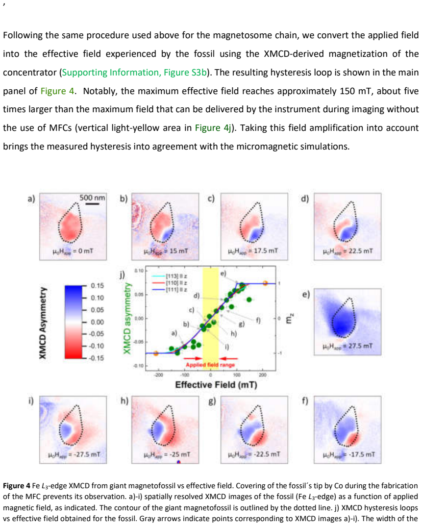
Authors: The 8 mT value is the applied field at which XMCD contrast reversal is first observed in the nanoparticle chain shown in Figure 2; the 50 mT prediction is obtained from micromagnetic simulations whose parameters and geometry are specified in the Methods section. The factor-of-five extension is quantified in Figure 4 by comparing the maximum field at which domain evolution can be imaged with and without the MFC. We will revise the abstract and main text to cite the relevant figures, state the number of particles and fields of view examined (multiple chains across 3–5 fields of view per sample type), and include error estimates on the extracted fields where appropriate. revision: yes
Circularity Check
No circularity: experimental demonstration with independent simulation baseline
full rationale
The paper reports direct experimental observation of magnetization reversal at 8 mT with sample-integrated MFCs versus a 50 mT simulation baseline without MFCs, plus a factor-of-five field-range extension for domain imaging. No equations, fitted parameters, or self-citations are invoked that define the reported amplification factors by construction or rename a known result. The central claims rest on fabrication, PEEM/XMCD measurements, and separate micromagnetic simulations whose inputs do not include the target experimental outcomes. This is a standard self-contained experimental result.
Axiom & Free-Parameter Ledger
free parameters (1)
- MFC geometry and material parameters
axioms (1)
- domain assumption Micrometer-sized MFCs can be fabricated directly on the samples without altering their intrinsic magnetic properties or the XMCD/PEEM signal.
Reference graph
Works this paper leans on
-
[1]
1 Jani, H. et al. Antiferromagnetic half-skyrmions and bimerons at room temperature. Nature 590 , 74-79 (2021). https://doi.org:10.1038/s41586-021-03219-6 2 Ruiz-Gómez, S. et al. Direct X-Ray Detection of the Spin Hall Effect in CuBi. Physical Review X 12 , 031032 (2022). https://doi.org:10.1103/PhysRevX.12.031032 3 Seemann, K. M. & Kronast, F. Artificial...
-
[2]
https://doi.org:10.1038/s43247-024-01540-2 45 Harrison, R
Communications Earth & Environment 5, 386 (2024). https://doi.org:10.1038/s43247-024-01540-2 45 Harrison, R. J. et al. Magnetic vector tomography reveals giant magnetofossils are optimised for magnetointensity reception. Commun Earth Environ 6, 810 (2025). https://doi.org:10.1038/s43247-025-02721-3 46 Heyen, U. & Schüler, D. Growth and magnetosome f ormat...
discussion (0)
Sign in with ORCID, Apple, or X to comment. Anyone can read and Pith papers without signing in.