REVIEW 4 major objections 5 minor 41 references
Batch Augmentation with Unimodal Fine-tuning for Multimodal Learning
T0 review · 4 major / 5 minor · reviewed 2026-08-15 · deepseek-v4-flash
Pith's one-line read Per-sample batch augmentation plus unimodal fine-tuning raises fetal organ detection to above 96 percent accuracy.
desk verdict FPU23 results are undermined by label leakage from text-derived auxiliary features; Food-101 results are plausible but unablated. read the letter →
The pith
A machine-rendered reading of the paper's core claim, the machinery that carries it, and where it could break.
The reading
What carries the argument
The mechanism that carries the argument is a multimodal dataloader and a three-stage training pipeline. The dataloader reads FPU23's .xaml text to find organ labels by word search, converts text fields such as fetal orientation, view direction, and collection invasiveness into numeric codes, and applies a fresh random augmentation independently to each image in a batch. The pipeline then fine-tunes the initial layers of a pre-trained ViT-L/16 or ResNet-50 on the target dataset, removes the model's head, extracts image features in batches, concatenates them with normalized text-derived numbers, and trains a new head on the combined vector. The paper argues that variable per-sample augmentation in each batch gives better weight updates and generalization than a constant batch-wide augmentation.
What would settle it
Rerun the proposed training on FPU23 with labels generated from the dataset's annotation boxes rather than from word search, keeping the image pipeline and the text-derived numeric features unchanged. If head, abdomen, arm, and legs accuracies fall substantially below the reported 96.90%, 91.51%, 93.21%, and 96.72%, the word-search labels or the text features were carrying information the images do not.
Extended reading notes
Core claim
The central claim is that batch augmentation plus unimodal fine-tuning of the initial layers, followed by late fusion, forms a generally better training procedure for image-text classification than either modality alone or standard multimodal training. The paper supports this with FPU23 results where the proposed ViT-L/16 configuration outperforms its own image-only and multimodal baselines on all four organ detection tasks by margins of roughly 4 to 11 percentage points, and with Food-101 results where the proposed approach reaches 92.63% against the 93.1% of a published ensemble built from multiple models. The authors further claim that this is the first use of batch augmentation in multimodal medical data and the first conversion of a vision transformer into an image-text model for fetal organ detection.
Load-bearing premise
The results stand on the assumption that searching the FPU23 text for the words 'Head', 'Abdomen', 'Arm', and 'Legs' produces correct binary organ labels, and that the text-derived features (fetal orientation, view, and collection method) are legitimate auxiliary inputs rather than a shortcut correlated with those labels.
Editorial extensions
If this is right
- On FPU23 head detection, the proposed ViT-L/16 reaches 96.90% test accuracy against 83.81% for image-only and 85.83% for standard multimodal training with the same backbone.
- The proposed training improves ResNet-50 on all four FPU23 organ tasks by roughly 7 to 10 percentage points over image-only training, so the gains are not specific to vision transformers.
- On Food-101, a single model trained with the proposed pipeline reaches 92.63% average accuracy, within 0.47 points of the 93.1% reported ensemble state of the art.
- Because the FPU23 gains from standard multimodal to proposed training are the largest (about 11 points on head detection), the initial-layer fine-tuning step carries most of the improvement on that dataset.
Reading between the lines
- The word-search labeling rule means the FPU23 accuracies are measuring how well the model reproduces organ mentions from the caption text given the image plus the auxiliary text fields; if those text fields are correlated with the labels by construction, the reported multimodal gains could partly be a text shortcut rather than image-based organ detection. This is the editor's inference, not a clai
- A direct test is to rerun the proposed training on FPU23 with labels taken from the dataset's annotation boxes instead of text search while keeping all other details fixed; a large drop in accuracy would confirm that the text-derived features carry label information.
- The same dataloader training recipe could be applied to other clinical multimodal tasks where structured text accompanies imaging, provided the numeric side features are checked to be independent of the label source.
- The Food-101 result suggests that the batch-augmentation component alone, without the unimodal fine-tuning step, may account for part of the gain there, since no medical fine-tuning is prescribed for that dataset; the paper does not ablate this separation.
Signed reviews
Editorial analysis
A structured set of objections, weighed in public.
Referee Report
Summary. The paper proposes a multimodal learning recipe composed of two interventions: (i) a dataloader that applies different random augmentations to different samples within a batch (batch augmentation), and (ii) a unimodal fine-tuning stage in which the initial layers of a pretrained vision model are further trained on the target medical images before multimodal head training. The method is evaluated on the UPMC Food-101 multimodal dataset and the FPU23 fetal ultrasound dataset using ResNet-50 and ViT-L/16 backbones. The best reported results are 92.63% average accuracy on Food-101 and up to 96.90% on FPU23 head detection with ViT-L/16. The authors also release scripts at a public GitHub repository.
Significance. If the reported gains were attributable to the proposed recipe, the paper would offer a simple and potentially useful prescription for improving multimodal medical image classification, and the public code release would be a practical strength. The Food-101 result (92.63% vs. a cited 93.1% ensemble SOTA) is credible as a near-SOTA unimodal-plus-text score. However, the significance is currently limited by three unresolved issues: the two components of the recipe are never ablated individually, the FPU23 auxiliary text features appear to be confounded with the label source, and the pseudocode does not actually describe the claimed unimodal fine-tuning stage. These issues must be settled before the FPU23 performance claims can be accepted as evidence for the method.
major comments (4)
- [Tables II and III; Section III-F] The proposed recipe combines per-sample random augmentation with a unimodal fine-tuning stage, but no experiment isolates these interventions. For example, Table III reports ViT-L/16 head accuracy jumping from 85.83% (multimodal) to 96.90% (proposed), yet the reader cannot tell whether this gain comes from batch augmentation, from unimodal fine-tuning of the initial layers, or from an interaction between the two. Without ablations such as image-only with batch augmentation, unimodal fine-tuning alone, and multimodal without fine-tuning, the central attribution claim that the full recipe is responsible for the gains is not supported.
- [Algorithm 1; Section III-F] Algorithm 1 as written trains only the newly declared head layer NN_H: the pretrained feature extractor NN is applied without weight updates (lines 12-13), and only the head is optimized (line 20). However, the abstract and Section III-F describe a separate prior stage that fine-tunes the initial layers on the unimodal image data with batch augmentation. This stage is absent from the pseudocode, and no hyperparameters, epoch count, or loss details are given for it. Consequently, the described method is not reproducible from the paper, and it is unclear whether the numbers in Tables II and III correspond to Algorithm 1 or to an undescribed procedure.
- [Section III-E; Algorithm 1 lines 10-11; Table I; Section IV-B] On FPU23, both the auxiliary Info vector and the binary labels are extracted from the same .xaml text. The label for head detection is produced by searching for the word 'Head' (Section IV-B), while Info encodes fetal orientation, view, and collection method (Section III-E). As Table I shows, the orientation categories literally include 'head up' and 'head down', so the substring 'head' in the Info source can make Info a deterministic or strongly predictive function of the label. The paper reports no control with Info removed, no Info-only model, and no validation of the word-search label rule against the dataset's annotation boxes. Therefore the multimodal and proposed accuracies in Table III cannot currently be attributed to image features or to the proposed training recipe.
- [Section IV-D vs. Table III] The prose in Section IV-D states that for arm detection the image-only ViT-L/16 model provides the best result (89.08±2.02%), whereas Table III reports the proposed ViT-L/16 model at 93.21±0.43% as the best value and marks it in bold. This is an internal contradiction in the paper's central results table. The authors must correct the inconsistency and state which value is correct and how it was obtained.
minor comments (5)
- [Abstract and throughout] The term 'multimodal large language model (LLM)' is inaccurate: the image backbone used is a vision transformer (ViT), not a language model. Consider using 'multimodal vision transformer' or 'multimodal model' consistently.
- [Tables II and III] The number of independent training runs used to compute the reported means and standard deviations is not stated. Please specify the number of seeds/repeats and the train-validation-test split protocol for both datasets.
- [Algorithm 1] The symbol BN is used both for the batch size in Eq. (2) and for the number of batches in Algorithm 1 line 8, which is confusing. Use distinct names for these quantities.
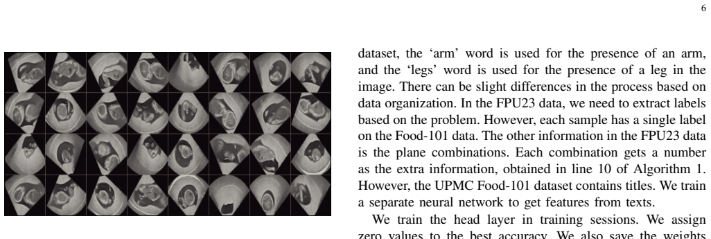
- [Section III-B] Please verify the resize size: the text says 'resize images to a size of 244 by 244' and then crops to 224 by 224; this is plausible but the figure captions refer to 32 images without stating the resolution after augmentation. Clarify the exact pipeline.
- [References] Reference [15] (Wightman et al., 'Resnet strikes back') is cited for the timm library, but the relevant citation for the timm model collection would be more appropriate. Please check reference accuracy.
Circularity Check
FPU23 head-detection Info and labels are co-extracted from the same .xaml annotation text, so the reported gain partly reduces to a text-search shortcut; the UPMC Food-101 benchmark remains independent.
-
self definitional
[Algorithm 1 lines 10-11; Sec. III-E; Sec. IV-B; Table I]
"Algorithm 1: 'Obtain Info [j] from texts.' / 'Extract Label[j,D ] from TexLabel [j]' / Sec. III-E: 'The Dataloader finds the presence of the label by searching for the word in the text. The Dataloader also processes several texts containing the orientation of the fetus, the sample collection process, and the direction of imaging.' / Sec. IV-B: 'To prepare labels for head detection, we search for the word ‘Head’ in the label containing text.' / Table I: 'head up (hu) or head down (hd)'."
FPU23's other input information Info[j] and the label Label[j,D] are both extracted from the same .xaml annotation text. For head detection, the label is defined by the presence of the word 'Head' in the label-containing text, while Table I shows that the Info orientation categories are literally 'head up'/'head down'. Thus the category name supplied to the head layer contains the same lexical item that defines the label; a head layer that reads Info can score the positive class without using image content. The reported 96.90% 'proposed' accuracy is therefore not a pure image-based prediction: the input and label are co-defined from one annotation source.
full rationale
The classic circularity failure modes are largely absent: Eqs. (2)-(4) in Sec. II-B are ordinary SGD and augmentation update rules, no fitted constant is later renamed as a prediction, and the UPMC Food-101 experiment is an external benchmark with independent labels and titles, so the 92.63% result is self-contained evidence for the late-fusion and batch-augmentation recipe. The circular-content signal is confined to the FPU23 evaluation. Algorithm 1 obtains Info and Label from the same texts; for head detection the label is defined by the word search 'Head', while Table I's Info orientation categories are literally 'head up' and 'head down'. The head layer can therefore exploit the annotation text itself, so the reported head accuracy (and, to a lesser extent, the other organ rows) is not guaranteed to measure image-based organ detection. Because no Info-only or no-Info control is reported, the proposed-vs-multimodal gap cannot be cleanly attributed to the proposed fine-tuning and augmentation either. Additionally, Sec. IV-D's prose contradicts Table III for arm detection (prose says image-only ViT-L/16 is best at 89.08%, while the table lists proposed ViT-L/16 at 93.21% as best), casting further doubt on the FPU23 reporting. No load-bearing self-citation chain was found; prior work by the authors is used only for general transfer-learning motivation. Overall, the central FPU23 claim partially reduces by construction, while the UPMC benchmark retains independent content; hence score 6.
Assumptions & free parameters
free parameters (4)
- Shared optimizer schedule (learning rate 5e-4, momentum 0.9, step size 7, gamma 0.1) =
5e-4, 0.9, 7, 0.1
- Epoch count for the unimodal fine-tuning stage =
Not stated
- Food-101 text score network (two hidden layers of 200 neurons, batch 128, LR 0.001, 10 epochs) =
200 neurons, 128, 0.001, 10 epochs
- Batch sizes =
64 (ResNet-50), 20 (ViT-L/16), 32 (figures)
assumptions (4)
- domain assumption Initial layers of pretrained vision models compute features generic enough that fine-tuning them on the target medical data improves downstream multimodal classification (Section II-A).
- ad hoc to paper A constant augmentation applied to all samples in a batch shifts the decision boundary and harms generalization, whereas per-sample variable augmentation brings the boundary closer to the ground truth (Section II-B, Fig. 2).
- domain assumption Word-search rules over the FPU23 text ('Head', 'Abdomen', 'Arm', 'Legs') produce correct binary labels for organ presence (Sections III-E, IV-B to IV-E).
- standard math Standard results: class activation map relation (Eq. 1) and SGD update equations (Eqs. 2-4).
Cite this review
Pith. "Pith review of Batch Augmentation with Unimodal Fine-tuning for Multimodal Learning." pith.science (2026). https://pith.science/paper/GQIO3GKR
@misc{pith2026250506592,
  author       = {Pith},
  title        = {Pith review of: Batch Augmentation with Unimodal Fine-tuning for Multimodal Learning},
  year         = {2026},
  howpublished = {\url{https://pith.science/paper/GQIO3GKR}},
  note         = {Machine review of arXiv:2505.06592}
}
read the original abstract
This paper proposes batch augmentation with unimodal fine-tuning to detect the fetus's organs from ultrasound images and associated clinical textual information. We also prescribe pre-training initial layers with investigated medical data before the multimodal training. At first, we apply a transferred initialization with the unimodal image portion of the dataset with batch augmentation. This step adjusts the initial layer weights for medical data. Then, we apply neural networks (NNs) with fine-tuned initial layers to images in batches with batch augmentation to obtain features. We also extract information from descriptions of images. We combine this information with features obtained from images to train the head layer. We write a dataloader script to load the multimodal data and use existing unimodal image augmentation techniques with batch augmentation for the multimodal data. The dataloader brings a new random augmentation for each batch to get a good generalization. We investigate the FPU23 ultrasound and UPMC Food-101 multimodal datasets. The multimodal large language model (LLM) with the proposed training provides the best results among the investigated methods. We receive near state-of-the-art (SOTA) performance on the UPMC Food-101 dataset. We share the scripts of the proposed method with traditional counterparts at the following repository: github.com/dipuk0506/multimodal
Figures


Figures from the paper (4 more)



Reference graph
Works this paper leans on
-
[1]
Augment your batch: Improving generalization through instance repeti- tion,
E. Hoffer, T. Ben-Nun, I. Hubara, N. Giladi, T. Hoefler, and D. Soudry, “Augment your batch: Improving generalization through instance repeti- tion,” in Proceedings of the IEEE/CVF Conference on Computer Vision and Pattern Recognition, 2020, pp. 8129–8138
2020
-
[2]
Image retrieval based on deep feature extraction and reduction with improved cnn and pca,
R. Chen, L. Pan, Y . Zhou, and Q. Lei, “Image retrieval based on deep feature extraction and reduction with improved cnn and pca,” Journal of Information Hiding and Privacy Protection , vol. 2, no. 2, p. 67, 2020
work page 2020
-
[3]
Dl-ids: Extracting features using cnn-lstm hybrid network for intrusion detection system,
P. Sun, P. Liu, Q. Li, C. Liu, X. Lu, R. Hao, and J. Chen, “Dl-ids: Extracting features using cnn-lstm hybrid network for intrusion detection system,” Security and communication networks , vol. 2020, no. 1, p. 8890306, 2020
work page 2020
-
[4]
The effectiveness of data augmentation in image classification using deep learning,
J. Wang, L. Perez et al. , “The effectiveness of data augmentation in image classification using deep learning,” Convolutional Neural Networks Vis. Recognit, vol. 11, no. 2017, pp. 1–8, 2017. 9
work page 2017
-
[5]
Generic database cost models for hierarchical memory systems,
S. Manegold, P. Boncz, and M. L. Kersten, “Generic database cost models for hierarchical memory systems,” in VLDB’02: Proceedings of the 28th International Conference on Very Large Databases . Elsevier, 2002, pp. 191–202
work page 2002
-
[6]
Torchvision the machine-vision package of torch,
S. Marcel and Y . Rodriguez, “Torchvision the machine-vision package of torch,” in Proceedings of the 18th ACM international conference on Multimedia, 2010, pp. 1485–1488
2010
-
[7]
F. Albardi, H. D. Kabir, M. M. I. Bhuiyan, P. M. Kebria, A. Khosravi, and S. Nahavandi, “A comprehensive study on torchvision pre-trained models for fine-grained inter-species classification,” in 2021 IEEE Inter- national Conference on Systems, Man, and Cybernetics (SMC) . IEEE, 2021, pp. 2767–2774
work page 2021
-
[8]
E. Abuelhia and A. Alghamdi, “Evaluation of arising exposure of ionizing radiation from computed tomography and the associated health concerns,” Journal of Radiation Research and Applied Sciences, vol. 13, no. 1, pp. 295–300, 2020
work page 2020
Show all 41 references
-
[9]
Fetal mri: Is it safe?
M. Bekiesinska-Figatowska, “Fetal mri: Is it safe?” Journal of Pediatric Neuroradiology, vol. 1, no. 03, pp. 155–159, 2012
2012
-
[10]
Lung ultra- sound vs. chest x-ray study for the radiographic diagnosis of covid-19 pneumonia in a high-prevalence population,
R. C. Gibbons, M. Magee, H. Goett, J. Murrett, J. Genninger, K. Mendez, M. Tripod, N. Tyner, and T. G. Costantino, “Lung ultra- sound vs. chest x-ray study for the radiographic diagnosis of covid-19 pneumonia in a high-prevalence population,” The Journal of emergency medicine,...
2021
-
[11]
Trends in ultrasound use in low and middle income countries: a systematic review,
K. A. Stewart, S. M. Navarro, S. Kambala, G. Tan, R. Poondla, S. Lederman, K. Barbour, and C. Lavy, “Trends in ultrasound use in low and middle income countries: a systematic review,” International Journal of Maternal and Child Health and AIDS , vol. 9, no. 1, p. 103, 2020
2020
-
[12]
Potential for use of portable ultrasound devices in rural and remote settings in australia and other developed countries: a systematic review,
L. Shaddock and T. Smith, “Potential for use of portable ultrasound devices in rural and remote settings in australia and other developed countries: a systematic review,”Journal of Multidisciplinary Healthcare, pp. 605–625, 2022
2022
-
[13]
Evaluation of deep convolutional neural networks for automatic classification of common maternal fetal ultrasound planes,
X. P. Burgos-Artizzu, D. Coronado-Guti ´errez, B. Valenzuela-Alcaraz, E. Bonet-Carne, E. Eixarch, F. Crispi, and E. Gratac ´os, “Evaluation of deep convolutional neural networks for automatic classification of common maternal fetal ultrasound planes,” Scientific Reports , vol....
2020
-
[14]
Aleatory-aware deep uncertainty quantification for transfer learning,
H. D. Kabir, S. Khanam, F. Khozeimeh, A. Khosravi, S. K. Mondal, S. Nahavandi, and U. R. Acharya, “Aleatory-aware deep uncertainty quantification for transfer learning,”Computers in Biology and Medicine, vol. 143, p. 105246, 2022
2022
-
[15]
Resnet strikes back: An improved training procedure in timm,
R. Wightman, H. Touvron, and H. J ´egou, “Resnet strikes back: An improved training procedure in timm,” arXiv preprint arXiv:2110.00476, 2021
2021 arXiv
-
[16]
Learning deep features for discriminative localization,
B. Zhou, A. Khosla, A. Lapedriza, A. Oliva, and A. Torralba, “Learning deep features for discriminative localization,” inProceedings of the IEEE conference on computer vision and pattern recognition, 2016, pp. 2921– 2929
2016
-
[17]
Deep inside convolutional networks: Visualising image classification models and saliency maps,
K. Simonyan, “Deep inside convolutional networks: Visualising image classification models and saliency maps,” arXiv preprint arXiv:1312.6034, 2013
2013 arXiv
-
[18]
Under- standing neural networks through deep visualization,
J. Yosinski, J. Clune, A. Nguyen, T. Fuchs, and H. Lipson, “Under- standing neural networks through deep visualization,” arXiv preprint arXiv:1506.06579, 2015
2015 arXiv
-
[19]
Visualizing and understanding convolutional networks,
M. Zeiler, “Visualizing and understanding convolutional networks,” in European conference on computer vision/arXiv , vol. 1311, 2014
2014
-
[20]
Spinalnet: Deep neural network with gradual input,
H. D. Kabir, M. Abdar, A. Khosravi, S. M. J. Jalali, A. F. Atiya, S. Nahavandi, and D. Srinivasan, “Spinalnet: Deep neural network with gradual input,” IEEE Transactions on Artificial Intelligence, vol. 4, no. 5, pp. 1165–1177, 2022
2022
-
[21]
Automatic detection of abnormal eeg signals using wavelet feature extraction and gradient boosting decision tree,
H. Albaqami, G. M. Hassan, A. Subasi, and A. Datta, “Automatic detection of abnormal eeg signals using wavelet feature extraction and gradient boosting decision tree,” Biomedical Signal Processing and Control, vol. 70, p. 102957, 2021
2021
-
[22]
Deep facial diagnosis: deep transfer learning from face recognition to facial diagnosis,
B. Jin, L. Cruz, and N. Gonc ¸alves, “Deep facial diagnosis: deep transfer learning from face recognition to facial diagnosis,” IEEE Access, vol. 8, pp. 123 649–123 661, 2020
2020
-
[23]
The effect of batch size on the generalizabil- ity of the convolutional neural networks on a histopathology dataset,
I. Kandel and M. Castelli, “The effect of batch size on the generalizabil- ity of the convolutional neural networks on a histopathology dataset,” ICT express, vol. 6, no. 4, pp. 312–315, 2020
2020
-
[24]
Fetal images: The power of visual culture in the politics of reproduction,
R. P. Petchesky, “Fetal images: The power of visual culture in the politics of reproduction,” in The medicalization of obstetrics . Routledge, 2021, pp. 361–390
2021
-
[25]
State abortion policies and maternal death in the united states, 2015–2018,
D. Vilda, M. E. Wallace, C. Daniel, M. G. Evans, C. Stoecker, and K. P. Theall, “State abortion policies and maternal death in the united states, 2015–2018,” American Journal of Public Health , vol. 111, no. 9, pp. 1696–1704, 2021
2015
-
[26]
Unified deep learning model for multitask rep- resentation and transfer learning: image classification, object detection, and image captioning,
L. Y . Bayisa, W. Wang, Q. Wang, C. C. Ukwuoma, H. K. Gutema, A. Endris, and T. Abu, “Unified deep learning model for multitask rep- resentation and transfer learning: image classification, object detection, and image captioning,” International Journal of Machine Learning and ...
2024
-
[27]
Artificial intelligence in obstetric ultrasound: A scoping review,
R. Horgan, L. Nehme, and A. Abuhamad, “Artificial intelligence in obstetric ultrasound: A scoping review,” Prenatal Diagnosis , vol. 43, no. 9, pp. 1176–1219, 2023
2023
-
[28]
Machine learning algorithms for classification of first-trimester fetal brain ultra- sound images,
S. Gofer, O. Haik, R. Bardin, Y . Gilboa, and S. Perlman, “Machine learning algorithms for classification of first-trimester fetal brain ultra- sound images,” Journal of Ultrasound in Medicine , vol. 41, no. 7, pp. 1773–1779, 2022
2022
-
[29]
Automatic fetal middle sagittal plane detection in ultrasound using generative adversarial network,
P.-Y . Tsai, C.-H. Hung, C.-Y . Chen, and Y .-N. Sun, “Automatic fetal middle sagittal plane detection in ultrasound using generative adversarial network,” Diagnostics, vol. 11, no. 1, p. 21, 2020
2020
-
[30]
Automatic detection of standard sagittal plane in the first trimester of pregnancy using 3-d ultrasound data,
S. Nie, J. Yu, P. Chen, Y . Wang, and J. Q. Zhang, “Automatic detection of standard sagittal plane in the first trimester of pregnancy using 3-d ultrasound data,” Ultrasound in medicine & biology , vol. 43, no. 1, pp. 286–300, 2017
2017
-
[31]
Ultrasound placental image texture analysis using artificial intelligence to predict hypertension in pregnancy,
K. Gupta, K. Balyan, B. Lamba, M. Puri, D. Sengupta, and M. Kumar, “Ultrasound placental image texture analysis using artificial intelligence to predict hypertension in pregnancy,” The Journal of Maternal-Fetal & Neonatal Medicine, vol. 35, no. 25, pp. 5587–5594, 2022
2022
-
[32]
No sonographer, no radiologist: New system for automatic prenatal detection of fetal biometry, fetal presentation, and placental location,
J. Arroyo, T. J. Marini, A. C. Saavedra, M. Toscano, T. M. Baran, K. Drennan, A. Dozier, Y . T. Zhao, M. Egoavil, L. Tamayo et al., “No sonographer, no radiologist: New system for automatic prenatal detection of fetal biometry, fetal presentation, and placental location,” PloS...
2022
-
[33]
An automated framework for image classification and segmentation of fetal ultrasound images for gestational age estimation,
J. C. Prieto, H. Shah, A. J. Rosenbaum, X. Jiang, P. Musonda, J. T. Price, E. M. Stringer, B. Vwalika, D. M. Stamilio, and J. S. Stringer, “An automated framework for image classification and segmentation of fetal ultrasound images for gestational age estimation,” in Medical I...
2021
-
[34]
Medical professional enhancement using explainable artificial intelligence in fetal cardiac ultrasound screening,
A. Sakai, M. Komatsu, R. Komatsu, R. Matsuoka, S. Yasutomi, A. Dozen, K. Shozu, T. Arakaki, H. Machino, K. Asada et al., “Medical professional enhancement using explainable artificial intelligence in fetal cardiac ultrasound screening,” Biomedicines, vol. 10, no. 3, p. 551, 2022
2022
-
[35]
Deep multimodal learning: A survey on recent advances and trends,
D. Ramachandram and G. W. Taylor, “Deep multimodal learning: A survey on recent advances and trends,” IEEE signal processing magazine, vol. 34, no. 6, pp. 96–108, 2017
2017
-
[36]
Multimodal deep learning,
J. Ngiam, A. Khosla, M. Kim, J. Nam, H. Lee, and A. Y . Ng, “Multimodal deep learning,” in Proceedings of the 28th international conference on machine learning (ICML-11) , 2011, pp. 689–696
2011
-
[37]
A survey on multimodal large language models for autonomous driving,
C. Cui, Y . Ma, X. Cao, W. Ye, Y . Zhou, K. Liang, J. Chen, J. Lu, Z. Yang, K.-D. Liao et al. , “A survey on multimodal large language models for autonomous driving,” in Proceedings of the IEEE/CVF Winter Conference on Applications of Computer Vision , 2024, pp. 958– 979
2024
-
[38]
Fpus23: an ultrasound fetus phantom dataset with deep neural network evaluations for fetus orientations, fetal planes, and anatomical features,
B. S. Prabakaran, P. Hamelmann, E. Ostrowski, and M. Shafique, “Fpus23: an ultrasound fetus phantom dataset with deep neural network evaluations for fetus orientations, fetal planes, and anatomical features,” IEEE Access, vol. 11, pp. 58 308–58 317, 2023
2023
-
[39]
Recipe recognition with large multimodal food dataset,
X. Wang, D. Kumar, N. Thome, M. Cord, and F. Precioso, “Recipe recognition with large multimodal food dataset,” in 2015 IEEE Interna- tional Conference on Multimedia & Expo Workshops (ICMEW) . IEEE, 2015, pp. 1–6
2015
-
[40]
Coca: Contrastive captioners are image-text foundation models,
J. Yu, Z. Wang, V . Vasudevan, L. Yeung, M. Seyedhosseini, and Y . Wu, “Coca: Contrastive captioners are image-text foundation models,” arXiv preprint arXiv:2205.01917, 2022
2022 arXiv
-
[41]
Stacking and voting ensemble models for improving food image recognition,
S. Suresh and A. Verma, “Stacking and voting ensemble models for improving food image recognition,” in 2024 IEEE 3rd International Conference on Computing and Machine Intelligence (ICMI) . IEEE, 2024, pp. 1–6
2024
Reviewed August 15, 2026 · model on record in the stance chip above.
Discussion (0). Continue with ORCID to comment.