REVIEW 4 major objections 7 minor 6 references
Cartilage-binding antibodies induce pain through immune complex-mediated activation of neurons
T0 review · 4 major / 7 minor · reviewed 2026-08-14 · deepseek-v4-flash
Pith's one-line read Cartilage-binding antibodies cause pain by directly activating Fcγ receptors on sensory nerve endings, independent of joint inflammation.
desk verdict A careful, well-controlled mouse study that convincingly shows cartilage-antibody pain depends on FcγRs, but stops short of proving those FcγRs act on neurons rather than other non-hematopoietic cells. read the letter →
The pith
A machine-rendered reading of the paper's core claim, the machinery that carries it, and where it could break.
The reading
What carries the argument
The load-bearing mechanism is FcγRI, the high-affinity receptor for the Fc portion of IgG, carried on peripheral endings of nociceptive neurons, and CII-IC, immune complexes made of collagen type II and anti-CII antibody. The paper argues that Fcgr1 mRNA is transported down sensory axons—it accumulates proximal to a sciatic-nerve ligature—so FcγRI can be locally expressed at terminals; when CII-IC engages it, the receptor drives excitatory signaling that raises intracellular calcium, generates an inward current, and releases the pain-associated neuropeptide CGRP. Every intervention that severs the Fc–FcγRI link—Fab fragments, EndoS deglycosylation, FcRγ-chain knockout, or loss of activating FcγRs from non-hematopoietic cells—also abolishes the pain behavior, which is what makes the receptor the pivot of the argument.
What would settle it
In mice with Fcgr1 deleted specifically from nociceptors, systemic anti-CII antibody injection should no longer produce mechanical hypersensitivity if the claim is correct; if such mice still develop hypersensitivity, the neuronal FcγRI requirement is not causal. A complementary observation would be fluorescently labeled CII immune complexes binding to joint-innervating nerve endings in wild-type but not neuron-specific Fcgr1 knockout mice.
Extended reading notes
Core claim
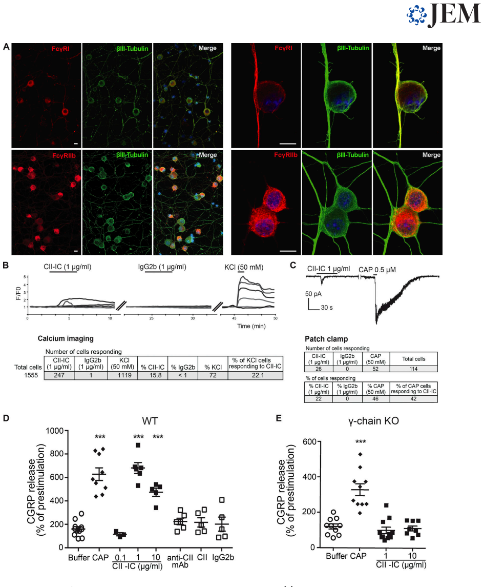
On its own terms, the paper's central discovery is that collagen type II–reactive antibodies evoke pain-like behavior in mice by forming immune complexes locally in cartilage and activating FcγRI on peripheral sensory-nerve terminals. Preformed CII immune complexes applied to cultured dorsal root ganglion neurons raise intracellular calcium, produce inward currents, and trigger CGRP release, whereas monomeric antibodies do not; neurons from FcRγ-chain-deficient mice do not respond. In vivo, intra-articular injection of CII immune complexes elicits mechanical hypersensitivity in wild-type but not FcRγ-chain-deficient mice, and systemic anti-CII antibodies lose their pronociceptive effect when their Fc interaction is disabled. Chimeric mice lacking activating Fcγ receptors on non-hematopoietic cells, including neurons, are protected from antibody-induced hypersensitivity, which places the relevant receptor on neurons rather than immune cells. The same logic extends to antibodies against cartilage oligomeric matrix protein, supporting the general claim that locally formed immune complexes can drive pain before inflammation begins.
Load-bearing premise
Joint-innervating sensory terminals actually carry functional FcγRI protein that local immune complexes can engage; the paper shows Fcgr1 mRNA moving toward peripheral axons and FcγRI immunoreactivity in skin nerve fibers, but it does not directly demonstrate FcγRI at joint terminals or immune-complex activation of those terminals in vivo.
Editorial extensions
If this is right
- Pain in autoantibody-driven arthritis can be separated from inflammation: measuring it in the first days after antibody transfer captures a distinct, FcγRI-dependent phase that standard arthritis scoring misses.
- Blocking FcγRI on peripheral nerve endings, or preventing immune-complex engagement of it, could relieve antibody-induced joint pain without general immunosuppression.
- The mechanism should generalize to other cartilage and joint antigens: the COMP antibody results show that any antibody that binds an antigen near joint afferents can produce pain once local immune complexes form.
- In humans the relevant neuronal Fc receptor may be FcγRIII rather than FcγRI, since human DRG neurons express FcγRIII; this makes FcγRIII a candidate target for autoimmune pain.
- FcRγ-chain-deficient mice become a screening platform for deciding whether other autoantibody-related pain phenotypes depend on activating Fcγ receptors.
Reading between the lines
- Beyond the paper, the same nerve-ending detector could operate in other conditions where antibodies meet local antigen without much inflammation, such as early seropositive arthralgia or crystal-associated arthropathies; this is a testable extension rather than a demonstrated result.
- The axonal Fcgr1 mRNA transport data suggest local translation at terminals: if true, injuring a nerve or blocking local protein synthesis should change how quickly CII antibodies produce hypersensitivity, a prediction the paper does not test.
- The roughly one-fifth of cultured neurons responding to CII-IC hints that immune-complex sensing may define a specific nociceptor subpopulation; single-cell profiling could identify molecular markers for that subset.
- Because the FcγR isoform on neurons differs between species, the mouse data may misidentify the therapeutic target in humans; monoclonal antibodies against human FcγRIII could be screened directly for blockade of IC-evoked human DRG excitation.
Signed reviews
Editorial analysis
A structured set of objections, weighed in public.
Referee Report
Summary. This manuscript investigates how cartilage-binding autoantibodies cause pain before joint inflammation. Using components of the collagen antibody-induced arthritis (CAIA) model, the authors report that anti-CII monoclonal antibodies elicit mechanical hypersensitivity from day 2 onward, before visual, histological, or molecular signs of inflammation, and that this is independent of complement C5 and cartilage destruction. They map Fcgr mRNA and protein in mouse DRG and peripheral nerve, show that CII immune complexes (CII-ICs) raise intracellular calcium, evoke inward currents, and release CGRP in WT but not FcRγ-chain-deficient DRG cultures, and show that anti-CII/anti-COMP antibodies and preformed ICs induce pain-like behavior in WT but not FcRγ-chain-deficient mice. Bone-marrow chimeras implicate non-hematopoietic FcγRs. The paper concludes that CII-ICs directly activate neuronal FcγRI to produce pain.
Significance. If the causal claim holds, this is a significant conceptual advance: it identifies a pre-inflammatory, non-canonical pathway for autoantibody-induced pain in rheumatoid arthritis and related autoimmune conditions, and it nominates neuronal FcγRs as a therapeutic target. The strengths are substantial: multiple independent readouts (behavior, CGRP release, calcium imaging, patch-clamp electrophysiology), blinded behavioral assessment, isotype and genetic controls including FcRγ-chain-, FcγRIII-, FcγRIV-, and C5-deficient mice, the use of both arthritogenic and non-arthritogenic CII-binding antibodies, Fab and EndoS treatments, and a bone-marrow chimera design. The microarray data are deposited, and public DRG transcriptome resources are used. The principal weakness is that the decisive in vivo genetic experiment does not isolate neurons from other non-hematopoietic cells, and the in vitro experiments do not establish cell-autonomous neuronal signaling; the authors partly acknowledge this in the Discussion.
major comments (4)
- [Fig. 8, H–J; Discussion] The in vivo evidence does not establish that sensory neurons, rather than other non-hematopoietic cells, are the FcγR-expressing cells that mediate anti-CII antibody pain. The WT-KO versus KO-WT bone-marrow chimera separates hematopoietic from non-hematopoietic compartments, but the non-hematopoietic compartment also includes keratinocytes, endothelial cells, synovial fibroblasts, and radio-resistant tissue-resident macrophages and mast cells, which can express FcγRs. The authors acknowledge in the Discussion that 'these results do not exclusively test the role of activating FcγRs on neurons,' and the abstract's phrase 'mice lacking activating FcγRs in neurons' is therefore an overstatement. A neuron-specific deletion of Fcgr1 or of the FcR γ-chain (for example with Advillin-Cre or Nav1.8-Cre) is needed to support the title's causal attribution.
- [Fig. 6, B–E; Fig. 4, E] The in vitro experiments do not prove that CII-ICs directly activate neurons. CGRP release and calcium imaging were conducted in mixed DRG cultures, and FcRγ-chain deletion removes activating FcγRs from macrophages and satellite glia as well as from neurons; the loss of CGRP release in FcRγ-chain-deficient cultures could therefore reflect loss of non-neuronal FcγR signaling and a secondary mediator. Because FcγRI immunoreactivity in intact DRG is restricted to resident macrophages (Fig. 5, A and B), the positive staining on cultured neurites (Fig. 6 A) needs to be supported by cell-autonomous experiments, such as purified or sorted neuronal cultures, conditioned-medium experiments, or in vitro neuron-specific ablation of FcγR signaling.
- [Fig. 5, E–I; Discussion] The paper does not demonstrate FcγRI protein at joint-innervating nerve terminals or local translation of Fcgr1 mRNA in nociceptor axons. FcγRI staining is shown in PGP9.5-positive fibers in glabrous skin, but not in joint afferents, and no FcRγ-chain-knockout negative control is shown for skin immunostaining. The sciatic-ligation smFISH shows accumulation of Fcgr1 and Fcgr2b mRNA proximal to the ligature, but because no axonal marker is provided, accumulation in Schwann cells or recruited macrophages is not excluded. The Discussion states that 'further work is necessary to determine if FcγRs are locally translated in nociceptors,' which is precisely the load-bearing step for the claim that locally formed CII-ICs engage neuronal FcγRI at peripheral terminals.
- [Fig. 6, D and E; Fig. 7, I–K] The attribution to FcγRI specifically is made by exclusion (FcγRIII deficiency does not alter calcium responses, and FcγRIV-deficient mice still develop pain), but no FcγRI-specific loss-of-function or blocking reagent is used. Since FcRγ-chain deletion removes FcγRI, FcγRIII, and FcγRIV together, the conclusion that FcγRI is the responsible receptor would be strengthened by an FcγRI-blocking antibody, Fcgr1 knockdown in cultured neurons, or conditional Fcgr1 deletion.
minor comments (7)
- [Abstract] The phrase 'mice lacking activating FcγRs in neurons' should be changed to 'mice lacking activating FcγRs in non-hematopoietic cells' until a neuron-specific manipulation is performed.
- [Fig. 5, E and F] Please add FcRγ-chain-knockout skin sections as a specificity control for the FcγRI and FcγRIIb immunoreactivity in nerve fibers.
- [Fig. 5, H and I] Please include a neuronal or axonal marker (for example neurofilament or SCG10) in the sciatic-ligation smFISH to support the interpretation of axonal mRNA transport.
- [Methods, Calcium imaging and Electrophysiology] Please report the number of independent cultures and animals, and the number of cells per group, for the calcium-imaging and patch-clamp experiments; the text gives aggregate cell counts but not the replicate structure.
- [Results, Fig. 6 B] The phrase 'directly activate cultured WT... DRG neurons' is too strong for a mixed-culture preparation; consider 'activate DRG neurons in mixed cultures' until cell-autonomy is demonstrated.
- [Discussion, human DRG] The human data show FcγRIII, not FcγRI, in DRG neurons; the translational discussion should more clearly separate the mouse FcγRI mechanism from the human FcγRIII observation, which is purely descriptive.
- [Fig. 8, H–J; Methods] The labels WT-KO and KO-WT are easy to misread; please spell out donor genotype into recipient genotype in the figure legend and define the direction of the bone-marrow transfer explicitly.
Circularity Check
No circularity found: the central claim is tested by independent genetic loss-of-function, behavioral, and in vitro assays, not derived from its own inputs.
full rationale
The load-bearing chain is: anti-CII/COMP antibodies form immune complexes in joints; FcγRI is present on peripheral sensory axons; CII-ICs excite WT but not FcRγ-chain-deficient DRG neurons; and anti-CII antibodies fail to induce mechanical hypersensitivity in FcRγ-chain-deficient mice and in chimeras lacking activating FcγRs on non-hematopoietic cells. Each link is supported by external readouts (von Frey thresholds, locomotion, CGRP release, calcium imaging, patch clamp) and by genetic loss-of-function models rather than by fitting a parameter to the outcome. The authors' prior work supplies reagents (anti-CII mAb cocktail, CAIA model, EndoS treatment, CIIF4 non-arthritogenic antibody) and background observations, but the present predictions do not reduce to those prior results by definition. The Discussion explicitly acknowledges the residual ambiguity of the chimera experiment ('these results do not exclusively test the role of activating FcγRs on neurons') and the lack of proof of local translation ('While further work is necessary to determine if FcγRs are locally translated in nociceptors'); these are honest scope limits, not circular reasoning. No equation, fitted parameter, or self-citation chain makes the conclusion equivalent to an input.
Assumptions & free parameters
assumptions (4)
- domain assumption FcR gamma-chain knockout eliminates cell surface expression and signaling of activating Fc gamma receptors I, III, and IV, while leaving Fc gamma receptor IIb intact.
- domain assumption Intravenously injected anti-CII antibodies reach the joint and form local immune complexes with CII fragments in the synovium or cartilage.
- domain assumption Bone-marrow chimeras separate hematopoietic from non-hematopoietic Fc gamma receptor expression without confounding radiation damage to peripheral neurons.
- domain assumption Mechanical withdrawal thresholds measured with von Frey filaments reflect pain-like behavior rather than motor impairment or general debilitation.
Cite this review
Pith. "Pith review of Cartilage-binding antibodies induce pain through immune complex-mediated activation of neurons." pith.science (2026). https://pith.science/paper/HIFGOHF7
@misc{pith2026190805298,
  author       = {Pith},
  title        = {Pith review of: Cartilage-binding antibodies induce pain through immune complex-mediated activation of neurons},
  year         = {2026},
  howpublished = {\url{https://pith.science/paper/HIFGOHF7}},
  note         = {Machine review of arXiv:1908.05298}
}
read the original abstract
Rheumatoid arthritis-associated joint pain is frequently observed independent of disease activity, suggesting unidentified pain mechanisms. We demonstrate that antibodies binding to cartilage, specific for collagen type II (CII) or cartilage oligomeric matrix protein (COMP), elicit mechanical hypersensitivity in mice, uncoupled from visual, histological and molecular indications of inflammation. Cartilage antibody-induced pain-like behavior does not depend on complement activation or joint inflammation, but instead on tissue antigen recognition and local immune complex (IC) formation. smFISH and IHC suggest that neuronal Fcgr1 and Fcgr2b mRNA are transported to peripheral ends of primary afferents. CII-ICs directly activate cultured WT but not FcR{\gamma} chain-deficient DRG neurons. In line with this observation, CII-IC does not induce mechanical hypersensitivity in FcR{\gamma} chain-deficient mice. Furthermore, injection of CII antibodies does not generate pain-like behavior in FcR{\gamma} chain-deficient mice or mice lacking activating Fc{\gamma}Rs in neurons. In summary, this study defines functional coupling between autoantibodies and pain transmission that may facilitate the development of new disease-relevant pain therapeutics.
Figures


Figures from the paper (4 more)


Reference graph
Works this paper leans on
-
[6]
https://doi .org/10.1002/1521-4141(200106)31:6<1847::AID-IMMU1847>3.0.CO;2-F Jonsson, R., A.L. Karlsson, and R. Holmdahl. 1989. Demonstration of immu- noreactive sites on cartilage after in vivo administration of biotinylated anti-type II collagen antibodies. J. Histochem. Cytochem. 37:265– 268. https://doi.org/10.1177/37.2.2911008 Klein, C.J., V.A. Lenno...
-
[1623]
https://doi.org/10.3389/fimmu.2018.01623 Nimmerjahn, F., P. Bruhns, K. Horiuchi, and J.V. Ravetch. 2005. Fcgam- maRIV: a novel FcR with distinct IgG subclass specificity. Immunity. 23: 41– 51. https://doi.org/10.1016/j.immuni.2005.05.010 Nimmerjahn, F., A. Lux, H. Albert, M. Woigk, C. Lehmann, D. Dudziak, P. Smith, and J.V. Ravetch. 2010. Fc γRIV deletion...
-
[1985]
Disappearing collagen antibodies in rheumatoid arthritis. Lancet. 2:501– 502. https://doi.org/10.1016/S0140-6736(85)90436-2 Price, T.J., and S.M. G ´eranton. 2009. Translating nociceptor sensitivity: the role of axonal protein synthesis in nociceptor physiology.Eur. J. Neurosci. 29:2253– 2263. https://doi.org/10.1111/j.1460-9568.2009.06786.x Qu, L., P. Zh...
arXiv 2009
-
[1992]
Induction of arthritis with monoclonal antibodies to collagen. J. Immunol. 148:2103– 2108. Tutt, A.L., S. James, S.A. Laversin, T.R. Tipton, M. Ashton-Key, R.R. French, K. Hussain, A.T. Vaughan, L. Dou, A. Earley, et al. 2015. Development and Characterization of Monoclonal Antibodies Specific for Mouse and Human Fc γ Receptors. J. Immunol. 195:5503– 5516....
work page 2015
-
[2010]
Patient perceptions concerning pain management in the treat- ment of rheumatoid arthritis. J. Int. Med. Res. 38:1213– 1224. https://doi .org/10.1177/147323001003800402 Terato, K., K.A. Hasty, R.A. Reife, M.A. Cremer, A.H. Kang, and J.M. Stuart
-
[2013]
Voluntary movements as a possible non-reflexive pain assay.Mol. Pain. 9:25. https://doi.org/10.1186/1744-8069-9-25 Clague, R.B., and L.J. Moore. 1984. IgG and IgM antibody to native type II col- lagen in rheumatoid arthritis serum and synovial fluid. Evidence for the presence of collagen-anticollagen immune complexes in synovial fluid. Arthritis Rheum.27:...
Reviewed August 14, 2026 · model on record in the stance chip above.
Discussion (0). Continue with ORCID to comment.