REVIEW 1 major objections 4 minor 100 references
Advanced encoding methods in diffusion MRI
T0 review · 1 major / 4 minor · reviewed 2026-08-14 · deepseek-v4-flash
Pith's one-line read Tensor-valued encoding separates microscopic diffusion anisotropy from orientation dispersion in diffusion MRI
desk verdict A genuinely useful tutorial review of tensor-valued diffusion MRI encoding, but with a concrete algebraic slip in the Sec. 7.3 orientational order parameter derivation that needs fixing before it can be trusted as a reference. read the letter →
The pith
A machine-rendered reading of the paper's core claim, the machinery that carries it, and where it could break.
The reading
What carries the argument
The load-bearing object is the b-tensor $B = \int_0^\tau q^2(t)\,n(t)n^{\mathsf T}(t)\,dt$, whose shape is set by the q-trajectory, paired with the Frobenius inner product $B:D$ that replaces the scalar $bD$ of conventional diffusion weighting. Because the shape of $B$ selects which projection of the diffusion tensor distribution is probed, two encodings with different shapes give variances whose difference isolates microscopic anisotropy from isotropic heterogeneity and orientation order. The diffusion tensor distribution $P(D)$, under the Gaussian and no-exchange assumptions, is the model that makes those projections interpretable.
What would settle it
Acquire linear- and spherical-encoding powder-averaged signals in a phantom of known pore size and in fixed neural tissue, over b-values up to about 10 ms/µm² and at two diffusion times. If the variance difference $\mu_2^{\mathrm{lin}}-\mu_2^{\mathrm{sph}}$ changes with diffusion time, or if the gamma-distribution fit of Eq. (205) deviates beyond noise at high b, the Gaussian, no-exchange tensor-distribution assumption behind the advanced-encoding metrics fails for that sample.
Extended reading notes
Core claim
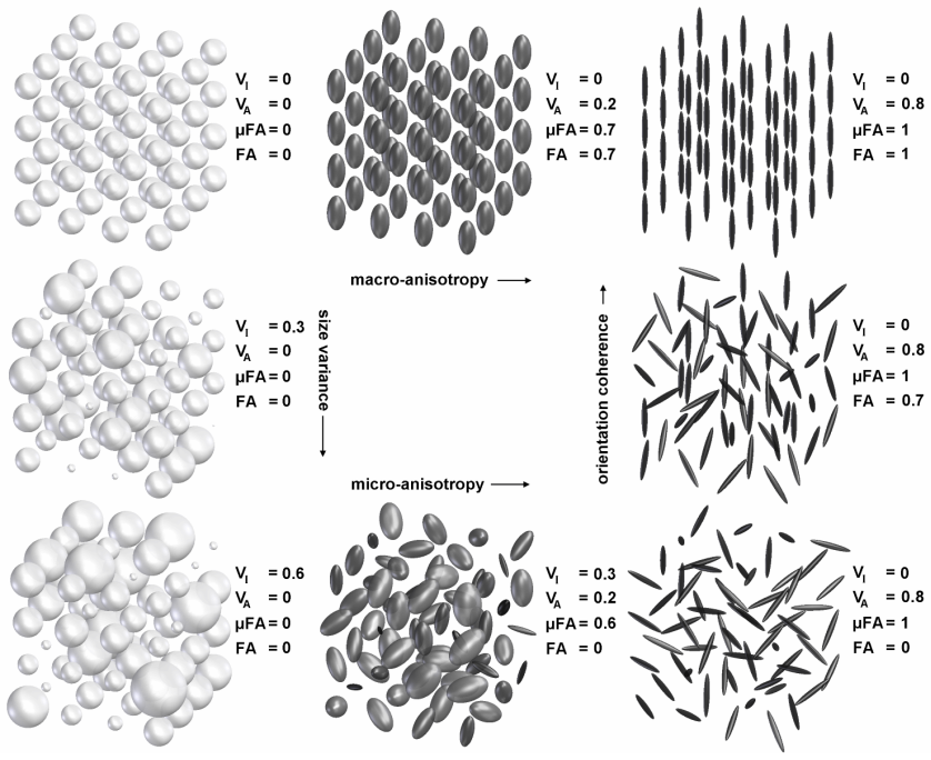
The central claim is that DTI's limitation is an encoding limitation, not just an inverse-problem nuisance. When the signal is written as $S=S_0\int P(D)e^{-B:D}dD$, the b-tensor $B$ determines which projection of the diffusion tensor distribution $P(D)$ is measured. A linear b-tensor, as in conventional DWI, mixes microscopic anisotropy with orientation dispersion, while a spherical b-tensor isolates the isotropic part, and the difference between the two variances is proportional to the mean squared microscopic anisotropy. The paper derives the powder-averaged signal, its cumulant expansion, and the variance decomposition that yields the microscopic fractional anisotropy and orientational order parameter, and it closes with the covariance-tensor approximation that avoids powder averaging altogether.
Load-bearing premise
The quantitative framework assumes that within each microscopic environment water diffusion is Gaussian and water does not move between environments during the measurement; the paper explicitly says this assumption must be validated in future studies.
Editorial extensions
If this is right
- Combining linear and spherical (or at least two distinct) encoding shapes yields separate estimates of mean diffusivity, isotropic variance, microscopic anisotropy, and orientational order from the same acquisition framework.
- The difference between linear and spherical second moments is proportional to the mean squared diffusion anisotropy of the microscopic tensors, giving a direct readout that does not require biophysical modeling.
- In the tumor application, meningiomas and glioblastomas, which both show low FA, become distinguishable through their microscopic fractional anisotropy.
- The gamma-distribution fit provides an analytic signal form that remains accurate beyond the cumulant expansion's range, extending usable b-values while staying within clinically feasible scan times.
- Because the encoding tensor's shape is set by the q-trajectory, arbitrary b-tensor shapes can be generated from simple axial waveforms, making the approach implementable on clinical scanners.
Reading between the lines
- Extension: combining linear and spherical encoding with oscillating or spectrally modulated gradients would add a time-dependence axis to the same variance decomposition, potentially estimating restriction sizes without biophysical modeling.
- Extension: the linear-versus-spherical variance difference is a candidate imaging biomarker that a prospective histology-calibrated study in one tumor type could validate directly; the paper shows feasibility but stops short of that validation.
- Extension: the solid-state NMR analogy suggests that optimal q-trajectory designs, not discussed quantitatively here, could shorten scans or sharpen the conditioning of the covariance-tensor estimate.
Signed reviews
Editorial analysis
A structured set of objections, weighed in public.
Referee Report
Summary. This manuscript is a self-contained tutorial/review of advanced encoding methods in diffusion MRI. It opens with NMR physics (spin, Bloch sphere, relaxation, spin echo), introduces diffusion and DWI/DTI, discusses tensor parameterizations, tensor-valued (b-tensor) encoding and waveform design, diffusion tensor distributions and ensemble statistics, and closes with methods for extracting microscopic anisotropy (powder averaging, cumulant expansion, Gamma fitting, DIVIDE, covariance tensor). The stated aim is to build a short but solid and logical path from spin to current limitations and advanced encoding methods such as tensor-valued encoding.
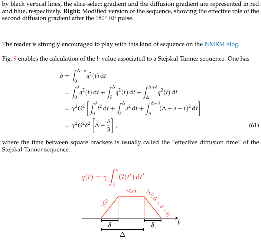
Significance. If the equations are correct, the paper fills a useful pedagogical gap: it collects derivations and recent tensor-valued-encoding concepts in one place, with clear notation (Haeberlen vs standard conventions, Frobenius product, b-tensor shapes) and honest statements of assumptions, notably Sec. 3.3, which explicitly flags that the diffusion tensor distribution model must be validated. Its strengths are the careful self-contained derivations of the b-value, the Frobenius inner product, the powder-averaged signal, and the DIVIDE metrics, as well as the effective visualizations. The paper does not contain new empirical results; its value is expository, so its significance for a research journal is moderate.
major comments (1)
- [Sec. 7.3, Eq. (190)] The angular-averaging identity used here is incorrect by a factor of two. For a uniaxial orientation distribution and a fixed radial direction, azimuthal averaging gives <P2(cos(θ+π/2))> = −1/2 <P2(cos θ)>, not −<P2(cos θ)>. Consequently Eq. (190) should read <D⊥> = <D> − (1/3)(D∥−D⊥)<P2(cos θ)>, and inserting the printed Eqs. (189)–(190) into Eq. (191) yields (4/3)<P2(cos θ)> rather than SZZ. The final expressions (195)–(196) are consistent with the corrected sign and factor, so the error is local, but it breaks the internal consistency of the orientational-order-parameter derivation and should be fixed.
minor comments (4)
- [Sec. 3.2.2] The citation "[9, 10, 11 ?]" contains a formatting artifact; it should read "[9, 10, 11]" since Ref. [11] is listed in the bibliography.
- [Sec. 5.3.1, Eq. (133)] In the expression for G(t), the third component of the second vector should be 0, not cos ζ, because it comes from the time derivative of n(t); the subsequent Eq. (135) is consistent with the corrected expression.
- [Sec. 7.3] The sentence introducing Eq. (189) contains a duplicated article: "the the macroscopic axial and radial diffusivity" should read "the macroscopic axial and radial diffusivity".
- [Sec. 7.3] The sentence preceding Eq. (195) reads "Eqs. (192) and (193) can now be combines to match" and should be corrected to "can now be combined to match".
Circularity Check
No significant circularity: the paper is a tutorial that re-derives known results from cited external literature; the authors' self-citation is transparent and not load-bearing.
full rationale
This manuscript is an educational review rather than an original research claim. Its derivations, such as the DWI signal attenuation in Sec. 3.1, the b-tensor encoding formalism in Sec. 5, and the powder-averaged cumulant expansion in Sec. 7.2, follow stated assumptions and cite independent prior work for each major step. The orientational order parameter derivation in Sec. 7.3 defines OP as <P2(cos theta)> and then algebraically relates it to the ratio of ensemble-averaged diffusivity differences; Eqs. (191), (195), and (196) are identities following from the definitions of mu2 and muFA2, not fitted parameters relabeled as predictions. The gamma-distribution fitting in Sec. 7.4.2 is explicitly presented as a convenient approximation with an analytic Laplace transform, and the DIVIDE decomposition is attributed to Refs. [61, 80, 6]. The only self-citation is Ref. [70], the first author's own book chapter, which is acknowledged at the start of Sec. 7 as the source of that section. That citation is transparent, the chapter is independently published, and the section's formulas are supported by additional external references such as [62, 71, 79]. No uniqueness theorem is imported from the authors' prior work, and no ansatz is smuggled in via a self-citation. Any possible algebraic slip in Sec. 7.3 would be a correctness issue, not circularity. The derivation chain is therefore self-contained relative to its tutorial purpose, and the circularity score is 0.
Assumptions & free parameters
assumptions (3)
- domain assumption Diffusion in each microscopic environment is approximately Gaussian.
- domain assumption Diffusing particles do not exchange between environments during encoding.
- domain assumption The DTD model is sufficiently accurate for the described tissue.
Cite this review
Pith. "Pith review of Advanced encoding methods in diffusion MRI." pith.science (2026). https://pith.science/paper/HNROFOYO
@misc{pith2026190804177,
  author       = {Pith},
  title        = {Pith review of: Advanced encoding methods in diffusion MRI},
  year         = {2026},
  howpublished = {\url{https://pith.science/paper/HNROFOYO}},
  note         = {Machine review of arXiv:1908.04177}
}
read the original abstract
Discovering a new field is not usually an easy process, especially when you choose magnetic resonance imaging (MRI). MRI is one of the few non-invasive medical procedures, if not the only one, that someone can receive in modern day hospitals. Its high sensitivity is a true prowess that is due to three things: subtle quantum physics, great engineering and clever imaging techniques from mathematics and computing science. However, the mathematics and physics of it are already daunting tasks by themselves, which makes MRI difficult to comprehend. This document paves a rather short, yet solid and logical, path from the subtle concept of spin to the current limitations of diffusion MRI. It then describes more advanced encoding methods designed to overcome diffusion MRI's limitations, such as tensor-valued encoding.
Figures


Figures from the paper (18 more)





Reference graph
Works this paper leans on
-
[15]
Szczepankiewicz, Imaging diffusional variance by MRI: The role of tensor-valued diffusion encoding and tissue heterogeneity, PhD thesis, Lund University, 2016
F. Szczepankiewicz, Imaging diffusional variance by MRI: The role of tensor-valued diffusion encoding and tissue heterogeneity, PhD thesis, Lund University, 2016
2016
-
[53]
Topgaard, Multidimensional diffusion MRI, Journal of Magnetic Resonance 275, 98 (2017)
D. Topgaard, Multidimensional diffusion MRI, Journal of Magnetic Resonance 275, 98 (2017). 64
work page 2017
-
[70]
A. Reymbaut, Chapter 3 - diffusion anisotropy and tensor-valued encoding, in Advanced Diffusion Encoding Methods in MRI, The Royal Society of Chemistry, 2019
work page 2019
-
[1]
Bloch, Nuclear Induction, Phys
F. Bloch, Nuclear Induction, Phys. Rev. 70, 460 (1946)
1946
-
[2]
E. L. Hahn, Spin Echoes, Phys. Rev. 80, 580 (1950)
1950
-
[3]
J. S. W. Campbell, Diffusion Imaging of White Matter Fibre Tracts, PhD thesis, McGill Univer- sity, 2004
2004
-
[4]
E. O. Stejskal and J. E. Tanner, Spin Diffusion Measurements: Spin Echoes in the Presence of a Time-Dependent Field Gradient, The Journal of Chemical Physics 42, 288 (1965)
1965
-
[5]
P . J. Basser, J. Mattiello, and D. LeBihan, MR diffusion tensor spectroscopy and imaging , Bio- phys J 66, 259 (1994)
1994
Show all 100 references
-
[6]
F. Szczepankiewicz et al., The link between diffusion MRI and tumor heterogeneity: Mapping cell eccentricity and density by diffusional variance decomposition (DIVIDE) , NeuroImage 142, 522 (2016)
2016
-
[7]
M. E. Moseley et al., Early detection of regional cerebral ischemia in cats: Comparison of diffusion- and T2-weighted MRI and spectroscopy, Magnetic Resonance in Medicine 14, 330 (1990)
1990
-
[8]
Lebel, L
C. Lebel, L. Walker, A. Leemans, L. Phillips, and C. Beaulieu, Microstructural maturation of the human brain from childhood to adulthood, NeuroImage 40, 1044 (2008)
2008
-
[9]
A. L. Alexander, K. M. Hasan, M. Lazar, J. S. Tsuruda, and D. L. Parker, Analysis of partial volume effects in diffusion-tensor MRI, Magnetic Resonance in Medicine 45, 770 (2001)
2001
-
[10]
A. L. Alexander, J. E. Lee, M. Lazar, and A. S. Field, Diffusion tensor imaging of the brain , Neurotherapeutics 4, 316 (2007)
2007
-
[11]
D. K. Jones and M. Cercignani, Twenty-five pitfalls in the analysis of diffusion MRI data, NMR in Biomedicine 23, 803 (2010)
2010
-
[12]
Pierpaoli, P
C. Pierpaoli, P . Jezzard, P . J. Basser, A. Barnett, and G. D. Chiro,Diffusion tensor MR imaging of the human brain., Radiology 201, 637 (1996)
1996
-
[13]
G. Douaud et al., DTI measures in crossing-fibre areas: Increased diffusion anisotropy reveals early white matter alteration in MCI and mild Alzheimer’s disease, NeuroImage 55, 880 (2011)
2011
-
[14]
Jeurissen, A
B. Jeurissen, A. Leemans, J.-D. Tournier, D. K. Jones, and J. Sijbers,Investigating the prevalence of complex fiber configurations in white matter tissue with diffusion magnetic resonance imaging , Human Brain Mapping 34, 2747 (2013)
2013
-
[16]
T. M. de Swiet and P . P . Mitra, Possible Systematic Errors in Single-Shot Measurements of the Trace of the Diffusion Tensor, Journal of Magnetic Resonance, Series B 111, 15 (1996)
1996
-
[17]
Beaulieu, The basis of anisotropic water diffusion in the nervous system a technical review , NMR in Biomedicine 15, 435 (2002)
C. Beaulieu, The basis of anisotropic water diffusion in the nervous system a technical review , NMR in Biomedicine 15, 435 (2002)
2002
-
[18]
D. A. Yablonskiy, G. L. Bretthorst, and J. J. Ackerman, Statistical model for diffusion attenuated MR signal, Magnetic Resonance in Medicine 50, 664 (2003). 62
2003
-
[19]
P . T. Callaghan, A. Coy, D. MacGowan, K. J. Packer, and F. O. Zelaya, Diffraction-like effects in NMR diffusion studies of fluids in porous solids, Nature 351, 467 (1991)
1991
-
[20]
Topgaard and O
D. Topgaard and O. Sderman, Experimental determination of pore shape and size using q-space NMR microscopy in the long diffusion-time limit, Magnetic Resonance Imaging 21, 69 (2003)
2003
-
[21]
Nilsson et al., Evaluating the accuracy and precision of a two-compartment Krger model using Monte Carlo simulations, Journal of Magnetic Resonance 206, 59 (2010)
M. Nilsson et al., Evaluating the accuracy and precision of a two-compartment Krger model using Monte Carlo simulations, Journal of Magnetic Resonance 206, 59 (2010)
2010
-
[22]
J. C. Gore et al., Characterization of tissue structure at varying length scales using temporal diffu- sion spectroscopy, NMR in Biomedicine 23, 745 (2010)
2010
-
[23]
D. E. Woessner, NMR spin-echo self-diffusion measurements on fluids undergoing restricted dif- fusion, The Journal of Physical Chemistry 67, 1365 (1963)
1963
-
[24]
R. E. Beck and J. S. Schultz, Hindered Diffusion in Microporous Membranes with Known Pore Geometry, Science 170, 1302 (1970)
1970
-
[25]
G. J. Stanisz, G. A. Wright, R. M. Henkelman, and A. Szafer, An analytical model of restricted diffusion in bovine optic nerve, Magnetic Resonance in Medicine 37, 103 (1997)
1997
-
[26]
Assaf, A
Y. Assaf, A. Mayk, and Y. Cohen, Displacement imaging of spinal cord using q-space diffusion- weighted MRI, Magnetic Resonance in Medicine 44, 713 (2000)
2000
-
[27]
M. D. Does, E. C. Parsons, and J. C. Gore, Oscillating gradient measurements of water diffusion in normal and globally ischemic rat brain, Magnetic Resonance in Medicine 49, 206 (2003)
2003
-
[28]
Assaf, T
Y. Assaf, T. Blumenfeld-Katzir, Y. Yovel, and P . J. Basser, Axcaliber: A method for measur- ing axon diameter distribution from diffusion MRI , Magnetic Resonance in Medicine 59, 1347 (2008)
2008
-
[29]
Lundell, C
H. Lundell, C. K. Snderby, and T. B. Dyrby,Diffusion weighted imaging with circularly polarized oscillating gradients, Magnetic Resonance in Medicine 73, 1171 (2015)
2015
-
[30]
L. M. Burcaw, E. Fieremans, and D. S. Novikov, Mesoscopic structure of neuronal tracts from time-dependent diffusion, NeuroImage 114, 18 (2015)
2015
-
[31]
C. A. Clark, M. Hedehus, and M. E. Moseley,Diffusion time dependence of the apparent diffusion tensor in healthy human brain and white matter disease , Magnetic Resonance in Medicine 45, 1126 (2001)
2001
-
[32]
Ronen, S
I. Ronen, S. Moeller, K. Ugurbil, and D.-S. Kim, Analysis of the distribution of diffusion co- efficients in cat brain at 9.4 T using the inverse Laplace transformation , Magnetic Resonance Imaging 24, 61 (2006)
2006
-
[33]
Nilsson et al., On the effects of a varied diffusion time in vivo: is the diffusion in white matter restricted?, Magnetic Resonance Imaging 27, 176 (2009)
M. Nilsson et al., On the effects of a varied diffusion time in vivo: is the diffusion in white matter restricted?, Magnetic Resonance Imaging 27, 176 (2009)
2009
-
[34]
S. D. Santis, D. K. Jones, and A. Roebroeck, Including diffusion time dependence in the extra- axonal space improves in vivo estimates of axonal diameter and density in human white matter , NeuroImage 130, 91 (2016)
2016
-
[35]
J. D. Quirk et al., Equilibrium water exchange between the intra- and extracellular spaces of mam- malian brain, Magnetic Resonance in Medicine 50, 493 (2003)
2003
-
[36]
Nilsson, Biophysical modelling in diffusion MRI: The role of tissue microstructure and water exchange, PhD thesis, Medical Radiation Physics, Lund, 2011
M. Nilsson, Biophysical modelling in diffusion MRI: The role of tissue microstructure and water exchange, PhD thesis, Medical Radiation Physics, Lund, 2011. 63
2011
-
[37]
Nilsson, D
M. Nilsson, D. van Westen, F. St ˚ahlberg, P . C. Sundgren, and J. L ¨att, The role of tissue mi- crostructure and water exchange in biophysical modelling of diffusion in white matter , Magnetic Resonance Materials in Physics, Biology and Medicine 26, 345 (2013)
2013
-
[38]
Nilsson et al., Noninvasive mapping of water diffusional exchange in the human brain using filter-exchange imaging, Magnetic Resonance in Medicine 69, 1572 (2013)
M. Nilsson et al., Noninvasive mapping of water diffusional exchange in the human brain using filter-exchange imaging, Magnetic Resonance in Medicine 69, 1572 (2013)
2013
-
[39]
B. Lampinen et al., Optimal experimental design for filter exchange imaging: Apparent exchange rate measurements in the healthy brain and in intracranial tumors , Magnetic Resonance in Medicine 77, 1104 (2017)
2017
-
[40]
Ltt et al., Diffusion-weighted MRI measurements on stroke patients reveal water-exchange mech- anisms in sub-acute ischaemic lesions, NMR in Biomedicine 22, 619 (2009)
J. Ltt et al., Diffusion-weighted MRI measurements on stroke patients reveal water-exchange mech- anisms in sub-acute ischaemic lesions, NMR in Biomedicine 22, 619 (2009)
2009
-
[41]
D. S. Novikov and V . G. Kiselev, Effective medium theory of a diffusion-weighted signal , NMR in Biomedicine 23, 682 (2010)
2010
-
[42]
V . G. Kiselev,Fundamentals of diffusion MRI physics, NMR in Biomedicine 30, e3602 (2017)
2017
-
[43]
D. S. Novikov, E. Fieremans, J. H. Jensen, and J. A. Helpern, Random walks with barriers , Nature Physics 7, 508 EP (2011)
2011
-
[44]
D. S. Novikov, J. H. Jensen, J. A. Helpern, and E. Fieremans, Revealing mesoscopic structural universality with diffusion, Proceedings of the National Academy of Sciences111, 5088 (2014)
2014
-
[45]
D. Topgaard, Chapter 7 - NMR methods for studying microscopic diffusion anisotropy, in Diffusion NMR of Confined Systems: Fluid Transport in Porous Solids and Heterogeneous Materi- als, pp. 226–259, The Royal Society of Chemistry, 2017
2017
-
[46]
Westin et al., Processing and visualization for diffusion tensor MRI, Medical Image Anal- ysis 6, 93 (2002)
C.-F. Westin et al., Processing and visualization for diffusion tensor MRI, Medical Image Anal- ysis 6, 93 (2002)
2002
-
[47]
Haeberlen, High resolution NMR in solids : selective averaging(Academic Press, New York, 1976)
U. Haeberlen, High resolution NMR in solids : selective averaging(Academic Press, New York, 1976)
1976
-
[48]
PB and M
K. PB and M. WG., Contrast-to-noise ratios of diffusion anisotropy indices, Magn Reson Med 2005;53:911-918., Magnetic Resonance in Medicine 54, 251 (2005)
2005
-
[49]
T. E. Conturo, R. C. McKinstry, E. Akbudak, and B. H. Robinson, Encoding of anisotropic diffusion with tetrahedral gradients: A general mathematical diffusion for- malism and experimental results , Magnetic Resonance in Medicine 35, 399 (1996), https://onlinelibrary.wiley.com/...
1996 doi
-
[50]
P . J. Basser and C. Pierpaoli, Microstructural and physiological features of tissues elucidated by quantitative-diffusion-tensor MRI, Journal of Magnetic Resonance 213, 560 (2011)
2011
-
[51]
A. L. Alexander, K. Hasan, G. Kindlmann, D. L. Parker, and J. S. Tsuruda, A geometric analysis of diffusion tensor measurements of the human brain, Magnetic Resonance in Medicine 44, 283 (2000)
2000
-
[52]
A. M. Ulug and P . C. van Zijl, Orientation-independent diffusion imaging without tensor diago- nalization: Anisotropy definitions based on physical attributes of the diffusion ellipsoid , Journal of Magnetic Resonance Imaging 9, 804 (1999)
1999
-
[54]
E. R. Andrew, A. Bradbury, and R. G. Eades, Removal of Dipolar Broadening of Nuclear Mag- netic Resonance Spectra of Solids by Specimen Rotation, Nature 183, 1802 (1959)
1959
-
[55]
Schmidt-Rohr and H
K. Schmidt-Rohr and H. Spiess, Multidimensional Solid-State NMR and Polymers (Academic Press, San Diego, 1994)
1994
-
[56]
H. C. Torrey, Bloch Equations with Diffusion Terms, Phys. Rev. 104, 563 (1956)
1956
-
[57]
W. S. Price, NMR studies of translational motion: principles and applications (Cambridge Uni- versity Press, 2009)
2009
-
[58]
S. Lasi, M. Nilsson, J. Ltt, F. Sthlberg, and D. Topgaard, Apparent exchange rate mapping with diffusion MRI, Magnetic Resonance in Medicine 66, 356 (2011)
2011
-
[59]
D. L. Bihan et al., MR imaging of intravoxel incoherent motions: application to diffusion and perfusion in neurologic disorders., Radiology 161, 401 (1986)
1986
-
[60]
Eriksson, S
S. Eriksson, S. Lasic, and D. Topgaard, Isotropic diffusion weighting in PGSE NMR by magic- angle spinning of the q-vector, Journal of Magnetic Resonance 226, 13 (2013)
2013
-
[61]
S. Lasi, F. Szczepankiewicz, S. Eriksson, M. Nilsson, and D. Topgaard,Microanisotropy imag- ing: quantification of microscopic diffusion anisotropy and orientational order parameter by diffu- sion MRI with magic-angle spinning of the q-vector, Frontiers in Physics 2, 11 (2014)
2014
-
[62]
Eriksson, S
S. Eriksson, S. Lasi, M. Nilsson, C.-F. Westin, and D. Topgaard, NMR diffusion-encoding with axial symmetry and variable anisotropy: Distinguishing between prolate and oblate microscopic diffusion tensors with unknown orientation distribution , The Journal of Chemical Physics ...
2015
-
[63]
Westin et al., Q-space trajectory imaging for multidimensional diffusion MRI of the human brain, NeuroImage 135, 345 (2016)
C.-F. Westin et al., Q-space trajectory imaging for multidimensional diffusion MRI of the human brain, NeuroImage 135, 345 (2016)
2016
-
[64]
D. G. Cory, A. N. Garroway, and J. B. Miller, Applications of spin transport as a probe of local geometry, 31, 149 (1990)
1990
-
[65]
E. C. Wong, R. W. Cox, and A. W. Song, Optimized isotropic diffusion weighting , Magnetic Resonance in Medicine 34, 139 (1995)
1995
-
[66]
Sjlund et al., Constrained optimization of gradient waveforms for generalized diffusion encoding, Journal of Magnetic Resonance 261, 157 (2015)
J. Sjlund et al., Constrained optimization of gradient waveforms for generalized diffusion encoding, Journal of Magnetic Resonance 261, 157 (2015)
2015
-
[67]
Szczepankiewicz, C.-F
F. Szczepankiewicz, C.-F. Westin, and M. Nilsson, Maxwell-compensated design of asymmetric gradient waveforms for tensor-valued diffusion encoding , Magnetic Resonance in Medicine 82, 1424 (2019)
2019
-
[68]
Lundell et al., Microscopic anisotropy with spectrally modulated q-space trajectory en- coding, in International Society for Magnetic Resonance Imaging (ISMRM), 2017
H. Lundell et al., Microscopic anisotropy with spectrally modulated q-space trajectory en- coding, in International Society for Magnetic Resonance Imaging (ISMRM), 2017
2017
-
[69]
S. Lasi, H. Lundell, D. Topgaard, and T. B. Dyrby, Effects of imaging gradients in sequences with varying longitudinal storage timeCase of diffusion exchange imaging , Magnetic Resonance in Medicine (2017)
2017
-
[71]
Saupe and G
A. Saupe and G. Englert, High-Resolution Nuclear Magnetic Resonance Spectra of Orientated Molecules, Phys. Rev. Lett. 11, 462 (1963). 65
1963
-
[72]
Schmidt-Rohr and H
K. Schmidt-Rohr and H. W. Spiess, Multidimensional Solid-State NMR and Polymers (Aca- demic Press, San Diego, 1994)
1994
-
[73]
Bloembergen and T
N. Bloembergen and T. Rowland, On the nuclear magnetic resonance in metals and alloys, Acta Metallurgica 1, 731 (1953)
1953
-
[74]
Herberthson, C
M. Herberthson, C. Yolcu, H. Knutsson, C.-F. Westin, and E. ¨Ozarslan, Orientationally- averaged diffusion-attenuated magnetic resonance signal for locally-anisotropic diffusion, Scientific Reports 9, 4899 (2019)
2019
-
[75]
Szczepankiewicz, C.-F
F. Szczepankiewicz, C.-F. Westin, F. Sthlberg, J. Ltt, and M. Nilsson, Minimum number of diffusion encoding directions required to yield a rotationally invariant powder average signal in single and double diffusion encoding, inInternational Society for Magnetic Resonance Imagi...
2016
-
[76]
Edn, Computer simulations in solid-state NMR
M. Edn, Computer simulations in solid-state NMR. III. Powder averaging, Concepts in Magnetic Resonance Part A 18A, 24 (2003)
2003
-
[77]
B. J. Frisken, Revisiting the method of cumulants for the analysis of dynamic light-scattering data, Appl. Opt. 40, 4087 (2001)
2001
-
[78]
D. E. Koppel, Analysis of Macromolecular Polydispersity in Intensity Correlation Spectroscopy: The Method of Cumulants, The Journal of Chemical Physics 57, 4814 (1972)
1972
-
[79]
D. L. VanderHart and H. S. Gutowsky, RigidLattice NMR Moments and Line Shapes with ChemicalShift Anisotropy, The Journal of Chemical Physics 49, 261 (1968)
1968
-
[80]
F. Szczepankiewicz et al., Quantification of microscopic diffusion anisotropy disentangles effects of orientation dispersion from microstructure: Applications in healthy volunteers and in brain tumors, NeuroImage 104, 241 (2015)
2015
-
[81]
Szczepankiewicz, J
F. Szczepankiewicz, J. Sjlund, F. Sthlberg, J. Ltt, and M. Nilsson, Tensor-valued diffusion encoding for diffusional variance decomposition (DIVIDE): Technical feasibility in clinical MRI systems, PLOS ONE 14, 1 (2019)
2019
-
[82]
J. H. Jensen, J. A. Helpern, A. Ramani, H. Lu, and K. Kaczynski, Diffusional kurtosis imag- ing: The quantification of non-gaussian water diffusion by means of magnetic resonance imaging , Magnetic Resonance in Medicine 53, 1432 (2005)
2005
-
[83]
R. N. Henriques, S. N. Jespersen, and N. Shemesh, Microscopic anisotropy misestimation in spherical-mean single diffusion encoding MRI , Magnetic Resonance in Medicine 81, 3245 (2019)
2019
-
[84]
Hong et al., PFG n.m.r
U. Hong et al., PFG n.m.r. study of diffusion anisotropy in oriented ZSM-5 type zeolite crystallites, Zeolites 11, 816 (1991)
1991
-
[85]
Lawrenz, M
M. Lawrenz, M. A. Koch, and J. Finsterbusch, A tensor model and measures of microscopic anisotropy for double-wave-vector diffusion-weighting experiments with long mixing times, Journal of Magnetic Resonance 202, 43 (2010)
2010
-
[86]
Lawrenz and J
M. Lawrenz and J. Finsterbusch, Detection of microscopic diffusion anisotropy on a whole-body MR system with double wave vector imaging, Magnetic Resonance in Medicine 66, 1405 (2011)
2011
-
[87]
Lawrenz and J
M. Lawrenz and J. Finsterbusch, Double-wave-vector diffusion-weighted imaging reveals micro- scopic diffusion anisotropy in the living human brain, Magnetic Resonance in Medicine 69, 1072 (2013). 66
2013
-
[88]
Lawrenz, S
M. Lawrenz, S. Brassen, and J. Finsterbusch, Microscopic diffusion anisotropy in the human brain: Reproducibility, normal values, and comparison with the fractional anisotropy, NeuroImage 109, 283 (2015)
2015
-
[89]
Lawrenz, S
M. Lawrenz, S. Brassen, and J. Finsterbusch, Microscopic diffusion anisotropy in the human brain: Age-related changes, NeuroImage 141, 313 (2016)
2016
-
[90]
Lawrenz and J
M. Lawrenz and J. Finsterbusch, Detection of microscopic diffusion anisotropy in human cortical gray matter in vivo with double diffusion encoding , Magnetic Resonance in Medicine 81, 1296 (2019)
2019
-
[91]
S. N. Jespersen, H. Lundell, C. K. Snderby, and T. B. Dyrby, Orientationally invariant metrics of apparent compartment eccentricity from double pulsed field gradient diffusion experiments, NMR in Biomedicine 26, 1647 (2013)
2013
-
[92]
J. a. P . de Almeida Martins and D. Topgaard, Two-Dimensional Correlation of Isotropic and Directional Diffusion Using NMR, Phys. Rev. Lett. 116, 087601 (2016)
2016
-
[93]
P . T. Callaghan and D. N. Pinder, Influence of polydispersity on polymer self-diffusion measure- ments by pulsed field gradient nuclear magnetic resonance, Macromolecules 18, 373 (1985)
1985
-
[94]
J. H. Jensen and J. A. Helpern, MRI quantification of non-Gaussian water diffusion by kurtosis analysis, NMR in Biomedicine 23, 698 (2010)
2010
-
[95]
Rding et al., The gamma distribution model for pulsed-field gradient NMR studies of molecular- weight distributions of polymers, Journal of magnetic resonance 222, 105111 (2012)
M. Rding et al., The gamma distribution model for pulsed-field gradient NMR studies of molecular- weight distributions of polymers, Journal of magnetic resonance 222, 105111 (2012)
2012
-
[96]
Rding, N
M. Rding, N. H. Williamson, and M. Nydn, Gamma convolution models for self-diffusion coef- ficient distributions in PGSE NMR, Journal of Magnetic Resonance 261, 6 (2015)
2015
-
[97]
N. H. Williamson, M. Nydn, and M. Rding, The lognormal and gamma distribution models for estimating molecular weight distributions of polymers using PGSE NMR , Journal of Magnetic Resonance 267, 54 (2016)
2016
-
[98]
Reisert, E
M. Reisert, E. Kellner, B. Dhital, J. Hennig, and V . G. Kiselev, Disentangling micro from mesostructure by diffusion MRI: A Bayesian approach, NeuroImage 147, 964 (2017)
2017
-
[99]
Coelho, J
S. Coelho, J. M. Pozo, S. N. Jespersen, D. K. Jones, and A. F. Frangi, Resolving degeneracy in diffusion MRI biophysical model parameter estimation using double diffusion encoding , Magnetic Resonance in Medicine 82, 395 (2019)
2019
-
[100]
Coelho, J
S. Coelho, J. M. Pozo, S. N. Jespersen, and A. F. Frangi, Optimal experimental design for bio- physical modelling in multidimensional diffusion MRI, arXiv e-prints , arXiv:1907.06139 (2019), 1907.06139. 67
2019 arXiv
Reviewed August 14, 2026 · model on record in the stance chip above.
Discussion (0). Continue with ORCID to comment.