Classification of IED-free EEG Responses for Assisted Epilepsy Diagnosis
Pith reviewed 2026-05-25 06:27 UTC · model grok-4.3
The pith
A stacked ensemble on multi-domain features classifies epilepsy from IED-free EEG during photic stimulation and hyperventilation.
A machine-rendered reading of the paper's core claim, the machinery that carries it, and where it could break.
Core claim
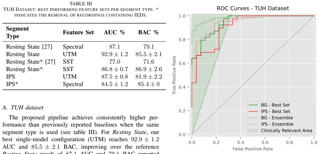
The central claim is that stimulation procedures, especially intermittent photic stimulation, evoke activity patterns that a multi-domain feature set and stacked ensemble can exploit to classify epilepsy in the complete absence of interictal epileptiform discharges, achieving leave-one-subject-out AUC values up to 94.1 percent on the TUH corpus and 79.4 percent on an external clinical cohort.
What carries the argument
The stacked ensemble that integrates complementary temporal, spectral, wavelet, and connectivity feature sets extracted from stimulation segments.
If this is right
- Intermittent photic stimulation supplies stronger discriminative signals than resting-state or hyperventilation for IED-free classification.
- Combining multiple feature domains via ensembling increases robustness across subjects and recording conditions.
- Hyperventilation yields usable discrimination once subjects are stratified by whether they show a physiological response.
- Leave-one-subject-out performance on two independent cohorts supports the reproducibility of the multi-domain pipeline.
Where Pith is reading between the lines
- If the same feature families prove stable across sites, the pipeline could be adapted to other provocation techniques such as sleep deprivation.
- The performance drop from TUH to the clinical cohort implies that site-specific factors will need explicit handling before routine clinical deployment.
- Future work that explicitly models medication load or arousal state could test whether the current separation truly isolates epilepsy-related physiology.
Load-bearing premise
The feature differences observed during photic stimulation and hyperventilation arise from epilepsy-specific physiology rather than from group differences in age, medication, arousal level, or recording artifacts.
What would settle it
A controlled replication on age- and medication-matched groups that finds classification performance dropping to chance levels would falsify the claim that the pipeline detects epilepsy-specific responses.
Figures

read the original abstract
Diagnosing epilepsy is challenging when routine EEGs lack interictal epileptiform discharges (IEDs). Intermittent photic stimulation (IPS) and hyperventilation (HV) can increase diagnostic yield, but their interpretation is subjective. We propose a reproducible pipeline that classifies EEG recordings acquired during stimulation procedures, using machine-learning features spanning temporal, spectral, wavelet, and connectivity domains, and a stacked ensemble to combine complementary feature sets. Performance is evaluated with leave-one-subject-out (LOSO) cross-validation on the TUH Epilepsy Corpus and a clinical Erasmus MC (EMC) cohort, including IED-free analyses on TUH. On TUH, ensembles achieve up to 97.8\% AUC / 93.1\% BAC on IED-free resting-state EEG and 94.1\% AUC / 86.8\% BAC on IED-free IPS. On EMC, IPS provides the strongest discrimination (79.4\% AUC / 73.9\% BAC), while HV performance benefits from stratifying subjects by responsiveness. These results indicate that stimulation-evoked activity, particularly IPS, contains meaningful discriminative information for IED-free epilepsy classification and that multi-domain ensembling improves robustness.
Editorial analysis
A structured set of objections, weighed in public.
Referee Report
Summary. The paper proposes a reproducible ML pipeline that extracts multi-domain features (temporal, spectral, wavelet, connectivity) from IED-free EEG segments recorded during resting-state, intermittent photic stimulation (IPS), and hyperventilation (HV), then applies a stacked ensemble for binary epilepsy classification. Performance is assessed via leave-one-subject-out (LOSO) cross-validation on the TUH Epilepsy Corpus (reporting up to 97.8% AUC / 93.1% BAC on resting-state and 94.1% AUC / 86.8% BAC on IPS) and an external Erasmus MC (EMC) cohort (79.4% AUC / 73.9% BAC on IPS), with a note that HV benefits from responsiveness stratification.
Significance. If the discriminative signal is shown to be epilepsy-specific rather than driven by cohort-level differences, the work would offer a concrete, objective aid for the clinically important subset of patients whose routine EEGs lack IEDs. The use of LOSO, multi-domain ensembling, and two-site evaluation are methodological strengths that increase credibility over single-cohort, non-held-out designs.
major comments (2)
- [Methods and Results] Methods and Results sections: No information is supplied on age, medication, arousal, or artifact matching between epilepsy and control groups, nor on any post-hoc exclusion criteria. Because the headline claim is that IPS/HV/resting-state features capture epilepsy physiology (rather than group differences), the absence of these controls is load-bearing; the reported LOSO AUCs are equally consistent with either interpretation.
- [Results] Results: The manuscript reports ensemble AUC/BAC values but supplies neither ablation results comparing the stacked ensemble to its constituent feature sets, nor any statistical tests (e.g., DeLong or permutation tests) on the AUC differences. Without these, it is impossible to determine whether the claimed improvement from multi-domain ensembling is robust or merely reflects the particular hyperparameter choices.
minor comments (1)
- [Abstract and Methods] Abstract and Methods: Feature definitions, exact hyperparameter search ranges, and the precise construction of the stacked ensemble are not described at a level that would allow immediate reproduction.
Simulated Author's Rebuttal
We thank the referee for the constructive feedback emphasizing the need for greater transparency on cohort characteristics and ensemble validation. We address each major comment below and will revise the manuscript to incorporate feasible improvements while noting limitations where data constraints prevent full resolution.
read point-by-point responses
-
Referee: [Methods and Results] Methods and Results sections: No information is supplied on age, medication, arousal, or artifact matching between epilepsy and control groups, nor on any post-hoc exclusion criteria. Because the headline claim is that IPS/HV/resting-state features capture epilepsy physiology (rather than group differences), the absence of these controls is load-bearing; the reported LOSO AUCs are equally consistent with either interpretation.
Authors: We agree that explicit reporting of demographic and clinical matching strengthens claims of epilepsy-specific signal. The TUH Epilepsy Corpus is a public dataset; we followed its published subject selection criteria with no additional post-hoc exclusions beyond the IED-free filter described in the Methods. For the EMC cohort we will add a supplementary table with available age, sex, and medication data. Standard artifact rejection (ICA and amplitude thresholding) was applied uniformly, but explicit per-group arousal or artifact matching was not performed. LOSO cross-validation and the external EMC validation provide some protection against subject-level confounds, yet we acknowledge this does not fully substitute for matched cohorts. We will expand the Limitations section accordingly. revision: partial
-
Referee: [Results] Results: The manuscript reports ensemble AUC/BAC values but supplies neither ablation results comparing the stacked ensemble to its constituent feature sets, nor any statistical tests (e.g., DeLong or permutation tests) on the AUC differences. Without these, it is impossible to determine whether the claimed improvement from multi-domain ensembling is robust or merely reflects the particular hyperparameter choices.
Authors: We concur that ablation experiments and formal statistical comparisons are required to substantiate the benefit of the stacked ensemble. In the revised manuscript we will report performance for each individual feature domain (temporal, spectral, wavelet, connectivity) alongside the ensemble, and we will add DeLong tests for AUC differences together with permutation tests (1000 iterations) to evaluate whether observed gains exceed chance. These results will appear in an expanded Results section with new tables and a supplementary figure. revision: yes
- Detailed per-subject arousal state and quantitative artifact burden metrics are not recorded in the TUH or EMC dataset metadata, preventing retrospective group matching on these variables.
Circularity Check
No circularity: empirical ML pipeline with held-out LOSO evaluation
full rationale
The paper describes a standard machine-learning classification pipeline (multi-domain features + stacked ensemble) whose performance metrics are obtained via leave-one-subject-out cross-validation on held-out subjects from two cohorts. No equations, fitted parameters, or self-citations are presented that would make the reported AUC/BAC values equivalent to the input labels by construction. The derivation chain consists of feature extraction followed by independent validation and contains no self-definitional, fitted-input-renamed-as-prediction, or load-bearing self-citation steps.
Axiom & Free-Parameter Ledger
free parameters (1)
- feature extraction and ensemble hyperparameters
axioms (1)
- domain assumption Stimulation-evoked EEG contains epilepsy-discriminative information independent of visible IEDs
Reference graph
Works this paper leans on
-
[1]
E. Hirsch et al., “ILAE definition of the Idiopathic Generalized Epilepsy Syndromes: Position statement by the ILAE Task Force on Nosology and Definitions,” en, Epilepsia, vol. 63, no. 6, pp. 1475–1499, Jun. 2022, ISSN: 0013-9580, 1528-1167.DOI: 10.1111/epi.17236
-
[2]
ILAE Official Report: A practical clinical definition of epilepsy,
R. S. Fisher et al., “ILAE Official Report: A practical clinical definition of epilepsy,” en,Epilepsia, vol. 55, no. 4, pp. 475–482, 2014,ISSN: 1528-1167.DOI: 10. 1111/epi.12550
work page 2014
-
[4]
Interictal EEG and the Diagnosis of Epilepsy,
J. Pillai and M. R. Sperling, “Interictal EEG and the Diagnosis of Epilepsy,” en,Epilepsia, vol. 47, no. s1, pp. 14–22, 2006,ISSN: 1528-1167.DOI: 10 . 1111 / j . 1528-1167.2006.00654.x
-
[5]
R. Basiri, A. Shariatzadeh, S. Wiebe, and Y . Aghakhani, “Focal epilepsy without interictal spikes on scalp EEG: A common finding of uncertain significance,”Epilepsy Research, vol. 150,ISSN: 0920-1211.DOI: 10.1016/j. eplepsyres.2018.12.009 [7]UC Davis Department of Neurology - Epilepsy FAQs. [Online]. Available: https : / / health . ucdavis . edu / neurol...
work page doi:10.1016/j 2018
-
[6]
Clinical correlations of photoparoxysmal responses,
P. Jayakar and K. H. Chiappa, “Clinical correlations of photoparoxysmal responses,”Electroencephalography and Clinical Neurophysiology, vol. 75, no. 3, pp. 251– 254, Mar. 1990,ISSN: 0013-4694.DOI: 10.1016/0013- 4694(90)90178-M
-
[7]
D. Kasteleijn-Nolst Trenit ´e et al., “Methodology of photic stimulation revisited: Updated European algo- rithm for visual stimulation in the EEG laboratory,” en, Epilepsia, vol. 53, no. 1, pp. 16–24, Jan. 2012,ISSN: 0013-9580, 1528-1167.DOI: 10 . 1111 / j . 1528 - 1167 . 2011.03319.x
-
[8]
Does Hyperventilation Elicit Epileptic Seizures?
M. D. Holmes, A. S. Dewaraja, and S. Vanhatalo, “Does Hyperventilation Elicit Epileptic Seizures?” en, Epilepsia, vol. 45, no. 6, pp. 618–620, 2004,ISSN: 1528-1167.DOI: 10.1111/j.0013-9580.2004.63803.x
-
[9]
M. S. B. Guaranha, E. Garzon, C. A. Buchpiguel, S. Tazima, E. M. T. Yacubian, and A. C. Sakamoto, “Hyperventilation Revisited: Physiological Effects and Efficacy on Focal Seizure Activation in the Era of Video-EEG Monitoring,” en,Epilepsia, vol. 46, no. 1, pp. 69–75, 2005,ISSN: 1528-1167.DOI: 10 . 1111 / j . 0013-9580.2005.11104.x
-
[10]
Sleep deprivation: A risk for epileptic seizures,
J. T. Dell’Aquila and V . Soti, “Sleep deprivation: A risk for epileptic seizures,”Sleep Science, vol. 15, no. 2, pp. 245–249, 2022,ISSN: 1984-0659.DOI: 10 . 5935 / 1984-0063.20220046 [13]Epilepsie, en. [Online]. Available: https : / / richtlijnendatabase . nl / richtlijn / epilepsie / elektrofysiologisch onderzoek bij epilepsie.html
-
[11]
R. M. Epstein, “Facing epistemic and complex uncer- tainty in serious illness: The role of mindfulness and shared mind,” eng,Patient Education and Counseling, vol. 104, no. 11, pp. 2635–2642, Nov. 2021,ISSN: 1873- 5134.DOI: 10.1016/j.pec.2021.07.030
-
[12]
Risk of recurrence after a first unprovoked seizure,
A. T. Berg, “Risk of recurrence after a first unprovoked seizure,” en,Epilepsia, vol. 49, no. s1, pp. 13–18, 2008, ISSN: 1528-1167.DOI: 10 . 1111 / j . 1528 - 1167 . 2008 . 01444.x
work page 2008
-
[13]
J. P. Carvajal-Dossman¹ et al., “Retraining and eval- uation of machine learning and deep learning models for seizure classification from EEG data,” en,Scientific Reports, vol. 15, no. 1, p. 15 345, May 2025,ISSN: 2045-2322.DOI: 10.1038/s41598-025-98389-y
-
[14]
Y . Shin et al., “Using spectral and temporal filters with EEG signal to predict the temporal lobe epilepsy outcome after antiseizure medication via machine learn- ing,” en,Scientific Reports, vol. 13, no. 1, p. 22 532, Dec. 2023,ISSN: 2045-2322.DOI: 10 . 1038 / s41598 - 023-49255-2
work page 2023
-
[15]
Expert level of detection of interictal discharges with a deep neural network,
M. C. Tjepkema-Cloostermans et al., “Expert level of detection of interictal discharges with a deep neural network,”Epilepsia, vol. 66, no. 1, pp. 184–194, Jan. 2025,ISSN: 0013-9580.DOI: 10.1111/epi.18164
-
[16]
Z. Zou, B. Chen, D. Xiao, F. Tang, and X. Li, “Accuracy of Machine Learning in Detecting Pediatric Epileptic Seizures: Systematic Review and Meta-Analysis,” EN, Journal of Medical Internet Research, vol. 26, no. 1, e55986, Dec. 2024.DOI: 10.2196/55986
-
[17]
An Attention- Enhanced 3D-CNN Framework for Spectrogram-Based EEG Analysis in Epilepsy Detection,
Z. Khan, A. Dayal, and H.-C. Kim, “An Attention- Enhanced 3D-CNN Framework for Spectrogram-Based EEG Analysis in Epilepsy Detection,”IEEE Access, pp. 1–1, 2025,ISSN: 2169-3536.DOI: 10 . 1109 / ACCESS.2025.3574646
-
[18]
Channel-annotated deep learning for enhanced interpretability in EEG-based seizure de- tection,
S. Wong et al., “Channel-annotated deep learning for enhanced interpretability in EEG-based seizure de- tection,”Biomedical Signal Processing and Control, vol. 103, p. 107 484, May 2025,ISSN: 1746-8094.DOI: 10.1016/j.bspc.2024.107484
-
[19]
S. A. Zendehbad, A. S. Razavi, N. Tabrizi, and Z. Sedaghat, “A systematic review of artificial intelligence techniques based on electroencephalography analysis in the diagnosis of epilepsy disorders: A clinical per- spective,”Epilepsy Research, vol. 215, p. 107 582, Sep. 2025,ISSN: 0920-1211.DOI: 10 . 1016 / j . eplepsyres . 2025.107582
-
[20]
Automated EEG analysis of epilepsy: A review,
U. R. Acharya, S. Vinitha Sree, G. Swapna, R. J. Martis, and J. S. Suri, “Automated EEG analysis of epilepsy: A review,”Knowledge-Based Systems, vol. 45, pp. 147–165, Jun. 2013,ISSN: 0950-7051.DOI: 10 . 1016/j.knosys.2013.02.014
work page 2013
-
[21]
EEG datasets for seizure detection and prediction— A review,
S. Wong et al., “EEG datasets for seizure detection and prediction— A review,”Epilepsia Open, vol. 8, no. 2, pp. 252–267, Feb. 2023,ISSN: 2470-9239.DOI: 10.1002/epi4.12704
-
[22]
Diagnosing Epilepsy with Normal Interictal EEG Us- ing Dynamic Network Models,
“Diagnosing Epilepsy with Normal Interictal EEG Us- ing Dynamic Network Models,” en,Annals of Neurol- ogy, vol. 97, no. 5, pp. 907–918, 2025,ISSN: 1531- 8249.DOI: 10.1002/ana.27168
-
[23]
Improving automated diagnosis of epilepsy from EEGs beyond IEDs,
P. Thangavel et al., “Improving automated diagnosis of epilepsy from EEGs beyond IEDs,” en,Journal of Neural Engineering, vol. 19, no. 6, p. 066 017, Dec. 2022,ISSN: 1741-2560, 1741-2552.DOI: 10 . 1088 / 1741-2552/ac9c93
work page 2022
-
[24]
Automated Epilepsy Diagnosis beyond IEDs by Multimodal Features and Deep Learning,
Y . Mirwani, “Automated Epilepsy Diagnosis beyond IEDs by Multimodal Features and Deep Learning,” M.S. thesis, TU Delft, 2024. [Online]. Available: https : / / resolver. tudelft . nl / uuid : c829feac - 3482 - 47a3 - 9c3e - 2e27e89056c0
work page 2024
-
[25]
Using machine learning models trained on IED-free EEGs to support epilepsy diagno- sis,
P. A. van der Kleij, “Using machine learning models trained on IED-free EEGs to support epilepsy diagno- sis,” M.S. thesis, TU Delft, 2025. [Online]. Available: https://repository.tudelft.nl/record/uuid:e89c0857-496b- 40a4-9361-c5a94680b908
work page 2025
-
[26]
The Temple University Hospital EEG Data Corpus,
I. Obeid and J. Picone, “The Temple University Hospital EEG Data Corpus,” English,Frontiers in Neuroscience, vol. 10, May 2016,ISSN: 1662-453X.DOI: 10.3389/ fnins.2016.00196
-
[27]
J. Thomas et al., “Automated Detection of Interictal Epileptiform Discharges from Scalp Electroencephalo- grams by Convolutional Neural Networks,”Interna- tional Journal of Neural Systems, vol. 30, no. 11, p. 2 050 030, Nov. 2020,ISSN: 0129-0657.DOI: 10 . 1142/S0129065720500306
work page 2020
-
[28]
XGBoost: A Scalable Tree Boosting System
T. Chen and C. Guestrin, “XGBoost: A Scalable Tree Boosting System,” inProceedings of the 22nd ACM SIGKDD International Conference on Knowledge Dis- covery and Data Mining, arXiv:1603.02754 [cs], Aug. 2016, pp. 785–794.DOI: 10.1145/2939672.2939785
work page internal anchor Pith review Pith/arXiv arXiv doi:10.1145/2939672.2939785 2016
discussion (0)
Sign in with ORCID, Apple, or X to comment. Anyone can read and Pith papers without signing in.